CSB012 WEEK 2 WED
1/33
There's no tags or description
Looks like no tags are added yet.
Name | Mastery | Learn | Test | Matching | Spaced | Call with Kai |
|---|
No analytics yet
Send a link to your students to track their progress
34 Terms
X-ray tube components
Cathode
Anode
Two types of filaments in x-ray tubes
fine focus
Broad focus
Fine focus function
Focus accurately on anode target from cathode
Limited by amount of current due to blowout
Broad focus functions
Allows for increase of current for higher exposure and intensity
Less focus- less spatial resolution
Focusing cup
Allows for a more accurate focus on anode target, effectiveness dependent on size, shape and position of cup
Anode functions
Electrical conductor
Mechanical support for the target
Thermal Dissipater
X-ray formation
Electrons from cathode filament through thermionic emission are accelerated to high speeds by a potential difference between the cathode and anode.
X-ray spectrum
X-ray beam typically consists of a continuous spectrum of x-ray photon energies (bremsstrahlung) with a peak (characteristic)
What can affect the x-ray spectrum?
Changing kVp or current
Removing low energy x-rays through filters
Explain Bremsstrahlung X-rays
Electrons travel close to atom nucleus
Protons in nucleus cause electrons to change directions, losing energy and speed (electrostatic force)
A loss of KE reappears as Bremsstrahlung x-rays
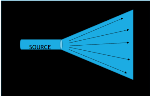
EM radiation travels how?
Straight lines (central beam) and diverge from their source in a fan beam (divergent beam). They continue to travel until they interact with matter.
Energy transfer during an x-ray
Electrical energy (x-ray machine) → EM energy (when x-rays are produced at the anode → Chemical energy (converted to electrical signal at IR)
What happens in medical imaging?
X-rays pass through patient and is captured on IR creating a latent image that is processed through chem energy to produce a 2D x-ray image.
Main X-ray interactions (3)
Compton Scatter
Photoelectric effect
Transmitted to IR
Other interactions (3)
Pair production
Coherent scattering
Photodisintegration
Explain Compton Scatter
Interaction between incident x-ray and outer shell electron, where the atom is ejected and the x-ray is redirected with less energy.
Wavelength of the scattered x-ray is longer than the incident
Negative impact for diagnostic imaging, creates image noise and reduces contrast for image
Photoelectric effect
interaction x-ray with inner shell electron, the atom is ejected and the x-ray is fully absorbed.
The x-ray does not reach IR therefore represents anatomical structures with high x-ray absorption - radiopaque
Transmitted xray
Penetrates the body and reaches the IR
Produces dark areas of x-ray with higher density than radiopaque
Anatomical structures are radiolucent, e.g. air in lungs
Primary radiation
Radiation existing in x-ray tube
Scatter radiation
Non diagnostic radiation (compton scatter)
Absorbed radiation
Radiation absorbed by the patient (photoelectric effect)
Remnant radiation
Radiation transmitted to IR
Prime exposure factors
kVp (kilovoltage peak)
mA (milliamperes)
S (variable seconds)
SID (source image distance)
Effect of kVp on resultant x-ray image (primary and secondary control)
Primary control (effect) - quality (kVp) energy/penetrability
Secondary control - Quality (KeV) - intensity of the beam
factors affected by kVp
Compton scatter interactions
Photoelectic effect (absorption) is less frequent
Affects image contrast
SID
Source image distance (from x-ray tube to IR)
mAs controls _____?
Radiographic density (blackening of image)
X-ray beam quantity (intensity)
X-ray intensity quantity) is directly proportional to mAs, explain?
2x mAs, 2x electrons at anode, 2× 2x x-ray emitted
mA and S are____ (relationship).
Inversely proportional, 2x mA and half seconds creates the same mAs (quantity)
What does quantum mottle produce, how is it created?
Image noise, grainy speckled images as a result of insufficient mAs.