A&P II- Respiratory I
1/6
There's no tags or description
Looks like no tags are added yet.
Name | Mastery | Learn | Test | Matching | Spaced | Call with Kai |
|---|
No analytics yet
Send a link to your students to track their progress
7 Terms
Respiratory System
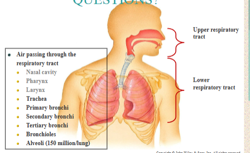
Structurally, upper and lower divisions or tracts.
upper respiratory tract nose, pharynx, larynx
lower respiratory tract larynx, trachea, bronchi and lungs
Functionally, conducting zone and the respiratory zone.
conducting zone: brings air to/from the site of gas exchange (external respiration): nose, pharynx, larynx, trachea, bronchi, bronchioles and terminal bronchioles.
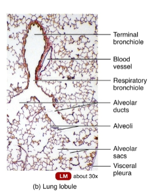
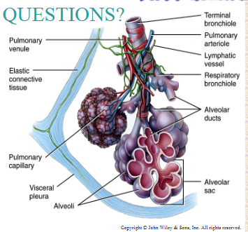
respiratory zone : main site of gas exchange : respiratory bronchioles, alveolar ducts, alveolar sacs, and alveoli
Air passing through the respiratory tract traverses the:
Nasal cavity
Pharynx
Larynx
Trachea
Primary (1o) bronchi
Secondary (2o) bronchi
Tertiary (3o) bronchi
Bronchioles
Alveoli (150 million/lung)
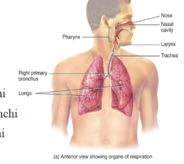
Respiratory structures
External nose is visible on the face.
Internal nose: cavity beyond the nasal vestibule. divided by nasal septum.
Function:
Filter, warm, moisten incoming air
Smell incoming air
Resonating chamber
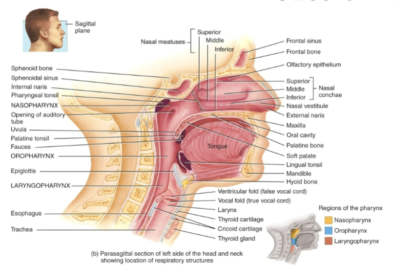
Three nasal conchae (or turbinates): protrude from each lateral wall.
Meatus under each nasal concha, for a duct that drains secretions of the sinuses and tears into the nose.
Increases turbulence mixing air for moisture and warmth
Pharynx: hollow tube starting posterior to the nasal cavity and descending to the opening of the larynx in the neck.
It functions as:
a passageway for air and food
a resonating chamber
a housing for the tonsils
3 anatomical regions:
The nasopharynx; oropharynx; and laryngopharynx
Upper Respiratory Tract
Most of the Respiratory tract is lined with ciliated pseudostratified columnar tissue
Cilia move mucous and trapped particles toward the pharynx and into the digestive tract
Larynx: composed of 9 pieces of cartilage: connects the laryngopharynx with the trachea (the “windpipe”). prevents food and water from entering the lower respiratory system
Resonating chamber
Epiglottis: flap of elastic cartilage covered with a mucus membrane, attached to the root of the tongue.
guards the entrance of the glottis, the opening between the vocal folds.
As air passes from the laryngopharynx into the larynx, it leaves the upper respiratory tract and enters the lower respiratory tract.
Lower Respiratory Tract
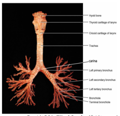
Trachea
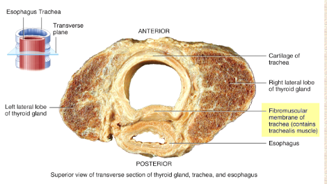
semi-rigid pipe of semi-circular cartilaginous rings, anterior to the esophagus. extends from the larynx into the mediastinum where it divides into right and left primary (1o, “mainstem”) bronchi.
It is composed of 4 layers:
a mucous secreting epithelium: mucosa
three layers of CT (submucosa, hyaline cartilage, and adventitia)
Tracheal cartilage rings are incomplete posteriorly, facing the esophagus.
Esophageal masses can press into this soft part of the trachea and make it difficult to breath, or even totally obstruct the airway
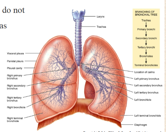
Primary (1o or “mainstem”) bronchi
emerge from the inferior trachea at the carina
go to the lungs, situated in the right and left pleural cavities
1o bronchi divide to form 2o (supply lobes) and 3o bronchi (supply segments) of each lung.
3o bronchi divide into bronchioles
branch through about 22 more divisions
The smallest are the terminal bronchioles.
As bronchi and bronchioles branch and become smaller.
mucous membrane changes from columnar to cuboidal
cartilaginous rings become more sparse, and eventually disappear altogether.
Smooth muscle increases.
Sympathetic stimulation: airway dilation
Parasympathetic stimulation: airway constriction
Lower Respiratory Tract 2
Trachea to the terminal bronchioles: conducting airways – they do not participate in gas exchange
Alveoli participate in gas exchange
Connected to respiratory bronchioles (simple cuboidal epithelium) and alveolar ducts (Simple squamous)
Pulmonary lobule.: alveoli, alveolar sacs, respiratory bronchioles, alveolar ducts and a terminal bronchiole
the functional unit of the lung
wrapped in elastic C.T.
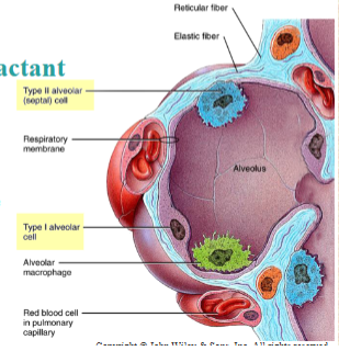
Alveoli: composed chiefly of type I alveolar cells, allowing for exchange of gases with pulmonary capillaries.
Type II cells: secrete surfactant that prevents collapse of the alveoli during exhalation.
Alveoli macrophages remove microscopic debris
The Lungs
The lungs receive blood via two sets of arteries
Pulmonary arteries carry deoxygenated blood from the right heart to the lungs for oxygenation
Bronchial arteries branch from the aorta and deliver oxygenated blood to the lungs (primarily the muscular walls of the bronchi and bronchioles)
Ventilation-perfusion coupling: matches perfusion (blood flow) of areas in the lungs to ventilation (airflow) in that area
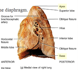
Lungs are divided into lobes by fissures.
The right lung: oblique and horizontal fissure: 3 lobes
The left lung: oblique fissure: 2 lobes
Each lobe receives it own 2o bronchus that branches into 3o segmental bronchi (which continue to further divide)
Apex of the lung: superior, extends slightly above the clavicles.
Base of the lungs: on the diaphragm.
Cardiac notch – (indentation for the heart) makes the left lung slightly smaller than the right lung.
Each lung is enclosed by a double-layered pleural membrane.
parietal pleura: line the walls of the thoracic cavity.
visceral pleura: adhere tightly to the lungs
On each side of the thorax, a pleural cavity is formed.
The integrity is crucial to the mechanism of breathing.
Pleural fluid: reduces friction.
Mechanical coupling: surface tension moves two layers together
