Pathology - GI/Urinary (Unit 1-4)
1/79
There's no tags or description
Looks like no tags are added yet.
Name | Mastery | Learn | Test | Matching | Spaced | Call with Kai |
|---|
No analytics yet
Send a link to your students to track their progress
80 Terms
Fluoro Contrast and Technique (3)
Contrast media uses higher atomic #
Higher kVp is used (110-125)
Double contrast uses 100
Short exposure times
Gallstones (Cholelithiasis)
Presence of one or more calculi (gallstones) in the gallbladder.
The majority of gallstones are radio-_____.
Lucent
What factors predispose a patient to gallstones?
Family hx
Over 40 years of age
Overweight
Female
What is the primary cause of Acute Cholecystitis?
Impacted gallstone in the cystic duct.
What are the symptoms of acute cholecystitis?
Abdominal pain
RUQ tenderness
Fever
Dysphagia
Difficulty swallowing due to a congential or acquired condition, a trapped bolus of food, paralysis of the pharyngeal or esophageal muscles, or inflammation.
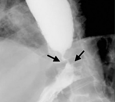
Achalasis
(aka Cardiospasm). Motor disorder of the esophagus in which peristalsis is reduced along the distal 2/3 of the esophagus.
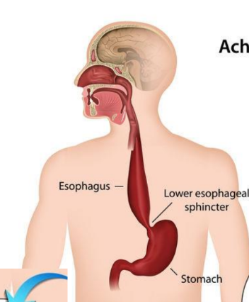
A patient with achalasia is unable to relax their _______, which causes:
Esophagogastric sphincter
Regurgitation and chest pain
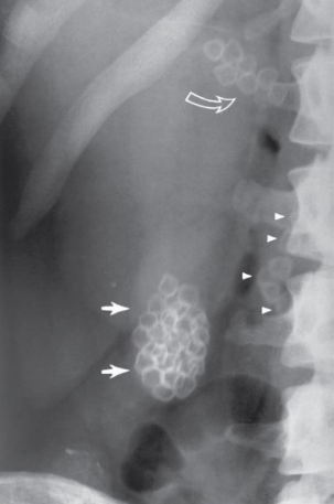
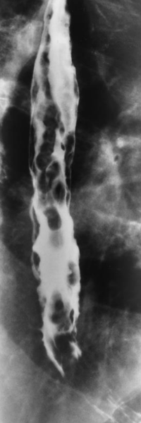
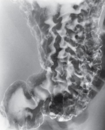
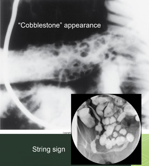
Esophageal Varices
Dilation of the veins in the wall of the distal esophagus; often asymptomatic until bleeding occurs.
What condition often accompanies esophageal varices?
Acute liver disease
What do esophageal varices look like on an esophagogram?
Cobblestone, wormlike appearance
GERD
Entry of gastric contents into the esophagus, irritating the lining of the esophagus, aka heartburn.
Valsalva Maneuver
Patient is asked to take a deep breath and while holding the breath in, to bear down.
Why is the Valsalva maneuver useful in evaluating GERD?
It distends the organs with air for more effective mucosal lining assessment.
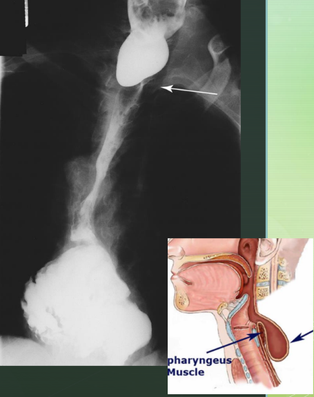
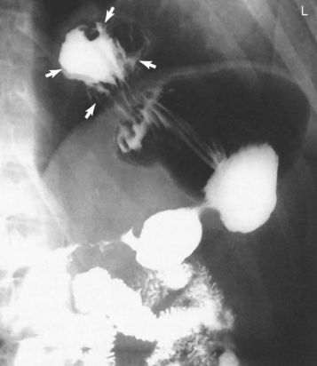
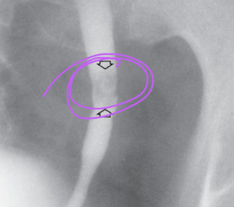
Zenker Diverticulum
Large outpouching of the esophagus just above the upper esophageal sphincter, potentially caused by weaking of the muscle wall.
Bezoar
A mass of undigested material that becomes trapped in the stomach.
Trichobezoar
Undigested hair
Gastritis
Inflammation of the stomach mucosa.
What causes gastritis?
Irritants
Alcohol
Corrosive agents
Infection
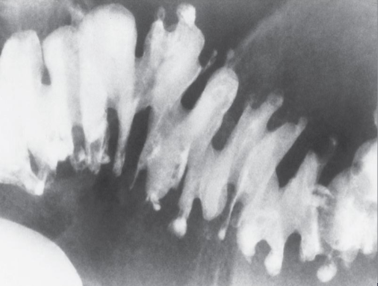
Peptic Ulcer Disease
Group of inflammatory processes involving the stomach and duodenum.
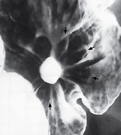
What are common causes of peptic ulcer disease?
Infection (H. pylori) and long-term use of NSAIDs
What type of ulcers can perforate when the patient has peptic ulcer disease?
Large, acute ulcers
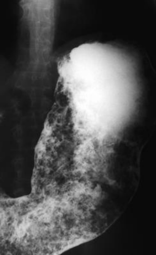
Hiatal Hernia
Portion of the stomach herniates though the diaphragmatic opening.
Commonly seen on Upper GI exams
What causes a hiatal hernia?
Congentially short esophagus or weakening of the muscle that surrounds the opening
Hiatal hernias are common in ____% of the US population over the age of ____.
50
50
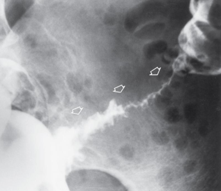
Crohn’s Disease (Regional Enteritis)
Idiopathic, chronic, inflammatory disorder involving any part of the gastrointestinal tract but commonly involving the terminal ileum.
Crohn's disease may lead to ___, ____, or the formation of an ____.
Obstruction
Fistula
Abscess
Diverticulosis
Condition of having numerous diverticula or small, outpouchings of colon.
Diverticulitis
Inflamed infected diverticula; leading to bleeding, necrosis, perforation, abscess, fistulas.
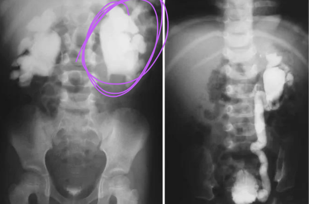
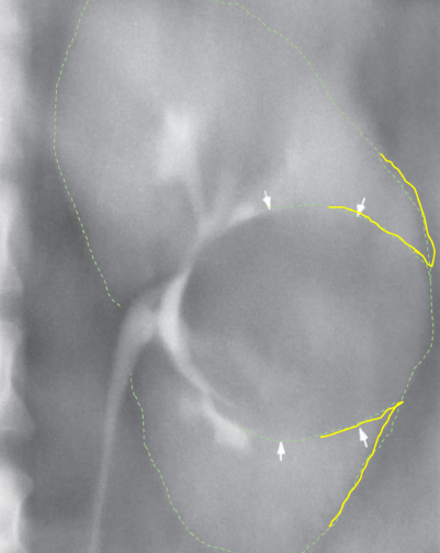
Meckel Diverticulum
Common birth defect of the ileum caused by the persistence of the yolk sac, resulting in a saclike outpouching of the intestinal wall.
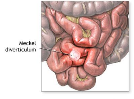
Surgical removal of a Meckel diverticulum is often recommended to prevent which conditions from developing?
Diverticulitis
Obstruction
Blood loss
What is the best modality to diagnose a Meckel diverticulum?
Nuc med
Malabsorption Syndrome
Conditions in which the gastrointestinal tract is unable to process and absorb certain nutrients.
Sprue
Inability to absorb certain proteins and dietary fats.
Celiac Disease
Sprue that affects the small bowel; gluten intolerance.
What does malabsorption syndrome look like on a SBS?
Mucosa may appear thickened as a result of constant irritation.
Ulcerative Colitis
Chronic idiopathic inflammatory disease of the colon.

UC primarily affects which age group?
Young adults
UC most frequently involves the _____ region of the colon.
Rectosigmoid
What conditions can UC cause?
Toxic megacolon or perforation
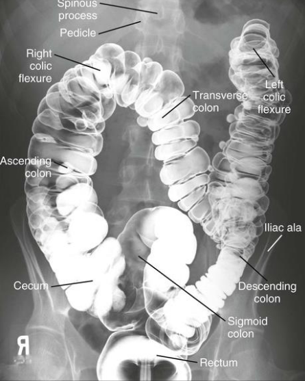
Where do most carcinomas of the large intestine (colon) occur?
Rectum and sigmoid colon
What are some predisposing conditions of cancer in the colon?
Ulcerative colitis
Hereditary polyposis
Peristalsis
Smooth muscle contractions that move the contents of the digestive system along the GI tract
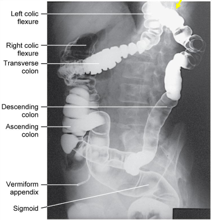
What position is the patient in? Air = black. Barium = white
Supine
What position is the patient in? Air = black. Barium = white
Prone
What position is the patient in? Air = black. Barium = white
Erect
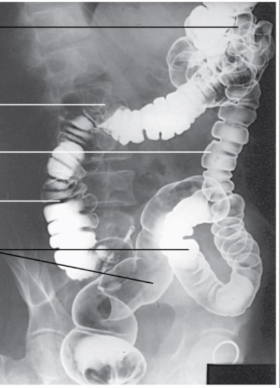
Identify the Position
RAO (40-70 degrees)
Identify the Position
Prone
Identify the Position
LPO (40-70 degrees)
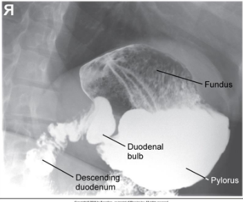
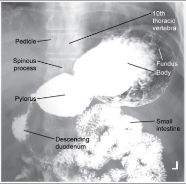
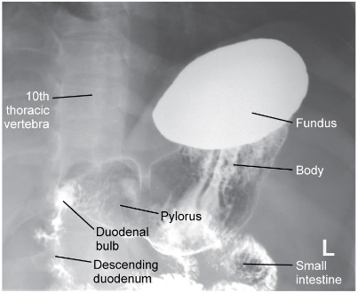
Small Bowel Series Timing and CRs
First 15-30 minutes view - CR is 2” above iliac crest to include the stomach since contrast has just entered the body
60+ minute radiographs - CR is at the iliac crest to include symphysis because the contrast has gone down further into the anatomy
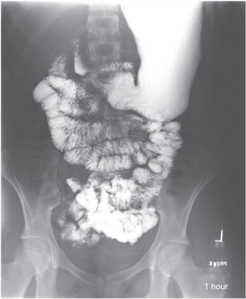
Small Bowel at 1 Hour: Is this Image Good?
This is at an hour, so the stomach does not need to be included, but the symphysis does (which it is). It is a little off-centered in terms of right and left, but it does not warrant a repeat since no barium filled anatomy was clipped.
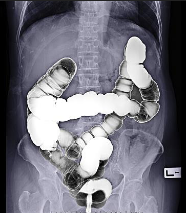
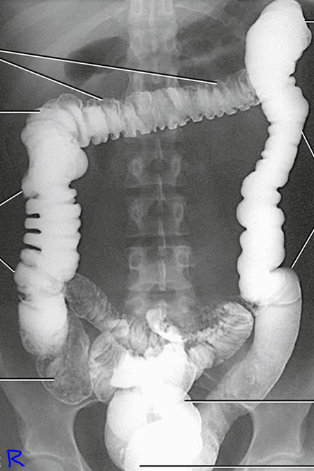
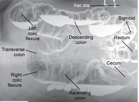
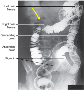
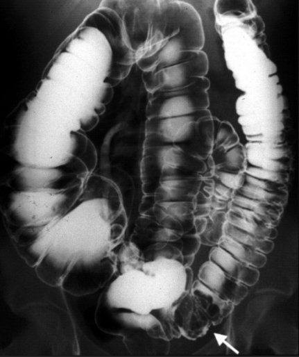
Large Intestine: Is this Image Good?
Proper positioning with the anatomy. CR is at the crests
What position is the patient in? Air = black. Barium = white
Supine
Air fills the anterior (stomach) portions while barium fills the posterior (back) portions.
What position is the patient in? Air = black. Barium = white
Prone
Air fills the posterior (back) portions while barium fills the anterior (stomach) portion
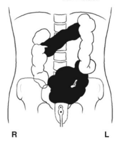
Barium Enema: Identify the Position
Prone (PA)
Barium Enema: Identify the Position
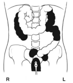
Supine (AP)
Barium Enema: Identify the Position
Right Lateral Decub
Barium in lateral ascending and medial descending
Barium Enema: Identify the Position
Left Lateral Decub
Barium in medial ascending and lateral descending
Barium Enema: Identify the Position
RAO or LPO
Barium Enema: Identify the Position
LAO or RPO
AP Barium Enema: Is this Image Good?
Overexposed (too dark)
Fix by lowering mAs
Clipped the Splenic Flexure and Transverse Colon
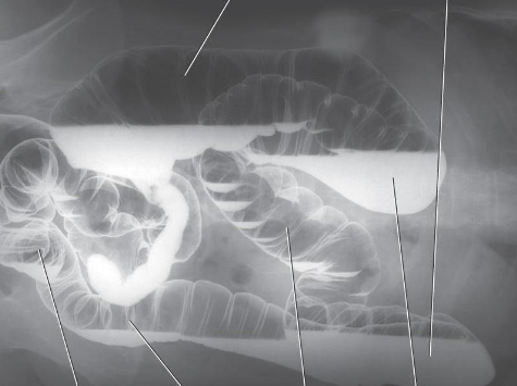
AP BE: Is this Image Good?
Hepatic Flexure is open and spine is off-centered
Spinous Processes are not aligned with the midline of the vertebral bodies
Patient is rotated LPO
Bring right side back toward IR
Urinary Calculi (kidney stones)
Formed in the kidneys
Asymptomatic (no pain) until they lodge in the ureter and cause partial obstruction (extreme pain)
Hydronephrosis
Distention of the renal pelvis and calyces of the kidneys that results from some obstruction of the ureters or renal pelvis.
What causes hydronephrosis?
Calculi
Tumors
Structural abnormalities
Hydronephrosis can occur along with ____.
Hydroureter
Renal Cyst
Most common unifocal mass of the kidney; fluid-filled.
“beak sign” on radiograph
Polycystic Kidney Disease
Hereditary disorder marked by multiple cysts of varying size scattered throughout one or both kidneys
“Bunch of grapes” scattered throughout the kidney
Acute Renal Failure
Rapid deterioration in kidney function
Results in accumulation of nitrogen-containing wastes
Caused by impaired blood flow, infection to kidneys, ureteral obstruction, Tylenol overdose
Malrotation
Abnormal position of the kidney in relationship to the psoas muscle or longitudinal or horizontal axis
Ectopic Kidney
Abnormal position, such as in the pelvis (pelvic kidney), or high near the diaphragm (intrathoracic kidney)
Horseshoe Kidney
Congenital fusion of the kidneys during development of the fetus, usually at the lower poles
Usually does not affect function
Duplication
Involves two ureters and/or the renal pelvis originating from the same kidney
Most common type of congenital anomaly of the urinary system
Cystitis
Inflammation of the urinary bladder caused by a bacterial or fungal infection
Common in females due to shorter urethra
Urinary frequency, urgency, burning sensation
Glomerulonephritis
Inflammation of the capillary loops of the glomeruli of the kidneys; sonography and nuc med
Causes oliguria
Acute - enlarged, darkened kidney
Chronic - small kidney size caused by fibrosis and cortex destruction from long-standing inflammation
Pyelonephritis
Inflammation of the kidney and renal pelvis caused by pyogenic bacteria
Primary affects the interstitial tissue between the nephron tubules
Renal Carcinoma
Most frequent type of malignant tumor of the kidney
3x more frequent in males
Carcinoma of the Bladder
Tumor usually is diagnosed after the age of 50 years, usually men
Vesicorectal Fistula
Abnormal communication between the bladder and rectum/aspects of colon