Chapter 25
1/79
There's no tags or description
Looks like no tags are added yet.
Name | Mastery | Learn | Test | Matching | Spaced | Call with Kai |
|---|
No analytics yet
Send a link to your students to track their progress
80 Terms
Function of Kidneys
The kidneys regulate water volume, solute concentration, ion concentrations, acid-base balance, excrete wastes, and produce erythropoietin and renin. As well as Activating vitamin D and carrying out gluconeogenesis.
Urinary System Components which involve the kidney
The urinary system includes ureters (transport), urinary bladder (storage), and urethra (transport) for urine transport and storage.
Location & External Anatomy of Kidneys
Located retroperitoneal and the right kidney will be crowded by the liver so it will be lower than the left. There are three layers of supportive tissue, Renal Fascia, Perirenal fat capsule and fibrous capsule
Renal Fascia (supportive tissue)
Anchoring outer layer of dense fibrous connective tissue

Perirenal fat capsule (supportive tissue)
Fatty cushion
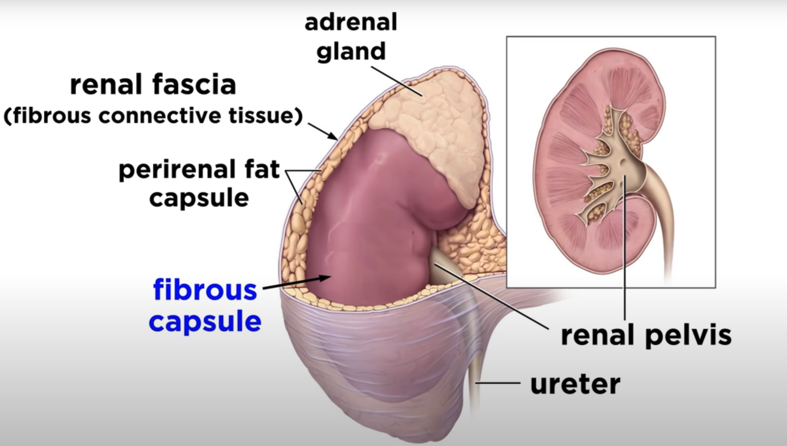
Fibrous capsule (supportive tissue)
Transparent capsule that prevents spread of infection to kidney
Gross Anatomy of Kidneys
The kidneys have a renal cortex, renal medulla with pyramids, renal pelvis, and calyces for urine collection and flow.
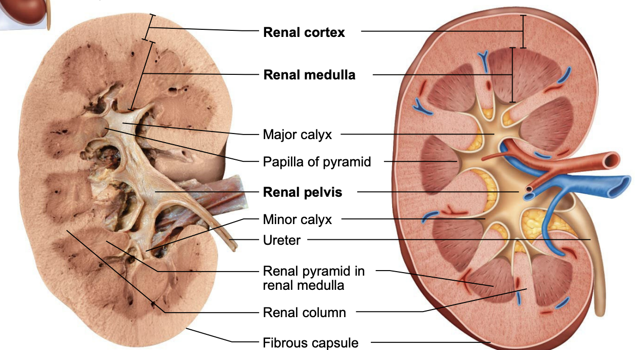
Minor calyces
Cup-shaped areas that collect urine draining from pyramidal papillae
Major Calyces
Areas that collect urine from minor calyces and Empty urine into renal pelvis
Blood Supply
Kidneys cleanse blood and adjust its composition, so it has a rich blood supply. Renal arteries deliver about one-fourth (1200 ml) of cardiac output to kidneys each minute
Arterial Flow
Renal → segmental → interlobar → arcuate → cortical radiate (interlobular)
Venous Flow
Cortical radiate → arcuate → interlobar → renal veins
No segmental veins
Nephrons
These are the functional units of the kidney, consisting of renal corpuscle and renal tubule for urine formation.

Renal Corpuscle
Two parts of renal corpuscle, the glomerulus and the glomerular capsule

Glomerulus
Tuft of capillaries composed of fenestrated endothelium
Highly porous capillaries
Allows for efficient filtrate formation
Filtrate: plasma-derived fluid that renal tubules process to form urine

Glomerular Capsule
Also called Bowman’s capsule: cup-shaped, hollow structure surrounding glomerulus. Has two layers parietal layer and visceral layer.
Parietal layer
Simple squamous epithelium
Visceral Layer
Clings to glomerular capillaries; branching epithelial podocytes
Renal Tubule (Description)
This is about 3 cm (1.2 in.) long
Consists of single layer of epithelial cells, but each region has its own unique histology and function
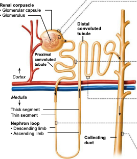
Renal Tubule (Three Major part)
Proximal Convoluted Tubule
Distal Convoluted Tubule
Nephron Loop

Proximal Convoluted Tubule
Closest to renal corpuscle
Cuboidal cells with dense microvilli that form brush border. Increase surface area and also have large mitochondria.
Functions in reabsorption and secretion
65% of Na+ and water reabsorbed, many ions, uric acids, half of urea
Confined to cortex
Nephron loop (Henle)
U-shaped structure consisting of both the descending and ascending limb.
Thin segment is passive to Na+ movement
Thick segment has Na+-K+-2Cl– symporters and Na+-H+ antiporters that transport Na+ into cell
Distal convoluted tubule and collecting duct (hormones)
Reabsorption is hormonally regulated in these areas
Antidiuretic Hormone
Aldosterone
Atrial natriuretic peptide
Parathyroid hormone
Antidiuretic Hormone (ADH)
Released by posterior pituitary gland and an increased ADH levels cause an increase in water reabsorption
Aldosterone
Targets collecting ducts (principal cells) and distal DCT and will cause little Na+ to leave the body. Without aldosterone, daily loss of filtered Na+ would be 2%, which is incompatible with life. Functions to increase blood pressure and decrease K+ levels.
Atrial natriuretic peptide
Reduces blood Na+, resulting in decreased blood volume and blood pressure. Released by cardiac atrial cells if blood volume or pressure elevated
Parathyroid hormone
Acts on DCT to increase Ca2+ reabsorption
Descending Limb
Proximal part of descending limb is continuous with proximal tubule. Distal portion also called descending thin limb; simple squamous epithelium. H2O cannot leave while solutes can leave.
Ascending Limb
Thick ascending limb, thin in some nephrons. H2O can leave however solutes can not.

Distal Convoluted Tubule (DCT)
Cuboidal cells with very few microvilli. Function more in secretion than reabsorption. This is also confined to cortex.
Collecting ducts
Two cell types principal cells and intercalated cells. These receive filtrate from many nephrons, Run through medullary pyramids, Give pyramids their striped appearance. The ducts fuse together to deliver urine through papillae into minor calyces.
Principal Cells (Collect duct)
Sparse with short microvilli
Maintain water and Na+ balance
Intercalated Cells (Collecting ducts)
Cuboidal cells with abundant microvilli
Two types of intercalated cells
A and B: both help maintain acid-base balance of blood
Classes of Nephrons
Two major groups of nephrons, cortical nephrons and juxtamedullary nephrons
Cortical Nephrons
Make up 85% of nephrons
Almost entirely in cortex
Juxtamedullary Nephrons
Long nephron loops deeply invade medulla
Ascending limbs have thick and thin segments
Important in production of concentrated urine
Nephron Capillary Beds
Renal tubules are associated with two capillary beds, the glomerulus and capillaries. Juxtamedullary nephrons are associated with the vasa recta
Glomerulus Capillaries
These are specialized for filtration. Different from other capillary beds because they are fed and drained by arteriole
Glomerulus (Afferent Arteriole)
Enters glomerulus and leaves via efferent arteriole
Arises from cortical radiate arteries

Glomerulus (Efferent Arteriole)
Feeds into either peritubular capillaries or vasa recta
Blood pressure is higher in the Glomerulus due to..
Afferent arterioles are larger in diameter than efferent arterioles
Arterioles are high-resistance vessels

Peritubular Capillaries
Low-pressure, porous capillaries adapted for absorption of water and solutes. Arise from efferent arterioles and cling to adjacent renal tubules in cortex. Empty into venules.

Vasa Recta
Long, thin-walled vessels parallel to long nephron loops of juxtamedullary nephrons. Arise from efferent arterioles serving juxtamedullary nephrons. Function in formation of concentrated urine.
Juxtaglomerular Complex (JGC)
Each nephron has one juxtaglomerular complex. Involves modified portions of:
Distal portion of ascending limb of nephron loop
Afferent (sometimes efferent) arteriole
Important in regulating rate of filtrate formation and blood pressure

3 cell populations are seen in JGC
Macula Densa
Granular cells
Extraglomerular mesangial Cells
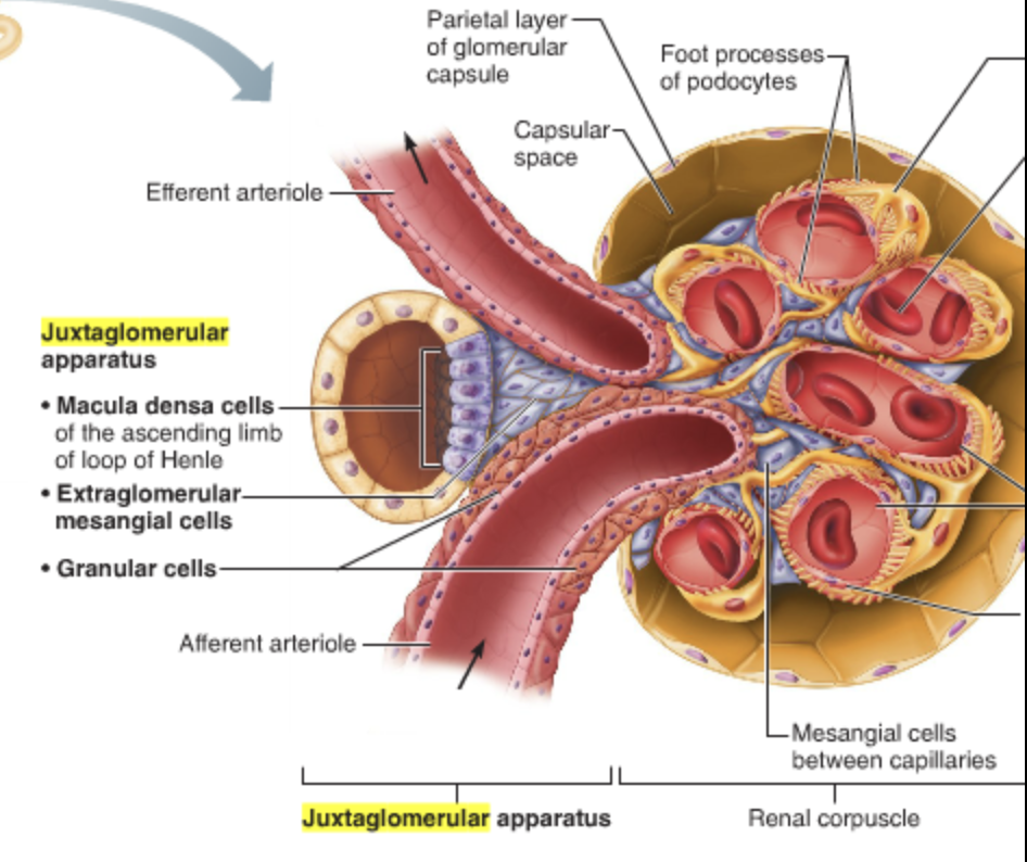
Macula Densa (cells in JGC)
Tall, closely packed cells of ascending limb, contain chemoreceptors that sense NaCl content of filtrate

Granular Cells/ Juxtaglomerular cells (JGC cells)
Enlarged, smooth muscle cells of arteriole. Act as mechanoreceptors to sense blood pressure in afferent arteriole. Contain secretory granules that contain enzyme renin.

Extraglomerular mesangial cells
Located between arteriole and tubule cells and interconnected with gap junctions. May pass signals between macula densa and granular cells
Physiology of Kidney
180 L of fluid processed daily, but only 1.5 L of urine is formed
Kidneys filter body’s entire plasma volume 60 times each day
Consume 20–25% of oxygen used by body at rest
Filtrate (produced by glomerular filtration) is basically blood plasma minus proteins
Urine is produced from filtrate
Urine
<1% of original filtrate
Contains metabolic wastes and unneeded substances

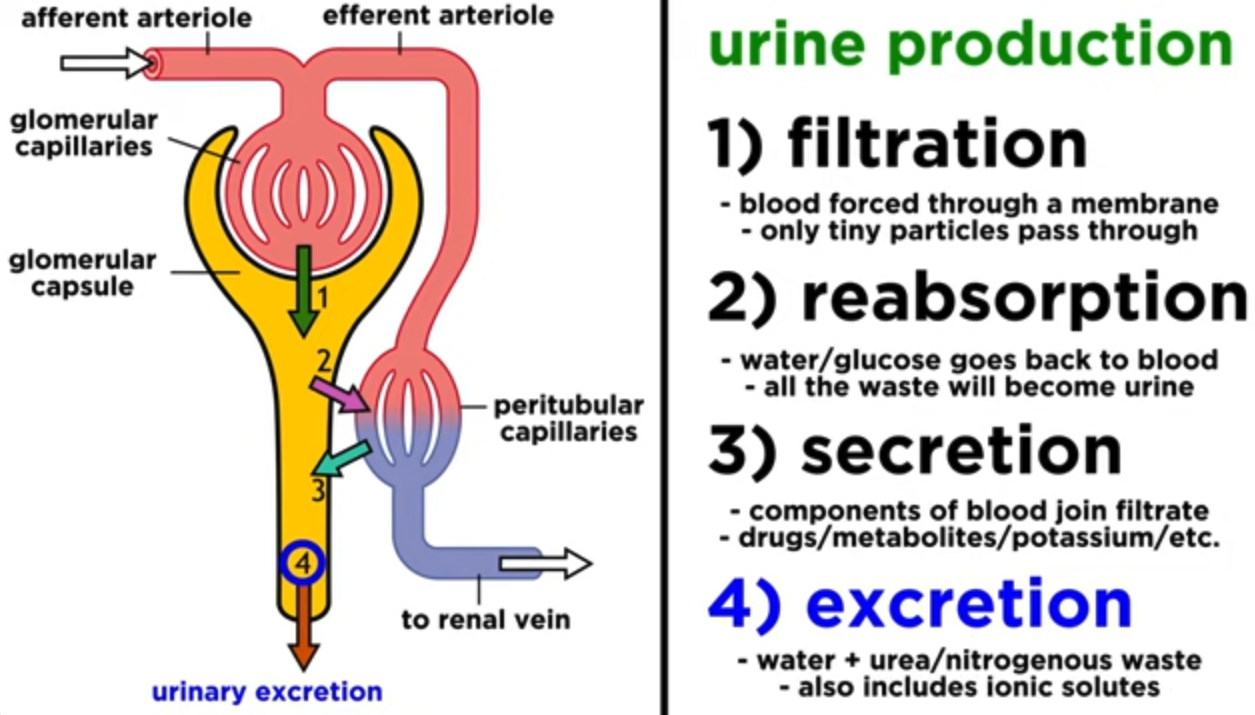
Glomerular Filtration
The glomerulus filters blood plasma to form filtrate in the Bowman's capsule through hydrostatic pressure.
Glomerular Filtration Rate (GFR)
This is the volume of filtrate formed per minute by both kidneys (normal = 120–125 ml/min)
GFR (Proportional to)
Net filtration pressure (NFP): Primary pressure is glomerular hydrostatic pressure
Total surface area available for filtration: Glomerular mesangial cells control by contracting
Filtration membrane permeability: Much more permeable than other capillaries
Regulation of Glomerular Filtration
Constant GFR is important as it allows kidneys to make filtrate and maintain extracellular homeostasis
Goal of local intrinsic controls (renal autoregulation): maintain GFR in kidney
GFR affects systemic blood pressure
Increased GFR causes increased urine output, which lowers blood pressure, and vice versa
Goal of extrinsic controls: maintain systemic blood pressure
Extrinsic controls
These are both neural and hormonal mechanisms. The purpose of extrinsic controls is to regulate GFR to maintain systemic blood pressure. These will override renal intrinsic controls if blood volume needs to be increased.
Renin-angiotensin-aldosterone mechanism
Main mechanism for increasing blood pressure
Three pathways to renin release by granular cells
Direct stimulation of granular cells by sympathetic nervous system
Stimulation by activated macula densa cells when filtrate NaCl concentration is low
Reduced stretch of granular cells
Tubular Reabsorption
Reabsorption process in renal tubules returns nutrients and ions to the blood, regulated by hormones like ADH and aldosterone.
Transcellular Route
Solute enters apical membrane of tubule cells. Travels through cytosol of tubule cells. Exits basolateral membrane of tubule cells. Enters blood through endothelium of peritubular capillaries
Paracellular Route
Between tubule cells. Limited by tight junctions, but leaky in proximal nephron. Water, Ca2+, Mg2+, K+, and some Na+ in the PCT move via this route
Sodium concentrate effects…
the water that is input into the cells
Tubular Secretion
Tubular secretion moves substances from blood to filtrate, including K+, H+, and creatinine, to adjust blood composition.
There are two types of countercurrent mechanisms
Countercurrent multiplier & countercurrent exchanger
Countercurrent multiplier
Interaction of filtrate flow in ascending/descending limbs of nephron loops of juxtamedullary nephrons.
Countercurrent exchanger
Blood flow in ascending/descending limbs of vasa recta.
Gradient in the Kidney
Runs from 300 mOsm in cortex to 1200 mOsm at the bottom of the medulla, allowing more water movement deeper into the kidneys.
Urine’s chemical composition
Consists of 95% water, nitrogenous wastes (urea, uric acid, creatinine), and normal solutes (Na+, K+, PO43–, SO42–, Ca2+, Mg2+, HCO3–).
Urine’s physical characteristics
Includes color (pale to deep yellow), transparency (should be clear), and odor (slightly aromatic when fresh, may develop ammonia odor upon standing).
pH of Urine
Slightly acidic (~pH 6, range of 4.5 to 8.0), influenced by diet and health conditions.
Specific gravity
Ratio of mass of substance to mass of equal volume of water, ranging from 1.001 to 1.035 due to water and solutes in urine.
Ureters
Slender tubes conveying urine from kidneys to bladder, preventing backflow as bladder pressure increases.
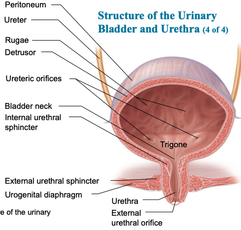
Urinary bladder anatomy
Muscular sac for temporary urine storage, located retroperitoneally, with specific structures in males and females.
Urethra anatomy
Includes internal urethral sphincter (involuntary) and external urethral sphincter (voluntary), with differences in male and female urethras.
Female urethra (3-4cm)
Tightly bound to anterior vaginal wall
External urethral orifice: anterior to vaginal opening; posterior to clitoris
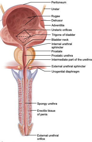
Male Urethra (three regions)
Prostatic urethra (2.5 cm): within prostate
Intermediate part of the urethra (membranous urethra) (2 cm): passes through urogenital diaphragm from prostate to beginning of penis
Spongy urethra (15 cm): passes through penis; opens via external urethral orifice
Trigone
Smooth triangular area outlined by openings for ureters and urethra. Infections tend to persist in this region
Internal urethral sphincter
Involuntary (smooth muscle) at bladder-urethra junction and contracts to open
External urethral sphincter
Voluntary (skeletal) muscle surrounding urethra as it passes through pelvic floor
Micturition
Also known as urination or voiding, the process of expelling urine from the bladder.