Muscular System - Final
1/80
There's no tags or description
Looks like no tags are added yet.
Name | Mastery | Learn | Test | Matching | Spaced | Call with Kai |
|---|
No analytics yet
Send a link to your students to track their progress
81 Terms
Fibrous Joint
Bones that are united by fibrous tissue
Ex: Sutures of the skull
Cartilaginous Joint
Bones that are connected by cartilage
Ex: Pubic symphysis
Synovial Joint
Articulating bones have a space (synovial cavity) between them.
Articulating Bone
The two(or more) bones that meet to form the joint
Articulating cartilage
The smooth, white tissue covering the ends of the ones to reduce friction.
Synovial Membrane
The inner lining of the capsule that secretes the “grease” for the joint
Synovial Fluid
The egg white like liquid that fills the cavity and lubricates movement
Articular capsule
The “envelope” surrounding the joint, made of an outer fibrous layer and the layer and the inner membrane
Collateral Ligaments
Strong bands of connective tissue located outside the capsule that prevent side to side instability
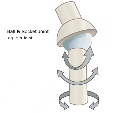
Ball and Socket joint
Ball-like surface of one bone fits who the cuplike depression of another bone
Ex: hip joints
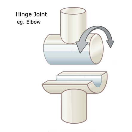
Hinge joint
Permits flexion and extension only
Ex: elbow
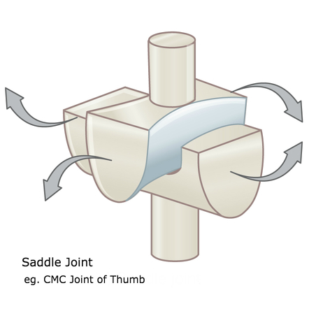
Saddle joint
The articular surface of one bone is saddle shaped and the articular surface of the other bone fits into
Ex: Thumb joint
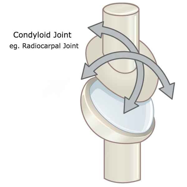
Condyloid joint
Reduced ball and socket joint
Ex: metacarpals
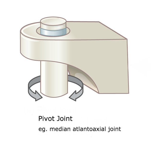
Pivot joint
Allows for rotation
Ex: between atlas and axis
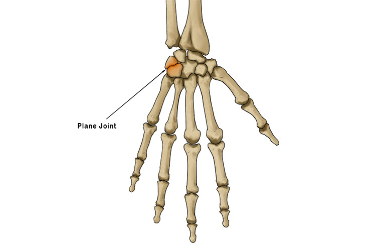
Planar joint
Bones move on a flat or slightly curved surface
Ex: Tarsals
Extension
Increase the angle at the joint
Ex: straightening the knee
Flexion
Decreases the angle at a joint
Ex: Bending the knee of elbow
Dorsiflexion
Standing on heels of foot
Plantarflexion
Standing on toes
Adduction
bringing the limbs toward the midline
Abduction
bringing the limbs away from the midline
Circumduction
Turning the arms in a circle
Rotation
Movement around the axis
Ex: Shaking head no
Supination
Movement so that the plans face up like in anatomical position
Pronation
Moving the palms so they face now. The radius crosses the ulna
Inversion
Turning sole in toward midline
Eversion
Turning sole outward away from midline
What are the functions of the muscular system (Get Some Muscles Please!)
Generated heat
stabilizes joints
Maintains posture
Produces movement (with skeletal system)
Is skeletal muscle straited or nonstraited, voluntary or nonvolunatry, Multinucleated or uninucleated, and location
It is straited, volunatary, multinucleated, and attached to bone
Is Smooth muscle straited or nonstraited, voluntary or involunatry, Multinucleated or uninucleated, and location
It is unstraited, involuntary, uninucleated, and lines the internal organs
Is cardiac muscle straited or nonstraited, voluntary or involunatry, Multinucleated or uninucleated, and location
It is straited, involuntary, uninucleated, and the heart
What are intercalated discs and what muscle tissue type are they found in?
They join the branching cells in cardiac muscle so the muscle acts as one unit. They are found on the heart
Origin
Point of attachment of muscle on immovable or less moveable bone
Insertion
Point of attachment of muscle on the more moveable bone
Prime mover
Muscles that provide the primary force for movement
Synergist
Muscles that help the prime movers by producing the same movement
Antagonist
Muscles that oppose a movement
Fixator
Muscles that stabilize the origin of the prime movers
Location
Relative to a bone region
Ex: temporalis
Action
Movement that muscle performs
Ex: Adductors
Direction
In which muscle fibers run
Ex: External oblique
Shape
Of the muscle
Ex: Trapezius
Number of origins
How many origins
Ex: biceps brachii
Origin & insertion
Location where muscles attaches
Ex: Sternocleidomastoid
Relative size
Of the muscle
Ex: Gluetus Maximus
Used in smiling
Zygomaticus
Used to suck in you cheeks
Buccinator
used in winking
Orbicularis oculi
Used to form the horizontal frown crease on the forehead
Frontalis
The “kissing” muscle
Orbicularis oris
Prime mover of jaw closure
masseter
Synergist muscle for jaw closure
Temporalis
Prime mover of head flexion: two-headed muscle
Sternocleidomastoid
Epimysium - Connective tissue layer
Covers entire muscle
Perimysium - ct layer
Surrounds fascicle
Endomysium - ct layer
Wraps individual muscle fibers
Fascicle
Bundle of muscle
Sarcolemma
Plasma membrane surrounding each muscle fiber
Sarcoplasm
Cytoplasm of the muscle cell
Myofibril
Inside muscle cells and is composed of myofilaments
Myofilament
Actin and myosin
Sarcomere
Contractile unit of muscle
Sliding Filament theory
Myosin heads pull on the actin causing the thin filaments to slide toward the center of the sarcomere. The filaments don’t change in length, the sarcomere shortens as the filaments overlap.
What happens to the I band and H zone
THe I band gets smaller and the H zone disappears
Motor unit
A neuron and all of the muscle cells it stimulates
What happens at the NMJ
Nerve impulses cause release of neurotransmitter Acj which is a chemical message sent across the synaptic cleft to the sarcolemma. This triggers a chain reaction, leading to muscle contraction.
How does Botox work
It blocks the release of neurotransmitter ach and temporarily paralyzes the muscle
The characteristic of muscle tissue that describes its ability to receive and respond to a stimulus is
excitability
Which muscles extends the forearm
Triceps Brachii
The “tailor muscle” that adducts the thigh/hip (crosses the legs")
sartorius
What is the muscle that is the prime mover for jaw closure
masseter
What muscle retracts and elevated the mandible and helps to close the jaw. It is the synergist to the masseter
Temporalis
What characteristic of muscle tissue allows it to return to its original size/shape after stretching
Elasticity
The ability of a muscle call to shorten in length
contractility
The ability of a muscle cells to stretch
Extensibility
T thick band of dense ct that forms most connections between muscle and bone
tendon
myofibrils
tiny fibers that run parallel along the entire length of the muscle cell
What does botulism poisoning do
it causes muscle paralysis because it blocks the release of ach from the axon terminal of the neuron so the muscle never gets the message to contract.
Skeletal muscle fiber contraction order
Ach is release from axon terminal
Action potential travels into T (Transverse) tubules
Sarcoplasmic reticulum releases calcium
Calcium combines with troponin
Energized myosin heads attach to active
What is the process of skeletal muscle fiber contraction order
Due to a nerve impulse, ACH is released from the axon terminal to the synaptic cleft to the sarcolema. From there, it binds and then we have action potential that travels down the transverse tubules. Then the sarcoplasmic reticulum relseases calcium that then binds with troponin so the troponin/tropomyosin complex binding sites on actin are exposed with the help of split ATP the myosin heads are now able to attach to actin creating a cross bridge and allowing for the powerstroke. The heads are able to attach because troponin/tripomyosin are not blocking the sites becayse troponin binded with calcium. For the heads to detach more ATP is needed and the myosin will recharge until the next contraction. The powerstroke is the myosin heads pulling actin filaments inward toward the sarcomere’s center (M-line).