Biology AS level
 Call Kai
 Learn
Learn Practice Test
 Spaced Repetition
 Match
Match Flashcards
 Knowt Play
1/32

Earn XP
Description and Tags
Name | Mastery | Learn | Test | Matching | Spaced | Call with Kai | Chat |
|---|
No analytics yet
Send a link to your students to track their progress
33 Terms
1
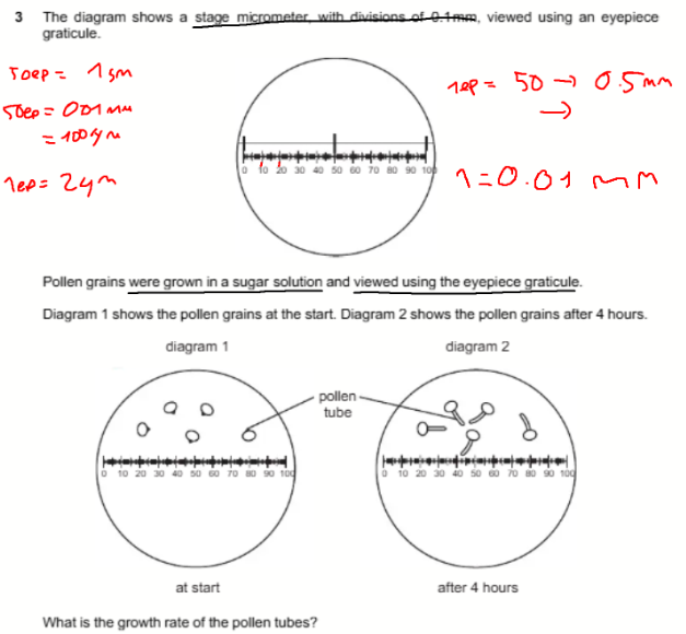
Q: What is the growth rate of the pollen tubes?
A: 5 μm h⁻¹
B: 10 μm h⁻¹
C: 5 mm h⁻¹
D: 10 mm h⁻¹
Answer: A — 5 μm h⁻¹
Explanation:
50 eyepiece divisions = 0.10 mm = 100 μm. Therefore, one eyepiece division = 2 μm. The tube grows 10 divisions:
10 × 2 = 20 μm
20 μm ÷ 4 hours = 5 μm h⁻¹.
Q: Which functions are carried out by the Golgi body?
Lysosome production
Polypeptide modification
Exocytosis
A: 1, 2 and 3
B: 1 and 2 only
C: 1 and 3 only
D: 2 and 3 only
Answer: B — 1 and 2 only
Explanation: The Golgi modifies polypeptides and produces vesicles that form lysosomes. Exocytosis occurs when a vesicle fuses with the cell-surface membrane, so the Golgi does not perform the fusion itself.
3
Q: Which cell structures contain ribosomal RNA?
Chloroplasts
Mitochondria
Nuclei
A: 1, 2 and 3
B: 1 and 2 only
C: 2 and 3 only
D: 3 only
Answer: A — 1, 2 and 3
Explanation: Chloroplasts and mitochondria contain their own ribosomes, which contain rRNA. The nucleus contains the nucleolus, where rRNA is produced and combined with proteins to form ribosomal subunits.
Q: Which cell structures contain nucleic acids?
Chloroplasts
Golgi bodies
Lysosomes
Ribosomes
A: 1, 2 and 3
B: 1, 2 and 4
C: 1 and 4 only
D: 2, 3 and 4
Answer: C — 1 and 4 only
Explanation: Chloroplasts contain DNA and RNA. Ribosomes contain rRNA. Golgi bodies and lysosomes do not contain nucleic acids.
5
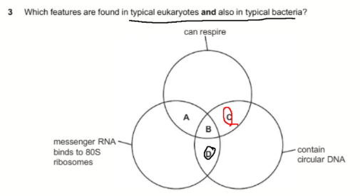
Q: Which region represents features found in both typical eukaryotes and typical bacteria?
The features are:
Can respire
Messenger RNA binds to 80S ribosomes
Contain circular DNA
A: Region A
B: Region B
C: Region C
D: Region D
Answer: C — Region C
Explanation: Both can respire and contain circular DNA. Eukaryotic cells contain circular DNA in mitochondria, while bacteria contain circular DNA in their main chromosome and plasmids. However, bacterial mRNA binds to 70S, not 80S, ribosomes.
6
Q: How many types of double-membrane structures found in animal cells are also found in plant cells?
A: 1
B: 2
C: 3
D: 4
Answer: B — 2
Explanation: The nucleus and mitochondria have double membranes and occur in both animal and plant cells. Chloroplasts also have a double membrane, but they are not found in animal cells.
7
Q: Which steps are needed to find the actual width of a xylem vessel using a ×10 objective lens?
Convert from mm to μm by multiplying by 10⁻³.
Calibrate the eyepiece graticule using a stage micrometer with a ×4 objective lens.
Measure the vessel’s width using an eyepiece graticule.
Multiply the number of eyepiece units by the calibration value.
A: 1, 2, 3 and 4
B: 1 and 2 only
C: 2, 3 and 4 only
D: 3 and 4 only
Answer: D — 3 and 4 only
Explanation: Measure the vessel in eyepiece units and multiply by the calibration value. Statement 1 is wrong because mm → μm requires multiplying by 10³. Statement 2 is wrong because calibration must use the same ×10 objective lens.
8
Q: Which statement about centrioles, cilia, microtubules and microvilli is correct?
A: Centrioles, cilia and microvilli are composed of microtubules.
B: Centrioles have roles in mitosis and semi-conservative DNA replication.
C: Cilia and microvilli both increase surface area for absorption.
D: Microtubules are made of protein and form the spindle in mitosis.
Answer: D — Microtubules are made of protein and form the spindle in mitosis.
Explanation: Microtubules are made of tubulin protein and form spindle fibres. Microvilli contain actin, not microtubules. Centrioles organise the spindle but do not replicate DNA. Microvilli increase surface area, while cilia usually move substances across a cell’s surface.
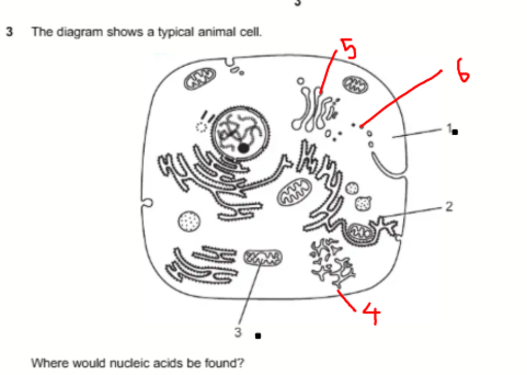
Q: Where would nucleic acids be found in the labelled animal cell?
A: 1, 2 and 3
B: 1 and 2 only
C: 1 and 3 only
D: 2 and 3 only
Answer: A — 1, 2 and 3
Explanation: Structure 1 is cytoplasm, which contains RNA. Structure 2 includes ribosomes containing rRNA. Structure 3 is a mitochondrion, not a chloroplast, and contains circular DNA and RNA.
Q: Which row correctly shows structures in typical plant and animal cells?
A: Plasmodesmata—present in both
B: Golgi body—present in plants only
C: Centriole—absent in plants; present in animals
D: Tonoplast—absent in both
Answer: C — Centriole absent in typical plant cells but present in typical animal cells
Explanation: Typical animal cells have centrioles, while typical plant cells do not. Plasmodesmata and tonoplasts are found in plants, and Golgi bodies occur in both cell types.
Q: What supports the fact that mature plant cells contain organelles performing the same role as lysosomes?
A: Mature plant vacuoles contain various hydrolytic enzymes.
B: Glycogen inside vesicles can be hydrolysed into glucose.
C: Double-membrane vesicles form from plant Golgi bodies.
D: Vesicles formed from the cell-surface membrane contain enzymes.
Answer: A — Mature plant vacuoles contain various hydrolytic enzymes
Explanation: Lysosomes use hydrolytic enzymes to digest substances. Mature plant vacuoles can contain similar enzymes, allowing them to perform a lysosome-like role.
Q: Which features identify a Golgi body in a transmission electron micrograph?
A: A stack of flattened, smooth, single-membrane cisternae without ribosomes
B: A double membrane with inner folds called cristae
C: Flattened membranes covered with ribosomes
D: Circular DNA surrounded by 70S ribosomes
Answer: A — A stack of flattened, smooth, single-membrane cisternae without ribosomes
Explanation: The Golgi consists of stacks of flattened membrane-bound sacs called cisternae. Its membranes are smooth because they have no attached ribosomes.
Q: Red light has a longer wavelength than green light. What happens when red light is used?
A: Magnification decreases; resolution remains the same
B: Magnification increases; resolution increases
C: Magnification remains the same; resolution decreases
D: Magnification remains the same; resolution increases
Answer: C — Magnification remains the same; resolution decreases
Explanation: Magnification is determined by the lenses, not wavelength. A longer wavelength produces poorer resolution, so red light gives lower resolution than green light.
Q: What is the function of the nucleolus?
A: Formation and breakdown of the nuclear envelope
B: Formation of rough endoplasmic reticulum
C: Synthesis of ribosomal proteins
D: Synthesis of rRNA
Answer: D — Synthesis of rRNA
Explanation: The nucleolus synthesises rRNA and assembles ribosomal subunits. Ribosomal proteins are produced by ribosomes in the cytoplasm and then transported into the nucleus.
Q: Which cell structures do not contain cristae?
Endoplasmic reticulum
Golgi body
Mitochondrion
Chloroplast
A: 1, 2, 3 and 4
B: 1, 2 and 4 only
C: 1 and 2 only
D: 3 and 4 only
Answer: B — 1, 2 and 4 only
Explanation: Cristae are folds of the inner mitochondrial membrane, so only mitochondria contain them. Chloroplasts contain thylakoids and grana instead.
1
Q: What is the best estimate for the diameter of the alveolus?
A: 0.960 mm
B: 3.84 mm
C: 240 μm
D: 384 μm
Answer: C — 240 μm
Explanation:
At ×10: 10 eyepiece units = 0.10 mm = 100 μm.
Therefore, 1 eyepiece unit = 10 μm.
At ×40, magnification is four times greater: 10 ÷ 4 = 2.5 μm per unit.
Diameter = 96 × 2.5 = 240 μm.
2
Q: What are functions of microtubules?
Allowing movement of cilia in a bronchus
Attachment to centromeres during metaphase
Moving secretory vesicles around a cell
A: 1, 2 and 3
B: 1 and 2 only
C: 1 and 3 only
D: 2 and 3 only
Answer: A — 1, 2 and 3
Explanation: Microtubules form cilia, form spindle fibres that attach to centromeres and provide tracks along which secretory vesicles are transported.
3
Q: Tay–Sachs disease causes lipids to accumulate inside cells. Which structure does not function correctly?
A: Golgi body
B: Lysosome
C: Mitochondrion
D: Smooth endoplasmic reticulum
Answer: B — Lysosome
Explanation: Lysosomes contain hydrolytic enzymes that digest substances, including lipids. In Tay–Sachs disease, a lysosomal enzyme is deficient, so particular lipids cannot be broken down and accumulate.
4
Q: Which sequence correctly orders stages in the production and secretion of an enzyme?
A: Golgi body → ribosome → rough endoplasmic reticulum → mRNA
B: mRNA → smooth endoplasmic reticulum → Golgi body → vesicle
C: Ribosome → rough endoplasmic reticulum → vesicle → Golgi body
D: Smooth endoplasmic reticulum → mRNA → vesicle → ribosome
Answer: C — Ribosome → rough endoplasmic reticulum → vesicle → Golgi body
Explanation: The enzyme is synthesised by a ribosome on the rough ER, enters the rough ER and is carried in a transport vesicle to the Golgi body for modification and packaging.
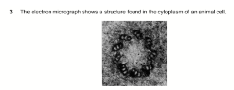
Q: The electron micrograph shows a circular structure containing nine groups of microtubules. What is it?
A: Centriole
B: Lysosome
C: Ribosome
D: Vesicle
Answer: A — Centriole
Explanation: A centriole contains nine microtubule triplets arranged in a ring. That means nine groups of three microtubules—not only nine individual microtubules.
6
Q: One hundred eyepiece-graticule units correspond to 0.1 mm. How should the value of one unit in μm be calculated?
A: Divide 100 by 0.1, then multiply by 1000
B: Divide 100 by 0.1, multiply by 1000, then divide by 100
C: Multiply 0.1 by 1000, then divide by 100
D: Multiply 0.1 by 1000, then divide twice by 100
Answer: C — Multiply 0.1 by 1000, then divide by 100
Q: Which statements about a stage micrometer scale are correct?
It can measure the actual length of cells directly.
It can calibrate the eyepiece graticule.
Less of the scale is visible when changing from ×10 to ×40.
A: 1, 2 and 3
B: 2 and 3 only
C: 1 only
D: 2 only
Answer: B — 2 and 3 only
Explanation: A stage micrometer calibrates the eyepiece graticule; it does not measure cells directly. Increasing magnification from ×10 to ×40 reduces the field of view, so less of the scale is visible.
Q: Thiomargarita namibiensis is 700 μm wide, contains a large vacuole and has circular DNA free in its cytoplasm. Which statement is correct?
A: It must be a eukaryote because 700 μm is too large for a prokaryote.
B: It must be a plant because it contains a vacuole.
C: It must be a plant because it has a cell wall.
D: It must be a prokaryote because its DNA is circular and located in the cytoplasm.
Answer: D — It must be a prokaryote
Explanation: Prokaryotes have circular DNA free in the cytoplasm because they lack a nucleus. Size, vacuoles and cell walls do not prove that an organism is a plant.
3
Q: A scale bar represents 2 μm. Which method calculates the magnification of the photograph?
A: Divide the cell diameter by the measured scale-bar length.
B: Measure the cell diameter in mm, multiply by 2000 and divide by the scale-bar length.
C: Measure the scale bar in mm, convert it to μm and divide by 2.
D: Measure the scale bar in mm, convert it to μm and multiply by 2.
Answer: C — Measure the scale bar, convert to μm and divide by 2
Explanation: Magnification = image size ÷ actual size. The measured scale-bar length is the image size, while 2 μm is its actual size.
Example:
Measured scale bar = 20 mm = 20 000 μm
Magnification = 20 000 ÷ 2 = ×10 000
4
Q: Which organelles are clearly visible using a light microscope at ×400?
A: Ribosomes and endoplasmic reticulum
B: Ribosomes and centrioles
C: Endoplasmic reticulum, centrioles and chloroplasts
D: Chloroplasts only
Answer: D — Chloroplasts only
Explanation: Chloroplasts are several micrometres in size and can be seen clearly. Ribosomes, endoplasmic reticulum and centrioles are too small to be seen clearly with a light microscope.

Q: Radioactively labelled amino acids are introduced into a secretory cell. Where will the radioactivity first become concentrated?
A: Structure A
B: Structure B
C: Structure C
D: Structure D
Answer: C — Structure C, the rough endoplasmic reticulum
Explanation: Amino acids are joined to form polypeptides by ribosomes attached to the rough ER. The labelled protein later travels to the Golgi body and secretory vesicles.
6
Q: What identifies a cell as a prokaryote?
A: Its DNA is associated with protein.
B: Its DNA is circular.
C: Its DNA forms a double helix.
D: Its DNA is surrounded by a membrane system.
Answer: B — Its DNA is circular
Explanation: A typical prokaryote has circular DNA free in its cytoplasm. Both prokaryotic and eukaryotic DNA form double helices, while prokaryotes lack a membrane-bound nucleus.
7
Q: What are the appropriate units for these structures?
A: Alveolus: mm; white blood cell: μm; cell wall: μm
B: Alveolus: μm; white blood cell: mm; cell wall: μm
C: Alveolus: μm; white blood cell: μm; cell wall: nm
D: Alveolus: mm; white blood cell: mm; cell wall: nm
Answer: C — μm, μm and nm
Explanation:
Alveolus diameter: hundreds of μm
White blood cell diameter: approximately 10–20 μm
Cell-wall thickness: appropriately measured in nm
Q: Where are digestive enzymes made in the illustrated secretory cell?
A: Structure A
B: Structure B
C: Structure C
D: Structure D
Answer: C — Structure C, the rough endoplasmic reticulum
Explanation: Digestive enzymes are proteins. They are synthesised by ribosomes attached to the rough ER before being transported to the Golgi body for modification and packaging.

1
Q: Which cell structure can be seen only with an electron microscope?
A: Cell-surface membrane
B: Cell wall
C: Chromosome
D: Nucleolus
Answer: A — Cell-surface membrane
Explanation: The cell-surface membrane is approximately 7–10 nm thick, which is below the resolution of a light microscope. Cell walls, condensed chromosomes and nucleoli can be seen using a light microscope.
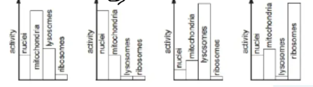
Q: Mammalian tissue is homogenised and separated into fractions containing nuclei, mitochondria, lysosomes and ribosomes. Which diagram shows the fraction with maximum mRNA synthesis?
A: Diagram A
B: Diagram B
C: Diagram C
D: Diagram D
Answer: A — Diagram A
Explanation: Most mRNA is synthesised by transcription of DNA inside the nucleus. Therefore, the fraction with the highest nuclear activity has the greatest mRNA synthesis. Ribosomes use mRNA for translation but do not synthesise it.
3
Q: Mitochondria may have evolved from prokaryotic cells. Which feature was lost during their evolution into mitochondria?
A: Cell wall
B: Circular chromosome
C: Endoplasmic reticulum
D: Ribosomes
Answer: A — Cell wall
Explanation: Mitochondria retain circular DNA and 70S ribosomes from their prokaryotic ancestors. The ancestral cell wall was lost. Prokaryotes never possessed endoplasmic reticulum, so it could not have been lost.
4
Q: A 2 cm scale line on a photomicrograph represents 5 μm. What is the magnification?
A: 1 × 10³
B: 2 × 10³
C: 4 × 10³
D: 5 × 10³
Answer: C — 4 × 10³
Explanation:
2 cm = 20 mm = 20 000 μm
Magnification = image size ÷ actual size
20 000 μm ÷ 5 μm = 4000 = 4 × 10³