distal limb
1/10
There's no tags or description
Looks like no tags are added yet.
Name | Mastery | Learn | Test | Matching | Spaced | Call with Kai |
|---|
No analytics yet
Send a link to your students to track their progress
11 Terms
What are the ddx of an aggressive digit lesion
primary neoplasia → soft tissue neoplasia (SCC)
Secondary neoplasia → metastasis to digits from primary lung neoplasia (mostly seen in cats)
Osteomyelitis due to bacterial infection
How do the ddx of aggressive digit lesions differ to an aggressive bone lesion elsewhere in the body?
Aggressive bone lesions are typically osteosarcomas elsewhere in the body OR osteomyelitis due to fungal
in aggressive bone lesions of the digits, we see soft tissue neoplasia (SCC), secondary digit lesions due to metastasis of primary lung neoplasia, or osteomyelitis due to bacterial infection
Explain lung digit syndrome in cats (pathogenesis, clinical signs, diagnostics, px)
secondary neoplasia from a primary lung carcinoma → metastasis occurs → aggressive digit lesion forms
Clinical signs → digit lesion noticed first
Diagnostics → thorax rads to look for primary neoplasm
Poor px → no amputation as other lesions quickly occur in other remaining digits
What is involved in the work up of an aggressive digit lesion?
FNA or amputate & histo (cannot determine the cause from radiographs)
Thorax radiographs
ID metastasis; do not do sx until thorax rads are taken
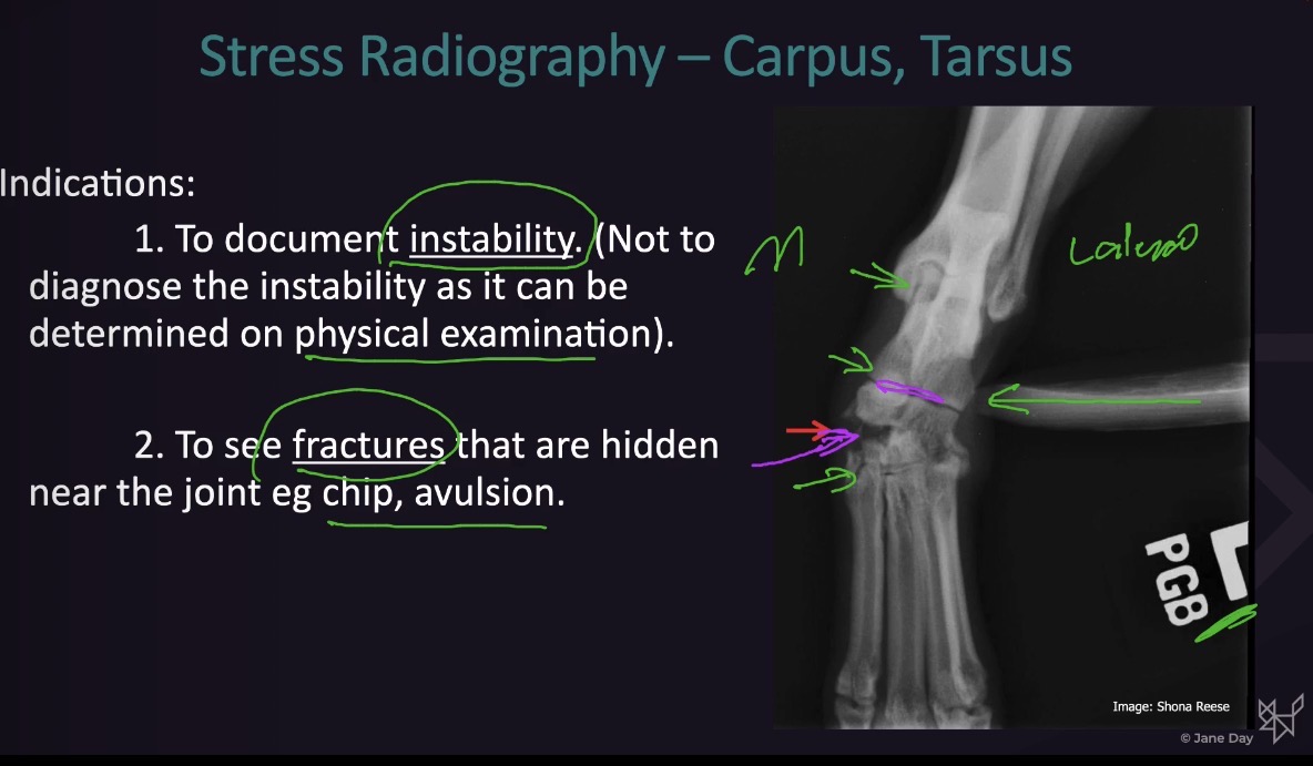
What are the indications for stress radiography?
Document instability via radiographs
See fractures that are hidden near the joint (chip, avulsion)
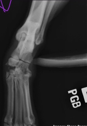
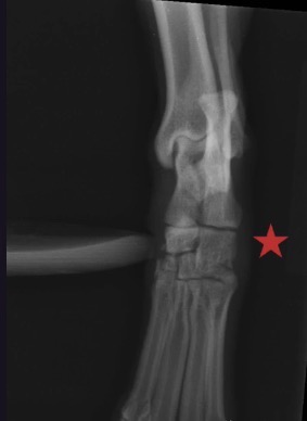
What view is this, what is being seen, what aspect of the joint is seen?
stress radiography on DP view
Opens up medial side of joint, medial intertarsal joint indicates trauma on medial side
Fracture fragments can be seen that are not seen when the joint is not opened up
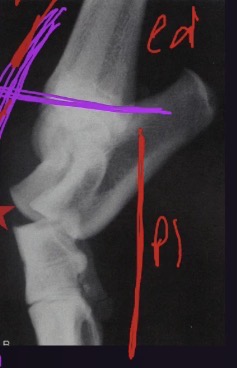
What view is this? What part of the joint can be seen?
stress radiography lateral view of tarsus
Stress applied to open up dorsal aspect of tarsus
Proximal intertarsal joint can be seen, it is unstable
What view is this? When stress is applied, what joint is opened up? Is this unstable or stable?
lateral view of tarsus
Stress applied to open up plantar aspect of tarsus, identifying that the proximal intertarsal joint is unstable
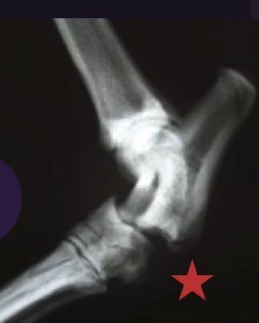
What view is this? When stress is applied, which joint opens up? Is this joint stable or unstable?
DP view of tarsus
Stress applied to open up lateral side of the joint → distal intertarsal joint visible
No instability seen on this side of the joint
Explain how stress radiography can be performed without holding
use sandbags or tape when doing radiographs → pull one piece of tape cranially or caudally depending on what joint you want to open up
What are common causes of aggressive bone lesions? Are they more common in dogs or cats?
common in dogs, uncommon in cats
Most common cause
Soft tissue neoplasia (SCC) - 2/3 of cases
Osteomyelitis from nailbed infection → 1/3 of cases (bacterial)