Midterms Histo — Catastrophe
1/4
There's no tags or description
Looks like no tags are added yet.
Name | Mastery | Learn | Test | Matching | Spaced | Call with Kai |
|---|
No analytics yet
Send a link to your students to track their progress
5 Terms
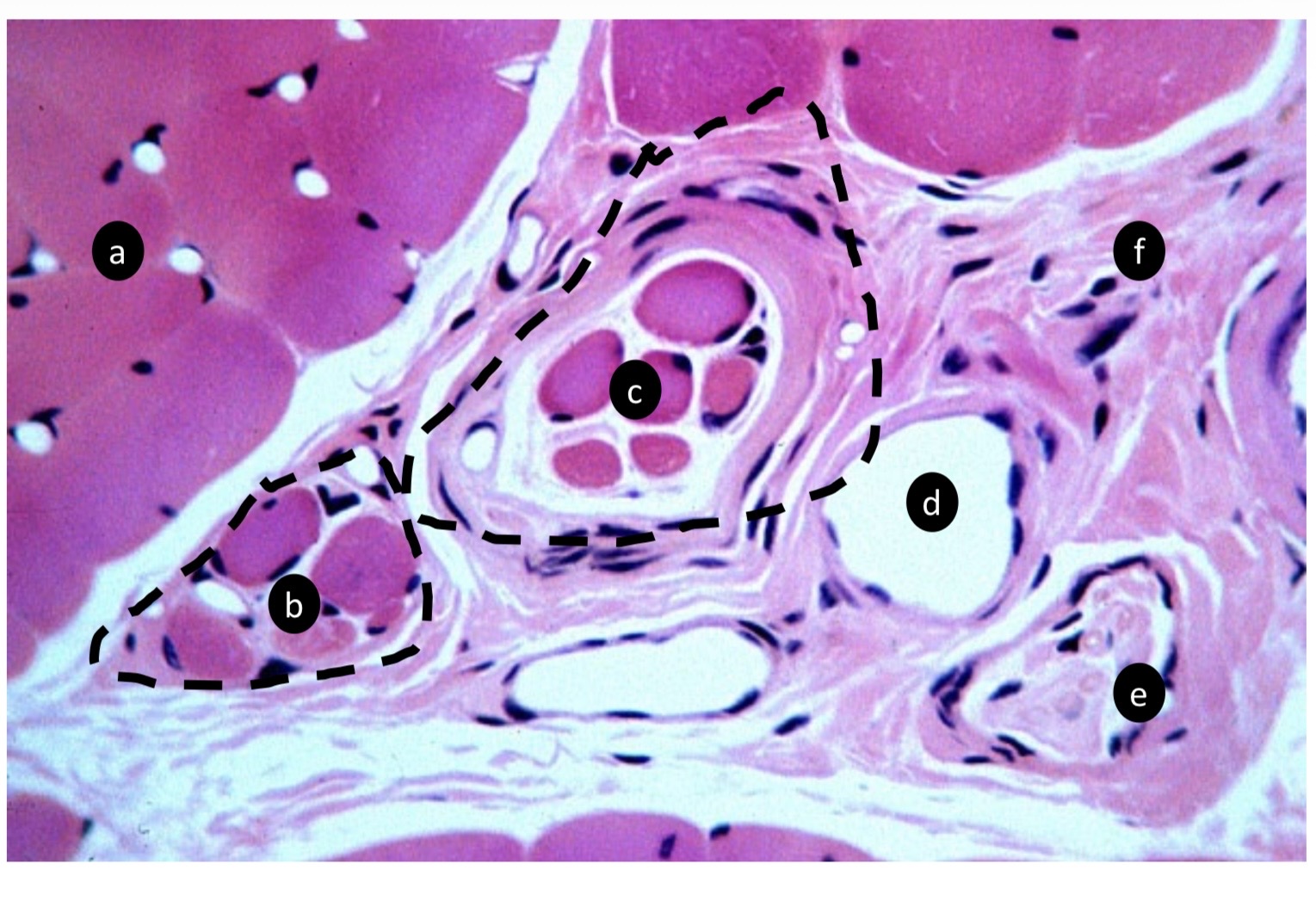
Label the image.
A. ID the specific tissue type
B. ID the specific tissue type
C. ID the specific tissue type
D. What is the function of the structure in ( c ) above?
E. ID the specific structure
F. ID the specific structure
G. ID the type of tissue
A. Extrafusal myofibers (skeletal muscle)
B. Intrafusal muscle fiber
C. Intrafusal muscle fiber (muscle spindle)
D. Stretch receptor (proprioception-detect length and velocity)
E. Vein
F. Nerve fibers
G. CT mainly collagen
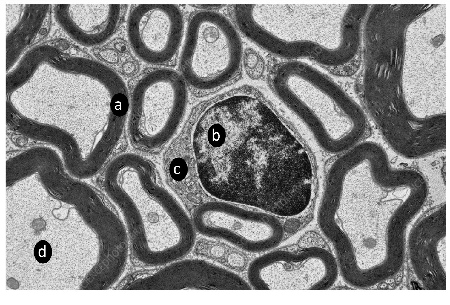
ID the labeled structures: A-C
D. Name two structures present at the labeled region.
A. Myelin Sheath
B. Schwann cell (nucleus)
C. Schwann cell cytoplasm
D. Microtubule, neurofilament, mitochondria
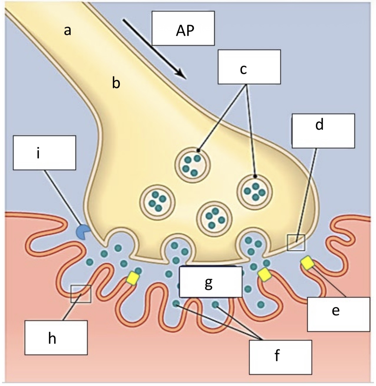
A. ID the labeled structure
B. Name the ions which stimulate action potential
C. ID the structure
D. ID the structure
E. ID the structure
F. ID the structure
G. ID the space
H. ID the labeled structure
I. ID the labeled compound
A. Axon
B. Calcium
C. Secretory vesicles
D. Axon terminal
E. Acetylcholine receptors (chemically gated ion channels)
F. Acetylcholine
G. Synaptic cleft
H. Sarcolemma
I. Acetylcholinesterase
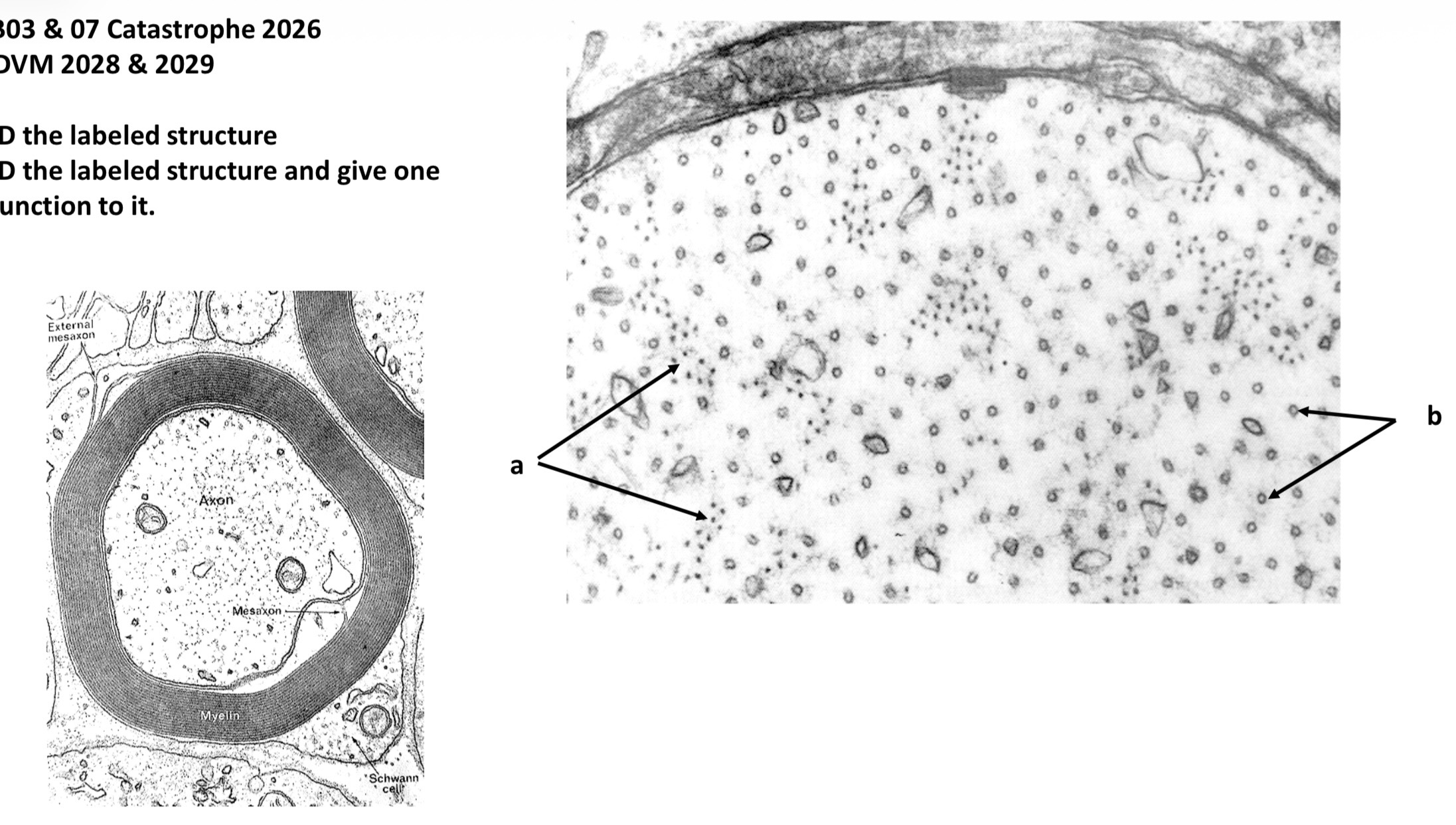
A. ID the labeled structure
B. ID the labeled structure and give one function to it
A. Neurofilament (microfilament)
B. Neurotubule (microtubule)
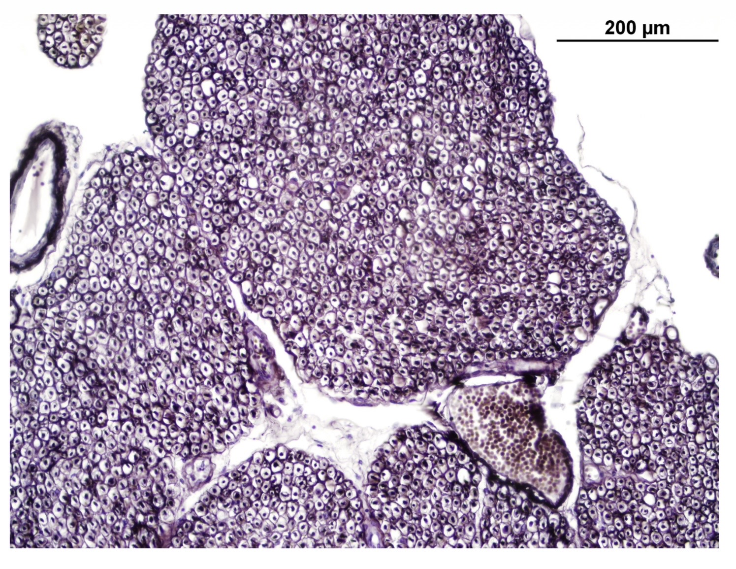
Describe in details the image.
Starts by type of tissue followed by the structures within it and the CT around and in between the structures.
Normal bundles of nerve fibers with blood vessels (artery bundles is surrounded by thin CT (epineurium) and CT between fascicles (perineurium)). The spaces within the individual axons are myelin sheath as it is removed during processing of the tissue. Schwann cells are not clear at this magnification.