NHA CCMA: EKG and other electrocardio tests
1/61
There's no tags or description
Looks like no tags are added yet.
Name | Mastery | Learn | Test | Matching | Spaced | Call with Kai |
|---|
No analytics yet
Send a link to your students to track their progress
62 Terms
Bipolar leads
Leads I, II, and III
Unipolar leads
AVL, AVR, AVF (must be augmented)
Lead I records
Right arm to left arm
Lead III records
Left leg to left arm
Lead II records
Right arm to left leg
aVL lead
left leg and right arm assist with the left arm tracing
aVR lead
left arm and left leg assist with the right arm
aVF lead
right arm and left arm assist with left leg tracing
Universal leads
RA, LA, LL, RL
precordial leads
V1-V6
V1 lead placement
right side of sternum, 4th intercostal space
V2 lead placement
Left side of sternum, 4th intercostal space, directly across from V1
V4 lead placement
Left side of the chest left, 5th intercostal space, midclavicular line
V3 lead placement
Left side of the chest, between V2 and V4
V5 lead placement
Left side of the chest, 5th intercostal space, anterior axillary line
V6 lead placement
Left side of the chest, 5th intercostal space, midaxillary line
P wave represents
atrial depolarization (atrial contraction)
QRS wave represents
ventricular depolarization
T wave represents
ventricular repolarization or relaxation
U wave represents
represents repolarization
P-R interval
Time it takes for the beginning of atrial depolarization to the beginning of ventricular depolarization
Q-T interval
Time from the beginning ventricular depolarization to ventricular repolarization
S-T segment
time from the end of the ventricular depolarization to the beginning of ventricular repolarization
Sinus Bradycardia
less than 60 bpm
Sinus Tachycardia
greater than 100 bpm
Sinus Arrest
SA node doesn't fire
Atrial Flutter
The atria is beating at an extremely rapid rate
Atrial Fibrillation
No organized contractions of the atrium
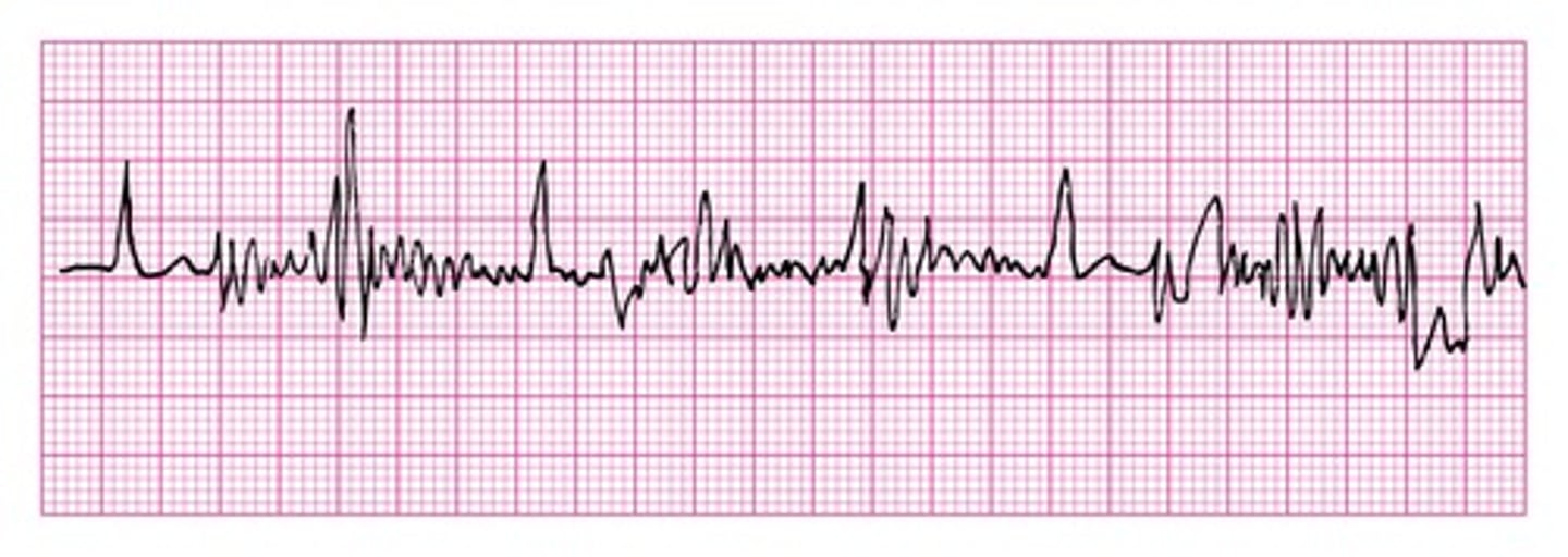
Ventricular Fibrillation
Ventricles don't contract but quiver, no waves noted
Somatic Tremor (artifact )
Irregular spike throughout the tracing, Muscle movement (shivering, Parkinson's disease)
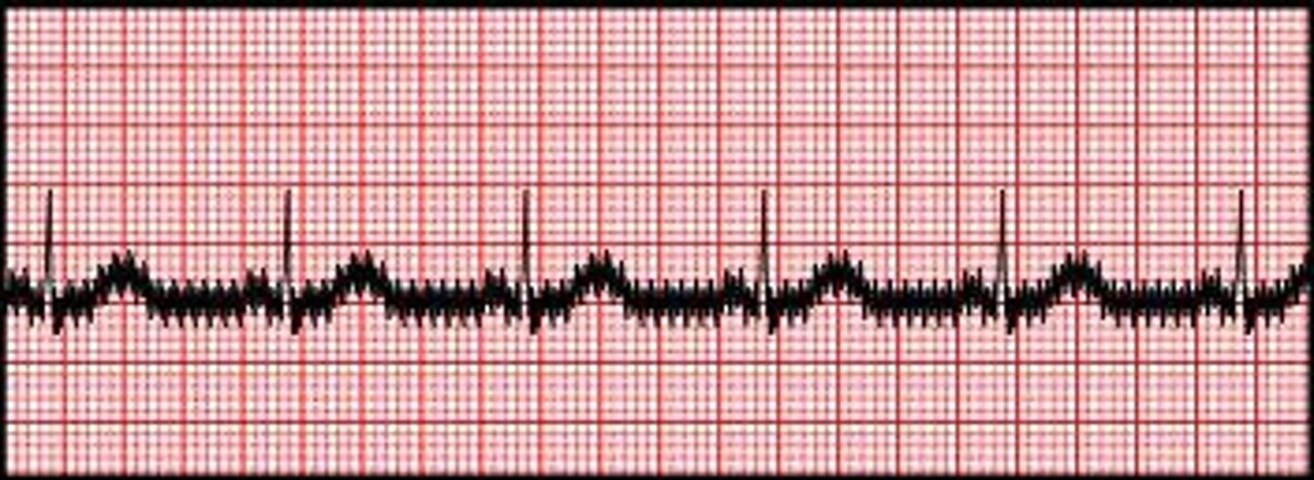
AC interference (artifact)
regular spikes, poor grounding, electrical interference (crossed wires)
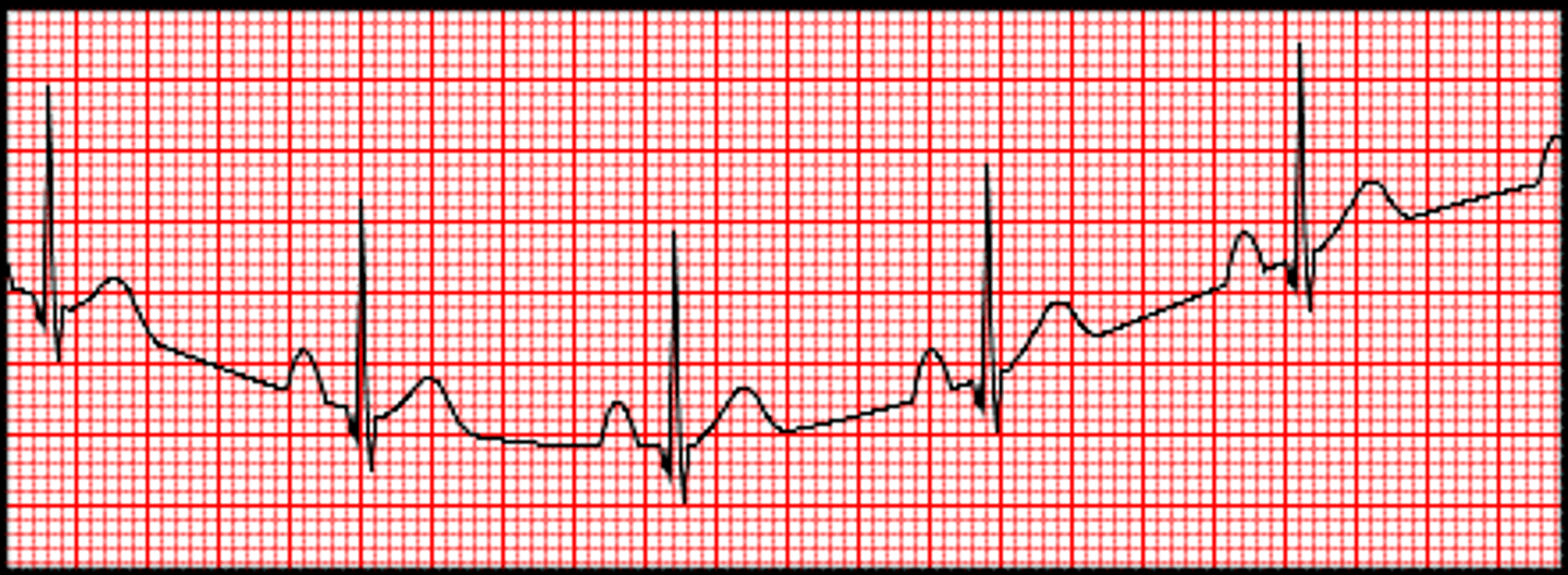
Wandering baseline (artifact)
move up and down rather than be straight, may be caused by: loose electrode; body creams, oils or lotions
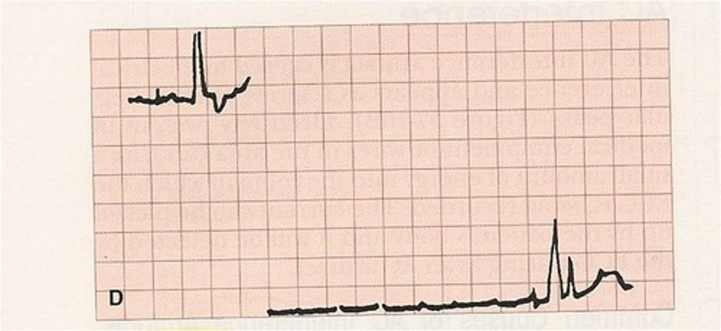
Interrupted baseline (artifact)
Break in tracing, may be caused by the metal tip of a lead wire becoming detached or by a broken pt. cable
EKG ratio
10:12
Widened QRS complex
PVC's
Polarization
Resting state of the myocardial wall, resulting in flatline or pause in EKG
25mm/sec
The normal running speed of the EKG paper
Heart rate calculation
divide 1500 by the number of small boxes between two R waves
asystole
absence of contractions of the heart
premature ventricular contraction
abnormal waves
P wave negative deflection
normally P waves should be positive deflected, a junctional dysrhythmia is likely present
LA
black lead
RA
white lead
LL
red lead
RL
green lead
v1
red
v2
yellow
v3
green
v4
blue
v5
orange
v6
purple
Holter monitor
a portable electrocardiograph that is worn by an ambulatory patient to continuously monitor the heart rates and rhythms over a 24-hour period
electrodes
sticky skin sensors that attach to the patient
vertical x-axis
amplitude
horizontal x-axis
time
how many seconds is each horizontal square
0.04 seconds
Paper speed
25 mm/sec
first 6 recorded leads
originate in the arms and legs
somatic tremor
AC interference
.
wandering baseline
.
interrupted baseline
.