breast pathology
1/28
There's no tags or description
Looks like no tags are added yet.
Name | Mastery | Learn | Test | Matching | Spaced | Call with Kai |
|---|
No analytics yet
Send a link to your students to track their progress
29 Terms
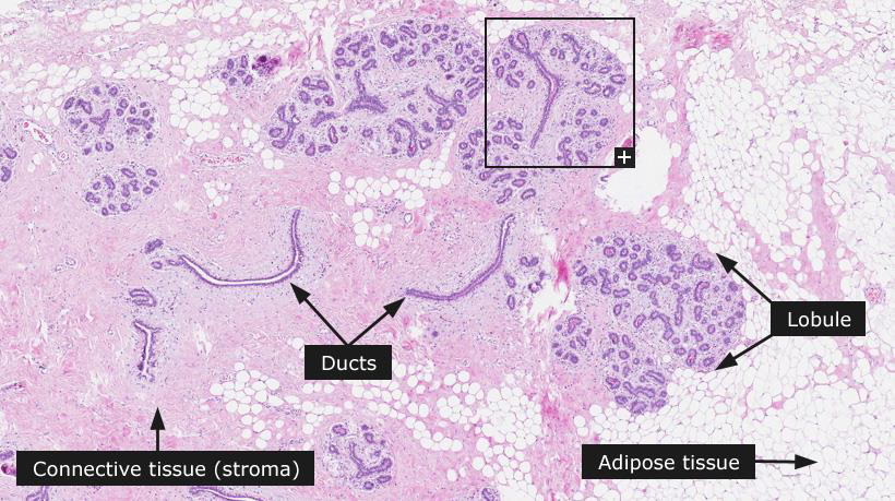
mammary gland normal composition
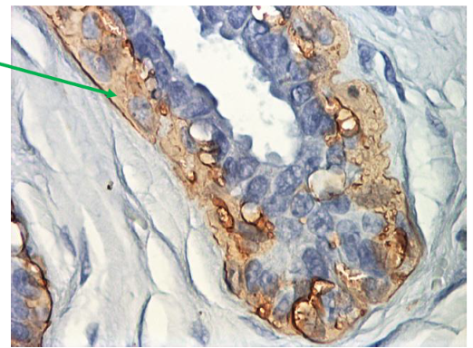
Myoepithelial cells: SMA+
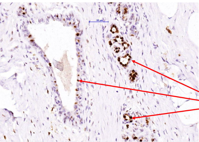
Luminal cells PR+
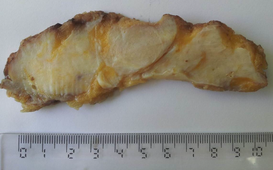
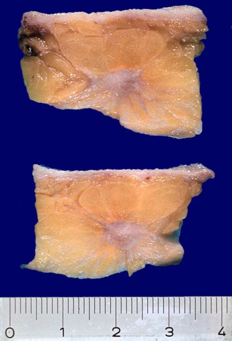
BREAST FIBROCYSTIC CHANGE
elastic, mobile, vaguely nodularity of breast tissue
On cut section an increase in dense, fibrous stroma and some degree of cystic
dilatation of the terminal ducts
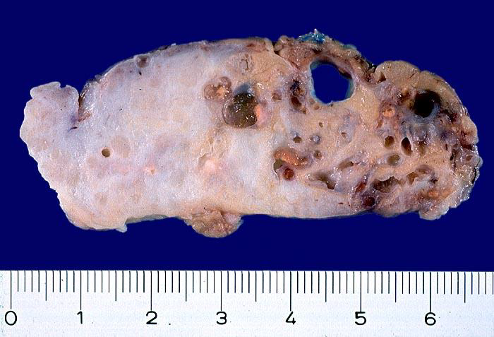
BREAST FIBROCYSTIC CHANGE
elastic, mobile, vaguely nodularity of breast tissue
On cut section an increase in dense, fibrous stroma and some degree of cystic
dilatation of the terminal ducts
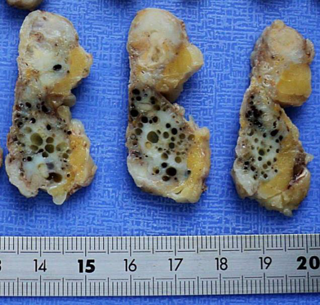
white areas are fibrosis, and black holes are cystic dilatation
BREAST FIBROCYSTIC CHANGE
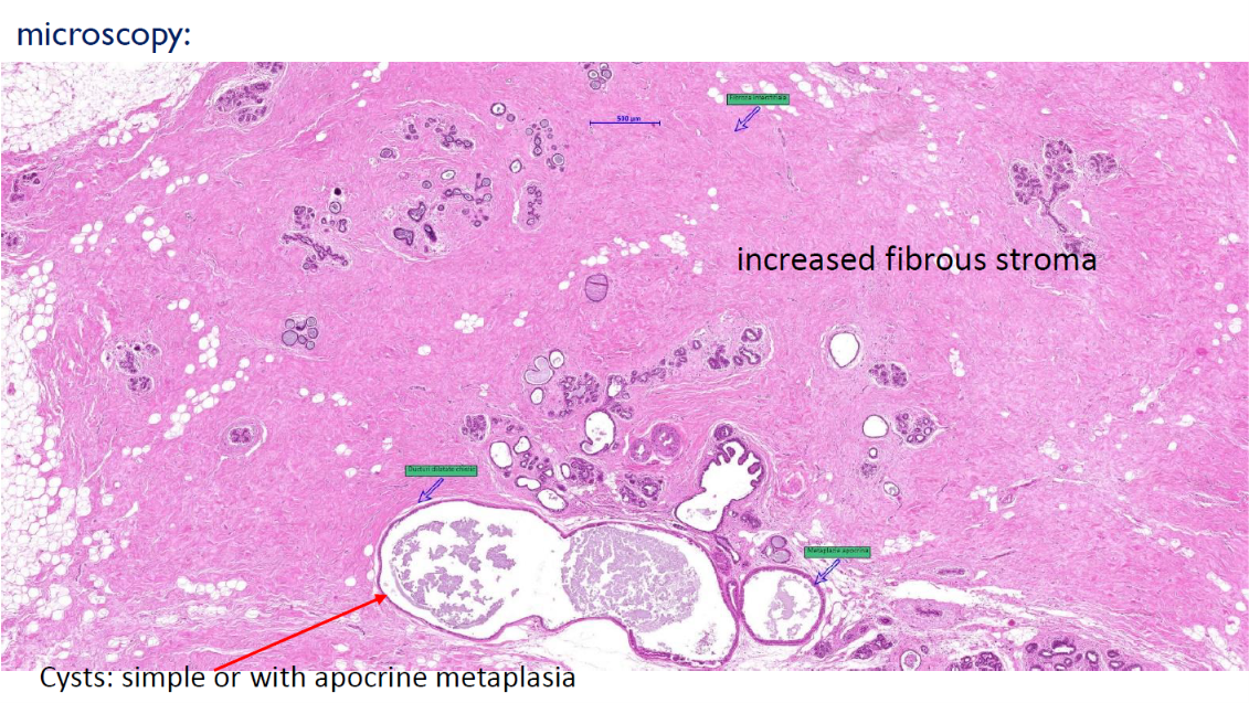
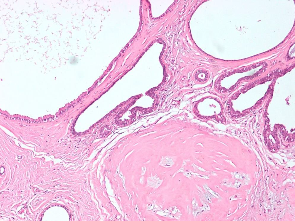
Cysts: simple or with
apocrine metaplasia
BREAST FIBROCYSTIC CHANGE
Cysts: simple or with
apocrine metaplasia
BREAST FIBROCYSTIC CHANGE
Cysts: simple or with
apocrine metaplasia
BREAST FIBROCYSTIC CHANGE
BREAST FIBROCYSTIC CHANGE
elastic, mobile, vaguely nodularity of breast tissue
On cut section an increase in dense, fibrous stroma and some degree of cystic
dilatation of the terminal ducts
white areas are fibrosis, and black holes are cystic dilatation
also some atrophic calcification
BREAST FIBROCYSTIC CHANGE
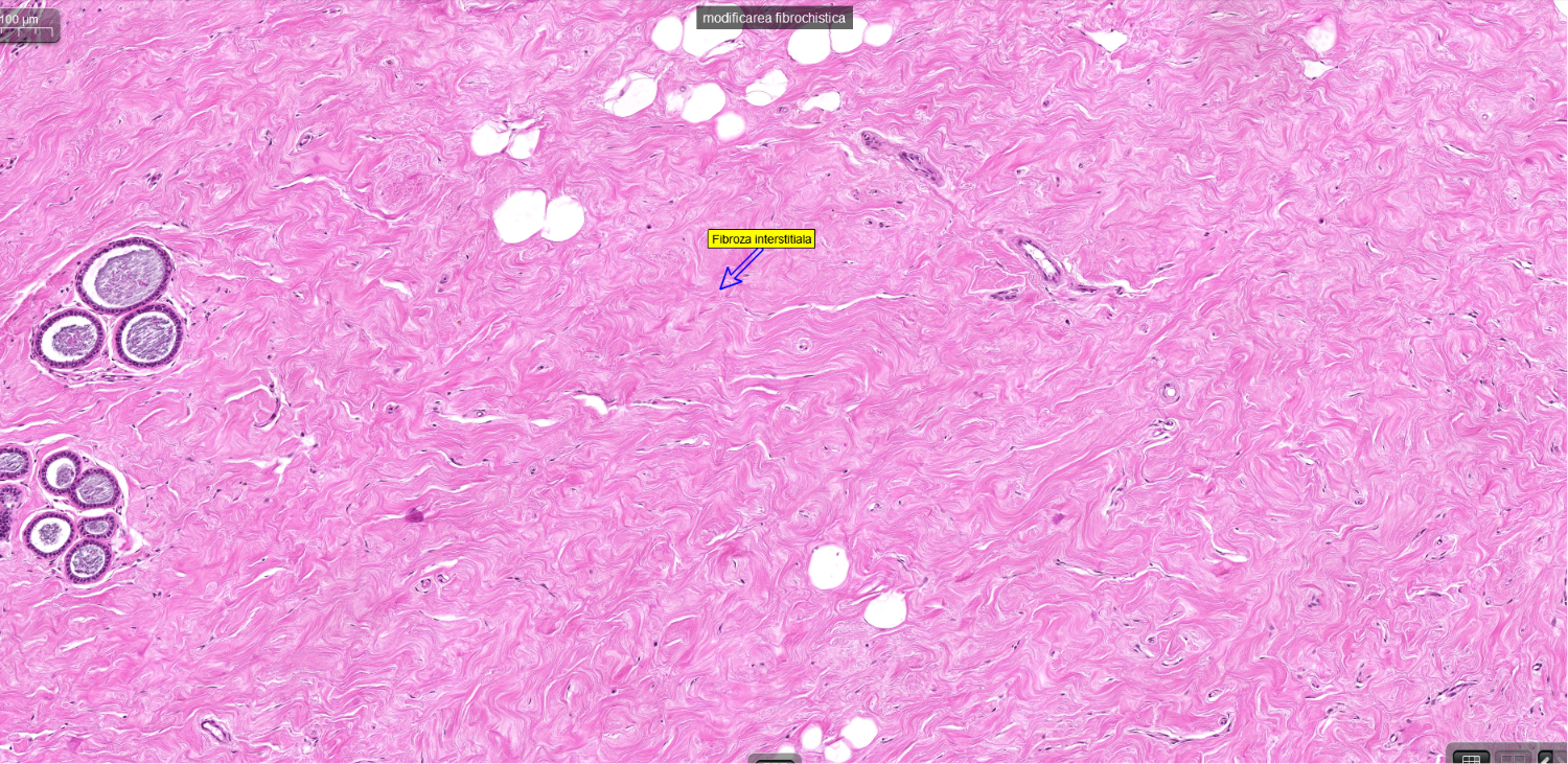
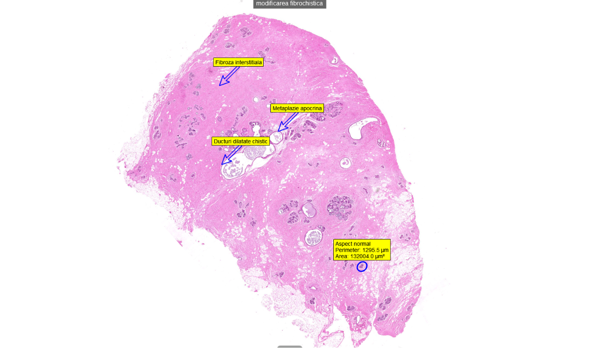
BREAST FIBROCYSTIC CHANGE
interstitial fibrosis
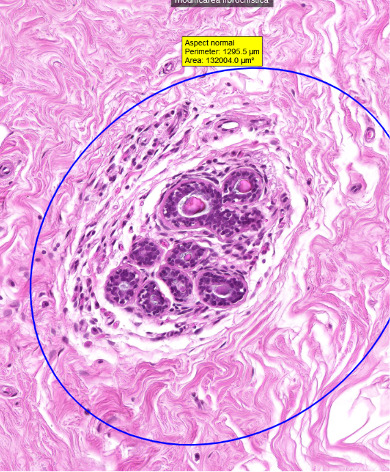
BREAST FIBROCYSTIC CHANGE
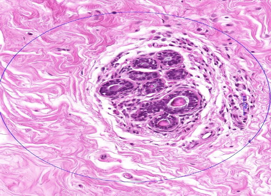
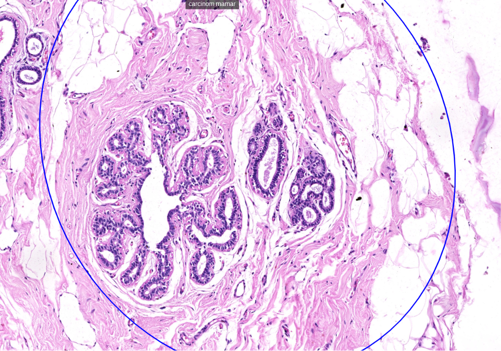
normal aspect of the mammary gland
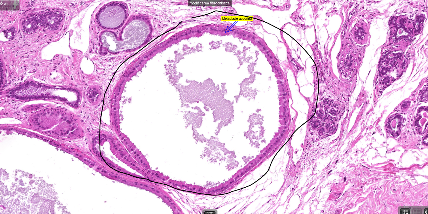
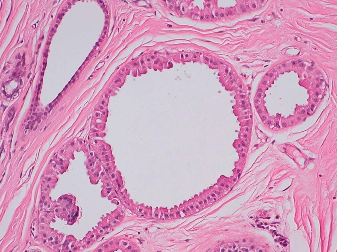
BREAST FIBROCYSTIC CHANGE
apocrine metaplasia of the duct
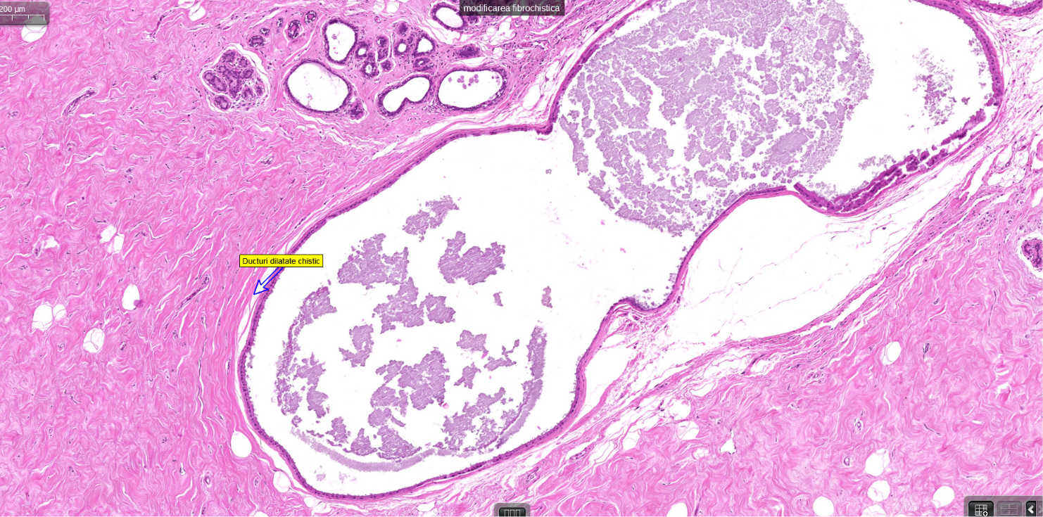
BREAST FIBROCYSTIC CHANGE
dilated cystic ducts
Breast carcinoma
most common malignant tumor of women
majorit y are carcinom as
types
•in situ
•infiltrative
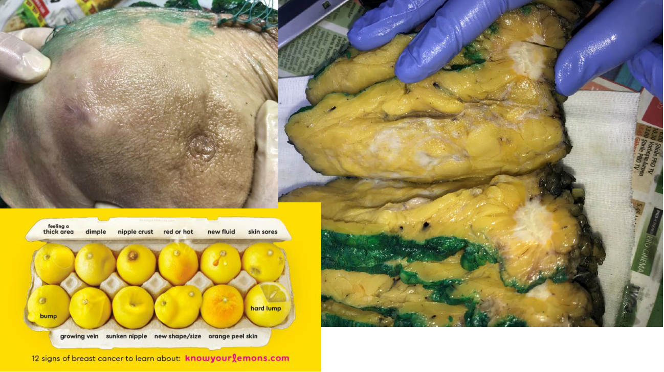
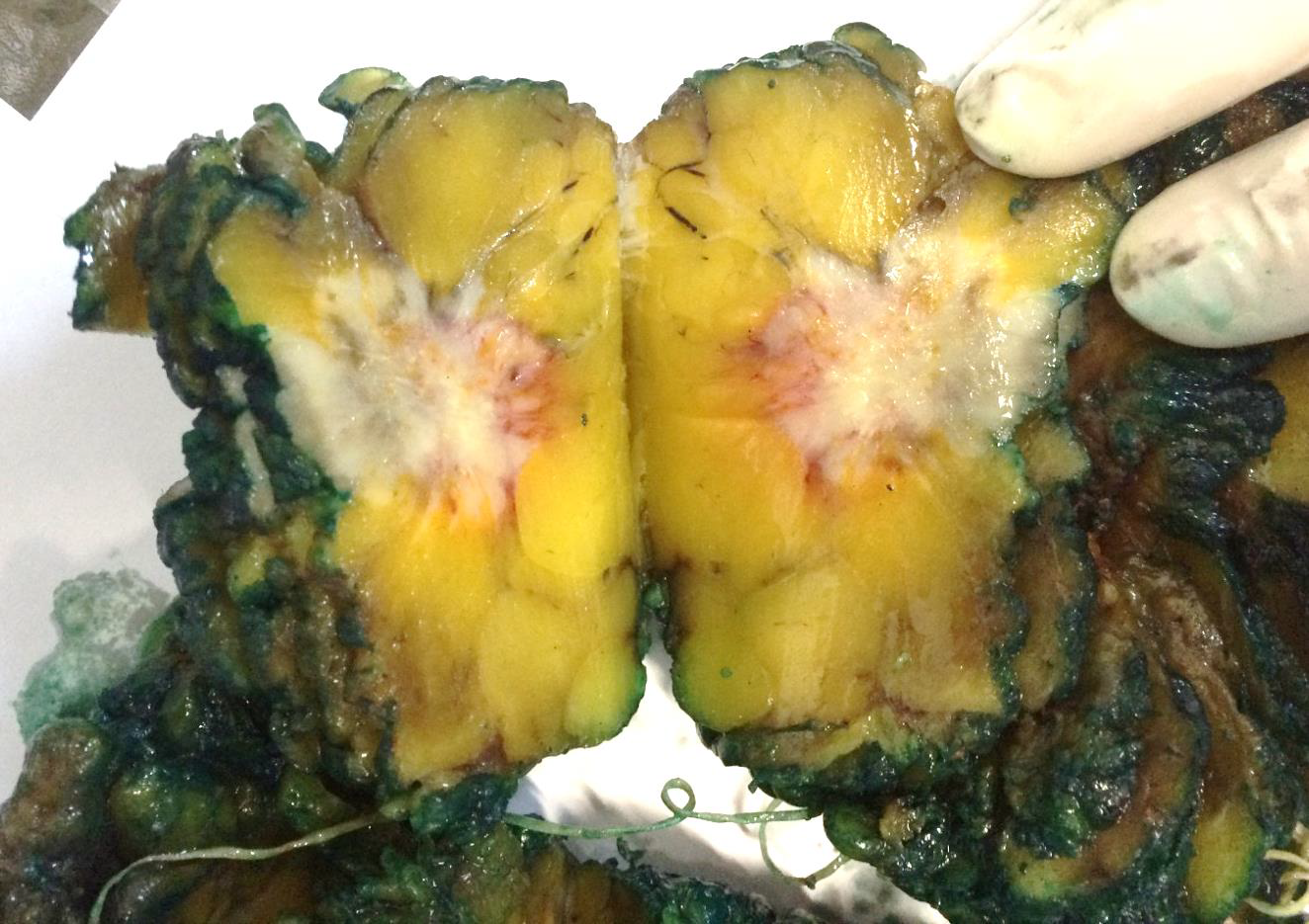
Gross
aspects of infiltrativ e carcinom a
Hard nodule 2 5 cm Ø, no define borders,
irregular contour, adherences with
sorrounding structures
On section tough firm, grey shiny,
sometimes with yellowish flecks like ripe
pear pulp)
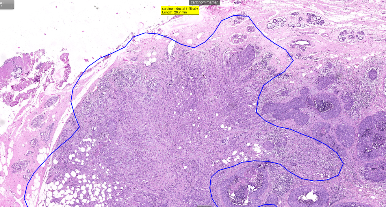
breast carcinoma
breast carcinoma
breast carcinoma
comedo
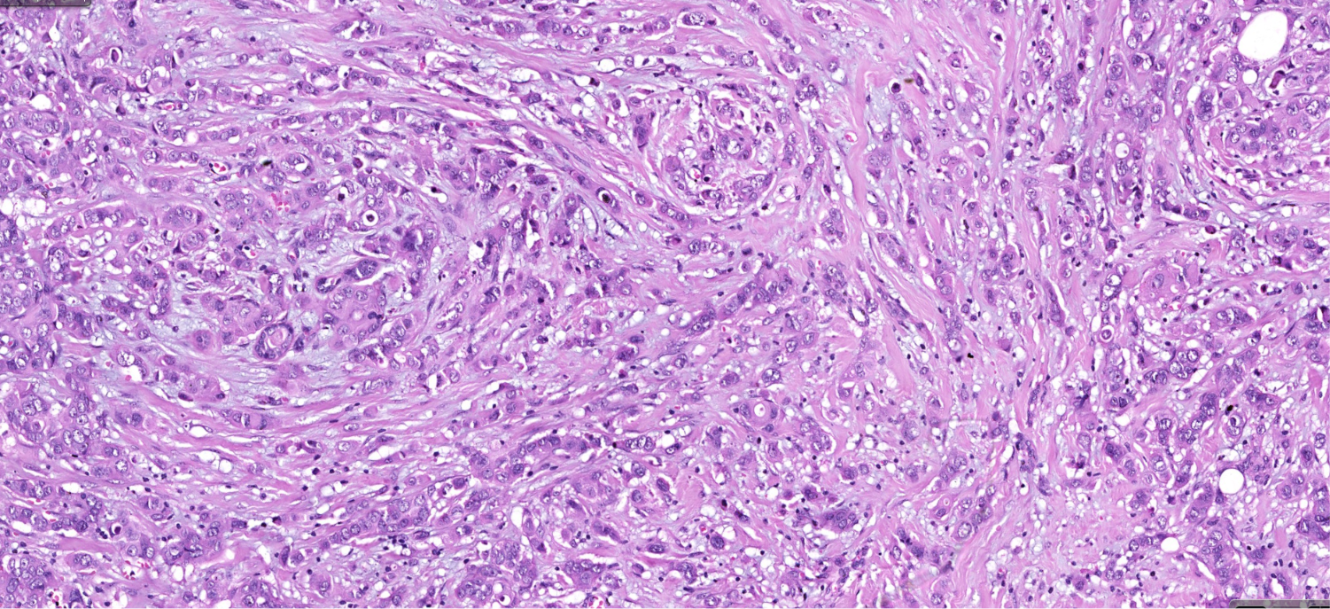
DCIS infiltrative
carcinoma NST ( ductal microscopy)
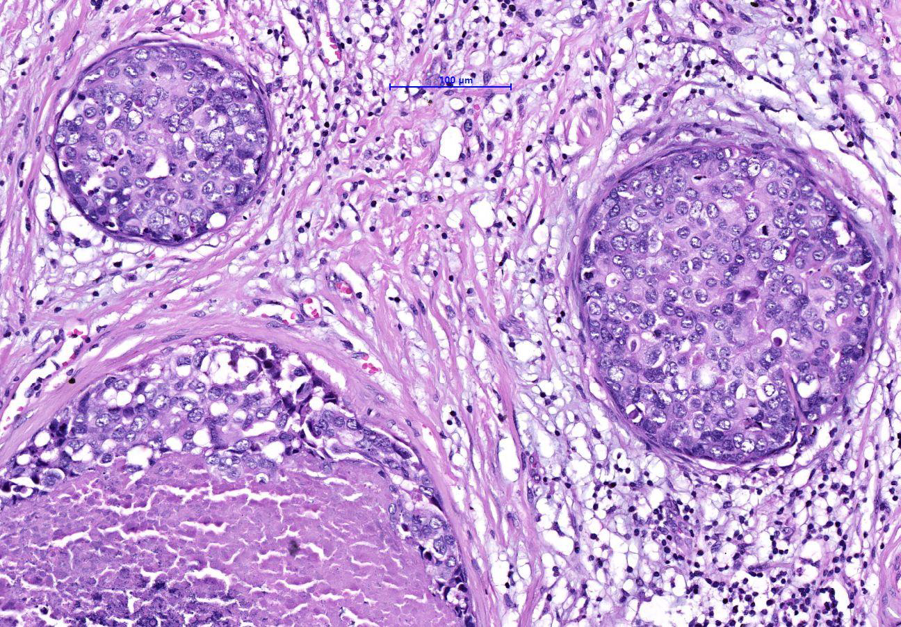
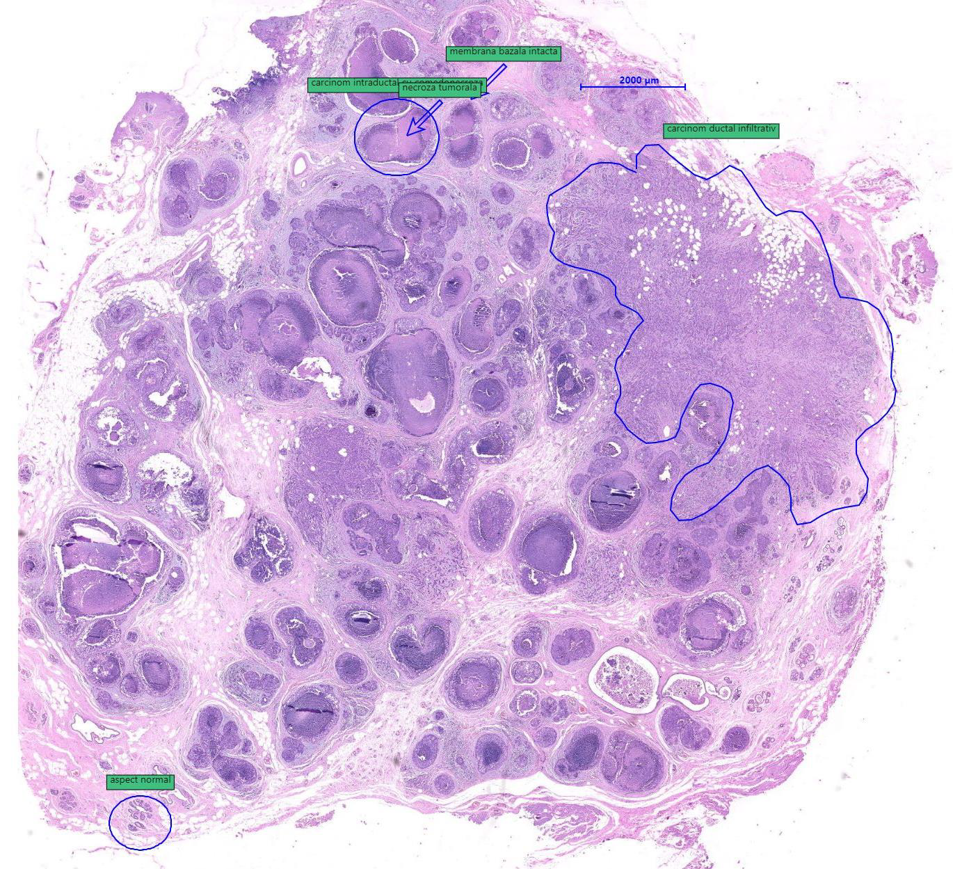
Breash carcinoma DCIS
intermingled with infiltrative carcinom a NST, poorly differentiated
•basement membrane of ducts: continuous / preserved
•central necrosis in dilated ducts
nests
ribbons of malignant
epithelial cells within a focally
dense fibrous stroma adipose
tissue infiltration
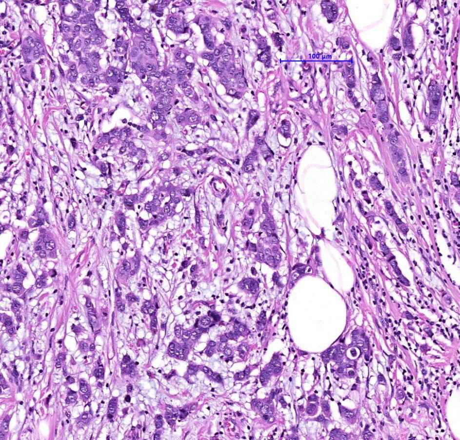
Breast carcinoma
invasive carcinoma
NST is
graded using the Nottingham
histologic score
•tubule formation (gland s
75% = scor e 1, 10 75% =
scor e 2, 10% = scor e 3),
•mitoses / 10HPF (scor e : 1
3)
•nuclear pleomorphism
(scor e : 1 3)
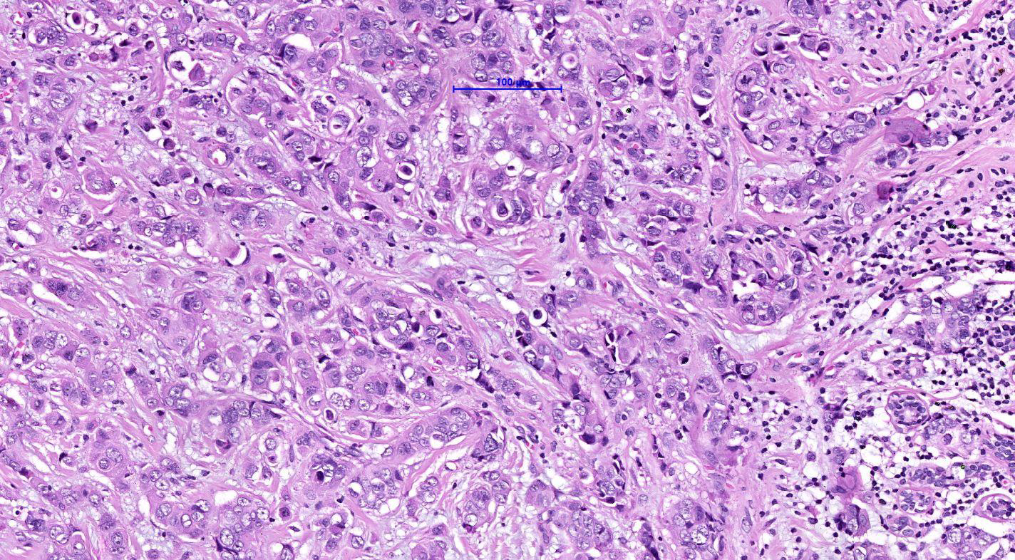
Grade I (G1): Score of \(3\), \(4\), or \(5\). These are considered well-differentiated; the cells closely resemble normal breast tissue and tend to grow slowly. [1]
Grade II (G2): Score of \(6\) or \(7\). These are considered moderately differentiated; the cells show more abnormalities and grow at a moderate rate. [1, 2]
Grade III (G3): Score of \(8\) or \(9\). These are considered poorly differentiated; the cells look very disorganized and tend to grow and divide more rapidly. [1, 2, 3, 4, 5]
infiltrative breast carcinoma
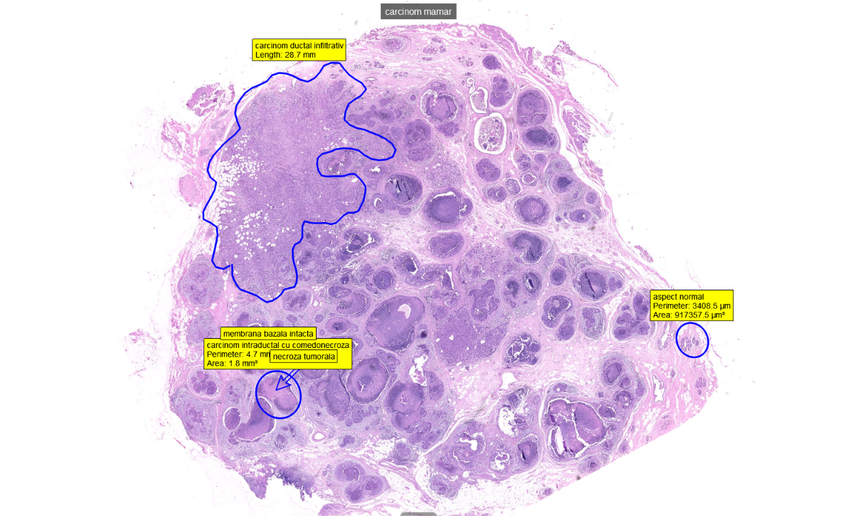
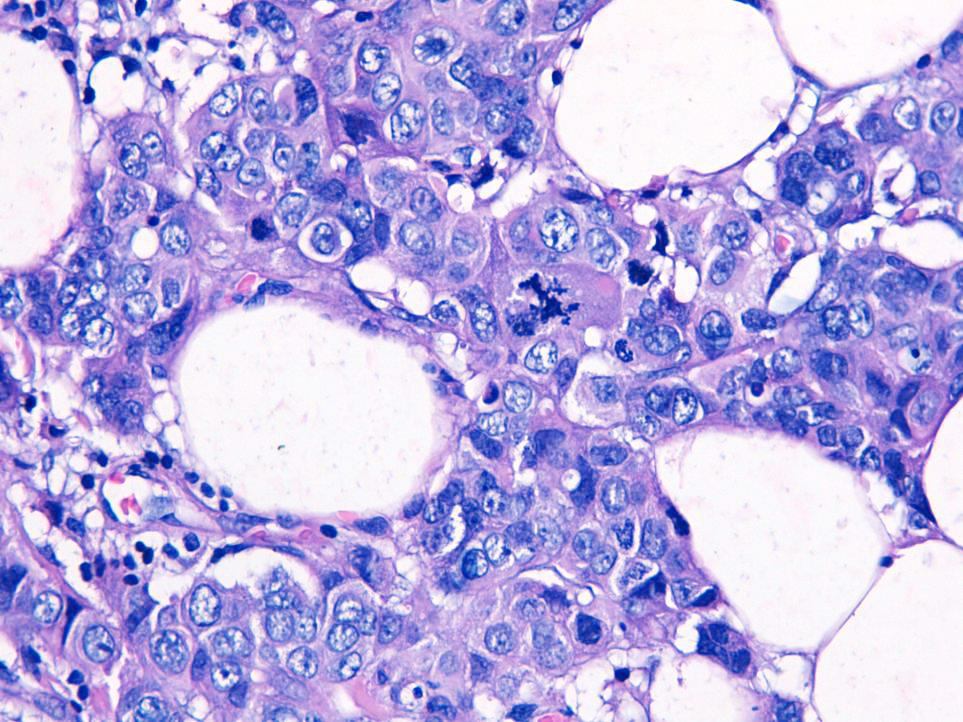
Breast carcinoma
breast carcinoma
normal aspect of glands
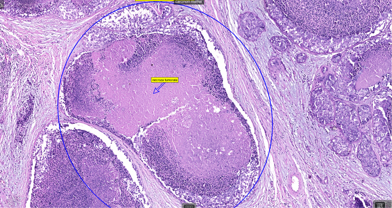
breast carcinoma DISC comedo
intruductal abundant pasty tumoral necrosis
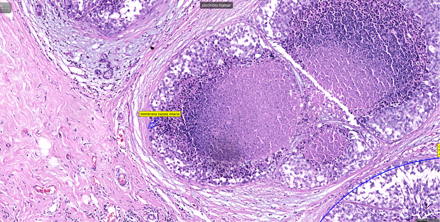
breast cancer
intact basal membrane

breast carcinoma
intact basal membrane
breast carcinoma
ductral infiltration
breast cancer
ductal infiltration