PEDIATRIC PROTOCOL S2 Lecture
1/69
There's no tags or description
Looks like no tags are added yet.
Name | Mastery | Learn | Test | Matching | Spaced | Call with Kai |
|---|
No analytics yet
Send a link to your students to track their progress
70 Terms
What frequency range is used for pediatric echo transducers?
→ 2.25 MHz to 12 MHz
What transducer frequency range is more commonly used in pediatric echo?
→ 12–8 MHz
Why are multiple transducers needed in pediatric echo?
→ Different patient sizes and imaging needs
What special transducer should be included in pediatric echo?
→ Non-imaging pedoff transducer
What is considered key in pediatric imaging?
→ Resolution
How long does a pediatric echocardiogram usually take?
→ 45–60 minutes
Can pediatric echo exams be extended longer than 60 minutes?
→ Yes
What should be explained thoroughly before the pediatric echo exam?
→ The exam to the family
What patient positions are commonly used during pediatric echo?
What type of room is preferred for pediatric echo?
What distraction methods may be used during pediatric echo?
What patient positions are commonly used during pediatric echo?
→ Left lateral decubitus (LLD) or supine reclined
What type of room is preferred for pediatric echo?
→ Dark room, warm gel,
What distraction methods may be used during pediatric echo?
→ Movies, books, and toys
What warming method may be used for infants during pediatric echo?
What sweet solution may help calm infants during pediatric echo?
What should be available if the baby becomes unstable?
What warming method may be used for infants during pediatric echo?
→ Warm blanket for swaddling
What sweet solution may help calm infants during pediatric echo?
→ Sugar water
What should be available if the baby becomes unstable?
→ Backup/help available
Why are longer what important in pediatric echo?
What may pediatric echo rely on instead of ECG?
What imaging mode evaluates anatomy in pediatric echo?
What echo mode measures chamber dimensions and motion?
Why are longer loops important in pediatric echo?
→ Better assessment due to motion/high heart rates
What may pediatric echo rely on instead of ECG timing?
→ Timing by imaging
What imaging mode evaluates anatomy in pediatric echo?
→ 2D imaging
What echo mode measures chamber dimensions and motion?
→ M-Mode
What Doppler evaluates blood flow velocities at a specific location?
What Doppler evaluates high velocity flow?
What Doppler evaluates direction and turbulence of flow?
What Doppler evaluates blood flow velocities at a specific location?
→ Pulsed wave Doppler
What Doppler evaluates high velocity flow?
→ Continuous wave Doppler
What Doppler evaluates direction and turbulence of flow?
→ Color flow Doppler
Why is flow direction important in pediatric echo?
What are the 3 possible shunt flow directions?
Why is flow direction important in pediatric echo?
→ To identify abnormal connections and shunts
What are the 3 possible shunt flow directions?
→ Left-to-right, right-to-left, or bidirectional
Pediatric echocardiograms evaluate what? (name 2 and assess for what?)
Do pediatric TTEs include standard adult TTE views?
What additional condition is commonly assessed in pediatric TTEs?
Pediatric echocardiograms evaluate what?
→ Normal and abnormal structures, flow jets, and pulmonary hypertension
Do pediatric TTEs include standard adult TTE views?
→ Yes
What additional condition is commonly assessed in pediatric TTEs?
→ Pulmonary hypertension
What is emphasized more in pediatric echo compared to adult echo?
What type of sweeps are commonly performed in pediatric echo?
What type of additional imaging planes are commonly used in pediatric echo?
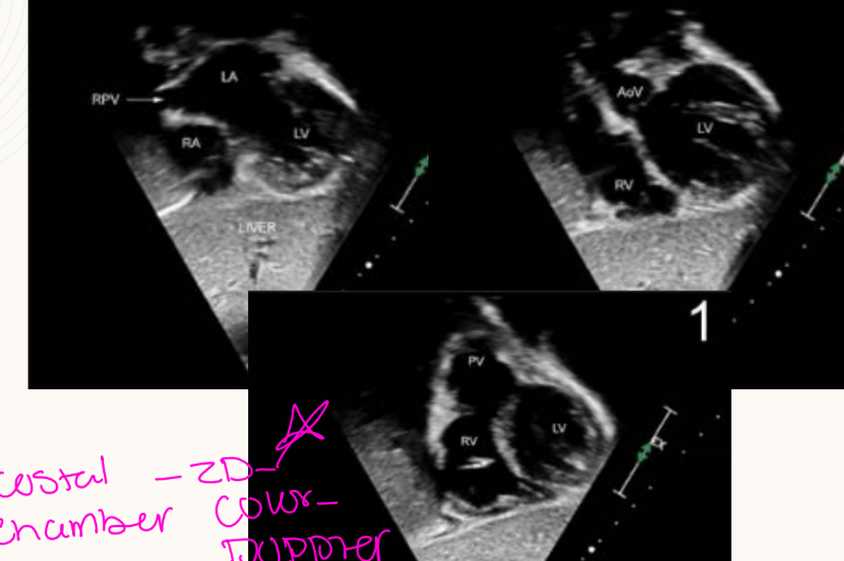
How are apical and subcostal views displayed in pediatric echo?
What is emphasized more in pediatric echo compared to adult echo?
→ Morphology, relationships, and sequential segmental analysis
What type of sweeps are commonly performed in pediatric echo?
→ Long sweeps in all views
What type of additional imaging planes are commonly used in pediatric echo?
→ Off-axis views
How are apical and subcostal views displayed in pediatric echo?
→ Anatomically correct orientation
What vessels are specifically evaluated in pediatric echo?
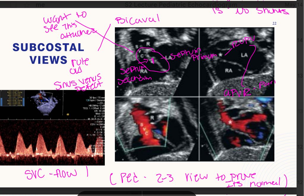
What special subcostal views are important in pediatric echo?
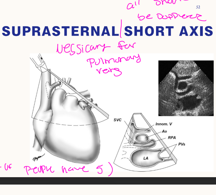
What structures are evaluated in suprasternal views?
What additional parasternal imaging windows are commonly used? that are the 2 new views
What vessels are specifically evaluated in pediatric echo?
→ Coronary arteries and pulmonary veins
What special subcostal views are important in pediatric echo?
→ Bicaval and short axis views
What structures are evaluated in suprasternal views?
→ Arch sidedness, PDA, pulmonary veins, SVC, and innominate vein
What additional parasternal imaging windows are commonly used?
→ High right and left parasternal views
SKIP
PEDIATRIC TTE ECHOCARDIOGRAM PROTOCOL
What views begin with in pediatric echo?
What direction should the sonographer sweep from in ^^^^?
What standard view is included in pediatric echo?
What high parasternal view is used to evaluate ____?
What additional major imaging windows are part of pediatric echo?
What other imaging borders are evaluated in pediatric echo? 2 new one
What do subcostal views begin with in pediatric echo?
→ Abdominal visceral situs
What direction should the sonographer sweep from in subcostals?
→ From abdomen to apex
What standard view is included in pediatric echo?
→ Parasternal long and short axis
What high parasternal view is used to evaluate PDA?
→ Ductal view
What additional major imaging windows are part of pediatric echo?
→ Apicals and suprasternal notch views
What other imaging borders are evaluated in pediatric echo?
→ Right and left sternal border
What is the purpose of the subcostal situs view? to determine what?
What structures’ relative locations are evaluated in subcostal situs? name 5
What venous connection should be displayed in subcostal situs?
What is evaluated in the abdominal aorta using color Doppler and PW Doppler?
What is the purpose of the subcostal situs view?
→ Determine abdominal visceral situs
What structures’ relative locations are evaluated in subcostal situs?
→ IVC, descending aorta, spine, liver, and stomach
What venous connection should be displayed in subcostal situs?
→ Hepatic segment of the IVC connecting to the RA
What is evaluated in the abdominal aorta using color Doppler and PW Doppler?
→ Pulsatility and flow
What venous structure is evaluated in subcostal coronal views?
What septal structure is evaluated in subcostal coronal views?
What valve connections are evaluated in subcostal coronal views?
What venous structure is evaluated in subcostal coronal views?
→ Coronary sinus
What septal structure is evaluated in subcostal coronal views?
→ Atrial septum
What valve connections are evaluated in subcostal coronal views?
→ AV connections
What LV structure is evaluated in subcostal coronal views?
What RV structure is evaluated in subcostal coronal views?
What septal region is evaluated in subcostal coronal views?
What arterial relationship is evaluated in subcostal coronal views?
What LV structure is evaluated in subcostal coronal views?
→ LV outflow tract
What RV structure is evaluated in subcostal coronal views?
→ RV outflow tract
What septal region is evaluated in subcostal coronal views?
→ Anterior muscular ventricular septum
What arterial relationship is evaluated in subcostal coronal views?
→ Ventriculoarterial connections
(The aorta arises from the morphologically left ventricle, and the pulmonary artery arises from the morphologically right ventricle)
simply means how the ventricles attach to the main arteries leaving the heart.
To understand the name, you can break it down into two parts:
Ventriculo refers to the ventricles (the lower, pumping chambers of the heart).
After sweeping the next step is to go step by step evluating wha and this the begining of the protocol. what dopplers are utilized?
What is the probe indicator direction in the subcostal Right Anterior Oblique view?
What structures are visualized simultaneously in the subcostal Right Anterior Oblique view?
What valve is displayed en face in the subcostal Right Anterior Oblique view?
Subcostal 4 chamber, 2 chamber AV opening, RVOT, utlizing 2D, Color, and doppler
What is the probe indicator direction in the subcostal Right Anterior Oblique view?
→ 2 o’clock
What structures are visualized simultaneously in the subcostal Right Anterior Oblique view?
→ RV inflow and RV outflow
What valve is displayed en face in the subcostal Right Anterior Oblique view?
→ Aortic valve
What is the probe indicator direction in subcostal SAX views?
What venous structures are evaluated in subcostal SAX views?
What pulmonary vein is specifically evaluated in subcostal SAX views?
What septal structure is evaluated in subcostal SAX views?
what view do you 1st start off with, what sweep is this? can you name the following views? Left to Right sweep or Right to Left sweep
what do you want to rule out in subcostal view?
What is the probe indicator direction in subcostal SAX views?
→ 6 o’clock
What venous structures are evaluated in subcostal SAX views?
→ SVC and IVC
What pulmonary vein is specifically evaluated in subcostal SAX views?
→ Right upper pulmonary vein
What septal structure is evaluated in subcostal SAX views?
→ Atrial septum
1st start off with the Bicaval view, AO, RVOT, The ventricle level
RIGHT to LEFT SWEEP
rule out sinus venous defects
What LV structure is evaluated in subcostal SAX views?
What RV structure is evaluated in subcostal SAX views?
What septal structure is evaluated in subcostal SAX views?
What LV assessment is performed in subcostal SAX views?
What LV structure is evaluated in subcostal SAX views?
→ LV outflow tract
What RV structure is evaluated in subcostal SAX views?
→ RV outflow tract
What septal structure is evaluated in subcostal SAX views?
→ Ventricular septum
What LV assessment is performed in subcostal SAX views?
→ LV cross-section
What fibrous continuity is evaluated in PLAX?
What mitral valve features are evaluated in PLAX?
What aortic valve features are evaluated in PLAX?
What fibrous continuity is evaluated in PLAX?
→ Mitral-to-aortic valve fibrous continuity
What mitral valve features are evaluated in PLAX?
→ Morphology and function
What aortic valve features are evaluated in PLAX?
→ Morphology and function
What aortic measurements are obtained in PLAX?
What coronary artery origin is evaluated in PLAX?
How is the RCA origin evaluated in PLAX?
What aortic measurements are obtained in PLAX?
→ Aortic annulus, aortic root, sinotubular junction, and ascending aorta diameters
What coronary artery origin is evaluated in PLAX?
→ RCA origin from the aortic root
How is the RCA origin evaluated in PLAX?
→ 2D and color mapping
What tricuspid valve features are evaluated in PLAX?
How is RV systolic pressure estimated in PLAX?
What pulmonary valve features are evaluated in PLAX?
What tricuspid valve features are evaluated in PLAX?
→ Morphology, function, and flow
How is RV systolic pressure estimated in PLAX?
→ Tricuspid regurgitation gradient
What pulmonary valve features are evaluated in PLAX?
→ Morphology, function, and flow
What pulmonary artery measurements are obtained in PLAX?
When are PV and MPA gradients measured?
What pulmonary artery measurements are obtained in PLAX?
→ PV annulus and MPA diameters
When are PV and MPA gradients measured?
→ If elevated
What valve morphology is evaluated in PSAX? name 2
What septal structure is evaluated in PSAX?
What valve morphology is evaluated in PSAX?
→ Aortic valve morphology
→ Mitral valve morphology
What septal structure is evaluated in PSAX?
→ Ventricular septum
What LV measurement is obtained in PSAX?
What LV function is evaluated in PSAX?
What left coronary artery is visualized in PSAX?
What LV measurement is obtained in PSAX?
→ LV size
What LV function is evaluated in PSAX?
→ LV systolic function
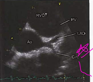
What left coronary artery is visualized in PSAX?
→ LMCA
What branches of the LMCA are visualized in PSAX?
How are coronary arteries evaluated in PSAX?
What coronary artery diameters are measured in PSAX?
What branches of the LMCA are visualized in PSAX?
→ Left circumflex and LAD
How are coronary arteries evaluated in PSAX?
→ 2D and color mapping
What coronary artery diameters are measured in PSAX?
→ LMCA, circumflex, LAD, and RCA
What tricuspid valve features are evaluated in PSAX?
How is RV systolic pressure estimated in PSAX?
What pulmonary valve features are evaluated in PSAX?
What tricuspid valve features are evaluated in PSAX?
→ Morphology, function, and flow
How is RV systolic pressure estimated in PSAX?
→ TR gradient
What pulmonary valve features are evaluated in PSAX?
→ Morphology, function, and flow
What pulmonary structures are measured in PSAX?
What Doppler is obtained in branch pulmonary arteries?
When do you measure the PV and MPA gradients?
What pulmonary structures are measured in PSAX?
→ PV annulus, MPA, and proximal branch pulmonary arteries
What Doppler is obtained in branch pulmonary arteries?
→ PW Doppler
When do you measure the PV and MPA?
→ measure PV and MPA gradient if elevated
What pulmonary arteries should both be Dopplered in pediatric echo?
→ Both branch pulmonary arteries
What tricuspid valve features are evaluated in apical views?
How is RV systolic pressure estimated in apical views?
What mitral valve features are evaluated in apical views?
What tricuspid valve features are evaluated in apical views?
→ Morphology, function, and flow
How is RV systolic pressure estimated in apical views?
→ TR gradient
What mitral valve features are evaluated in apical views?
→ Morphology, function, and flow
What septal structure is evaluated in apical views?
What LV measurement is obtained in apical views?
What LV functions are evaluated in apical views?
What septal structure is evaluated in apical views?
→ Ventricular septum
What LV measurement is obtained in apical views?
→ LV size
What LV functions are evaluated in apical views?
→ Systolic and diastolic function
What atrial measurement is obtained in apical views?
What pulmonary veins should be evaluated in apical views?
What fibrous continuity is evaluated in apical views?
What atrial measurement is obtained in apical views?
→ LA size
What pulmonary veins should be evaluated in apical views?
→ At least one right and/or one left pulmonary vein entering the LA
What fibrous continuity is evaluated in apical views?
→ Mitral-to-aortic valve fibrous continuity
What outflow structures are evaluated in apical views?
What aortic valve features are evaluated in apical views?
When should LVOT and AoV gradients be measured?
What outflow structures are evaluated in apical views?
→ LVOT and proximal aorta
What aortic valve features are evaluated in apical views?
→ Morphology, function, and flow
When should LVOT and AoV gradients be measured?
→ If elevated
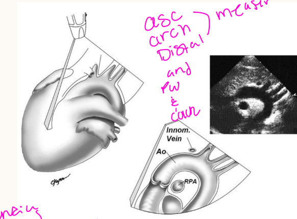
What arch structure is evaluated in suprasternal notch long axis?
What arch branch vessels are evaluated in suprasternal long axis?
What arch structure is evaluated in suprasternal long axis?
→ Aortic arch
What arch branch vessels are evaluated in suprasternal long axis?
→ Right innominate, left common carotid, and left subclavian arteries
What aortic measurements are obtained in suprasternal long axis?
What flow is evaluated in suprasternal long axis?
What aortic measurements are obtained in suprasternal long axis?
→ Proximal and distal transverse arch and aortic isthmus diameters
What flow is evaluated in suprasternal long axis?
→ Flow along the aortic arch and proximal descending aorta
What is the first branch of the aortic arch seen in suprasternal SAX?
What does the right innominate artery bifurcate into?
What is the second branch of the aortic arch?
What is the first branch of the aortic arch seen in suprasternal SAX?
→ Right innominate artery
What does the right innominate artery bifurcate into?
→ Right subclavian and right common carotid arteries
What is the second branch of the aortic arch?
→ Left common carotid artery
What is the third branch of the aortic arch?
How many pulmonary veins should drain into the LA normally?
How are pulmonary veins evaluated in suprasternal SAX?
What is the third branch of the aortic arch?
→ Left subclavian artery
How many pulmonary veins should drain into the LA normally?
→ Two right and two left pulmonary veins
How are pulmonary veins evaluated in suprasternal SAX?
→ 2D, color Doppler, and PW Doppler
crab view caution:
What pulmonary vein may be confused with the RUPV?
What structure may be confused with the LUPV?
What pulmonary vein may be confused with the RUPV?
→ Right middle pulmonary vein
What structure may be confused with the LUPV?
→ Left atrial appendage
What is the probe indicator direction in right parasternal long? and pt postion?
What venous flows are evaluated in right parasternal long?
How are SVC and IVC flow evaluated in right parasternal long?
What is the probe indicator direction in right parasternal long?
→ 12 o’clock, pt RLD
What venous flows are evaluated in right parasternal long?
→ SVC and IVC flow into the RA
How are SVC and IVC flow evaluated in right parasternal long?
→ Color mapping
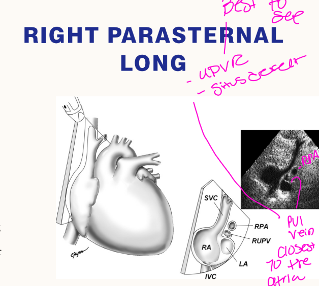
What septal structure is evaluated in right parasternal long?
What defect should be excluded in right parasternal long?
What septal structure is evaluated in right parasternal long?
→ Atrial septum
What defect should be excluded in right parasternal long?
→ Superior sinus venosus defect
What pulmonary veins are evaluated in right parasternal long?
What aortic pathology may require AoV flow evaluation in right parasternal long?
What pulmonary veins are evaluated in right parasternal long?
→ Right pulmonary veins entering the LA
What aortic pathology may require AoV flow evaluation in right parasternal long?
→ Subvalvar, valvar, or supravalvar aortic stenosis
What pulmonary vein is evaluated in right parasternal SAX?
How is RUPV flow evaluated in right parasternal SAX?
What pulmonary vein is evaluated in right parasternal SAX?
→ Right upper pulmonary vein (RUPV) flow into LA
How is RUPV flow evaluated in right parasternal SAX?
→ Imaging and color mapping
Are pediatric echo measurements the same as adult echo measurements?
Name all the new Rt side measuremnets name 2
Are pediatric echo measurements the same as adult echo measurements?
→ Mostly yes, with additional measurements
RV inflow (E and A) and TDI E ‘
RV MPI (RIMP)
What does RIMP stand for?
What does S/D ratio stand for?
How is the S/D ratio calculated?
What does RIMP stand for?
→ RV myocardial performance index
What does S/D ratio stand for?
→ Systolic-to-diastolic duration ratio
How is the S/D ratio calculated?
→ Duration of TR divided by the duration of the rest of the cardiac cycle
name the new measuremnets in peds
on the left side name 3
Pveins (S, D, Ar) - All 4 pveins dopplered
LIMP
Measure all cardiac valve annuli (PA, AO, TV, MV)
What LV dimensions are measured in PSAX?
What does LIMP stand for? ***
What valve structures should all be measured in pediatric echo?
What LV dimensions are measured in PSAX?
→ IVS, posterior wall, LVEDD, and LVESD
What does LIMP stand for?
→ LV myocardial performance index
What valve structures should all be measured in pediatric echo?
→ Cardiac valve annuli
What is the normal fractional shortening range in pediatric echo?
What is the normal M-mode fractional shortening range?
What is the normal EF range in pediatric echo?
What is the normal fractional shortening range in pediatric echo?
→ 25–46%
What is the normal M-mode fractional shortening range?
→ 28–44%
What is the normal EF range in pediatric echo?
→ 48–78%
What is the normal stroke volume range in pediatric echo?
What is the normal cardiac output range in pediatric echo?
What is the normal cardiac index range in pediatric echo?
What is the normal stroke volume range in pediatric echo?
→ 30–65 mL
What is the normal cardiac output range in pediatric echo?
→ 4–8 L/min
What is the normal cardiac index range in pediatric echo?
→ 2.8–4.2 L/min/m²
Why are Z scores important in pediatrics?
What do Z scores help provide?
What does a Z score determine?
Why are Z scores important in pediatrics?
→ Large variation in BSA/BMI due to growth
What do Z scores help provide?
→ Normalized comparison data sets
What does a Z score determine?
→ Whether a structure is above or below the mean
What does a Z score of 0 represent?
What does a Z score of +2 represent?
What does a Z score of -2 represent?
What does a Z score of 0 represent?
→ Measurement equals the population mean
What does a Z score of +2 represent?
→ Large structure above the 95th percentile
What does a Z score of -2 represent?
→ Small structure below the 5th percentile
What does retrograde filling of the ascending aorta from the PDA suggest? name 3
What may be present if the aortic arch cannot be visualized? name 4
What does retrograde filling of the ascending aorta from the PDA suggest?
→ HLHS, critical AS, or ductal-dependent lesion
What may be present if the aortic arch cannot be visualized?
→ Hypoplastic arch, interrupted arch, coarctation, or left heart lesions
What does exclusive right-to-left shunting at the atrial level suggest?
What lesions can cause R-to-L shunting at the atrial level? name 5
What does exclusive right-to-left shunting at the atrial level suggest?
→ Not enough blood entering the LA
What lesions can cause R-to-L shunting at the atrial level?
→ TAPVR, pulmonary vein stenosis/atresia, TV atresia, or PV atresia
What does right-to-left shunting at the ductal level indicate?
What lesions can cause R-to-L shunting at the ductal level? name 5
What does right-to-left shunting at the ductal level indicate?
→ Pulmonary circulation supplying systemic circulation
What lesions can cause R-to-L shunting at the ductal level?
→ AS, HLHS, coarctation, interrupted aortic arch, or severe pulmonary hypertension
If the apical 4 chamber does not appear normal, what may be present?
What does one underdeveloped ventricle suggest?
What syndrome is associated with single ventricle physiology?
If the apical 4 chamber does not appear normal, what may be present?
→ Congenital heart disease
What does one underdeveloped ventricle suggest?
→ HLHS or HRHS
What syndrome is associated with single ventricle physiology?
→ Heterotaxy syndrome
What can mitral regurgitation suggest in pediatrics? name 4
What can tricuspid regurgitation suggest in pediatrics? name 3
What can mitral regurgitation suggest in pediatrics?
→ Cardiomyopathy, severe AS, L-TGA, or AV canal defects
What can tricuspid regurgitation suggest in pediatrics?
→ Ebstein anomaly, cardiomyopathy, or AV canal defect
What does a midline or rightward apex suggest?
What syndromes are associated with abnormal apex position?
What does a midline or rightward apex suggest?
→ Complex heart disease or abnormal organ arrangement
What syndromes are associated with abnormal apex position?
→ Heterotaxy or situs inversus totalis
If a normal PLAX of the LV and aorta cannot be demonstrated, what lesions may be present?********
name 5
If a normal PLAX of the LV and aorta cannot be demonstrated, what lesions may be present?
→ L-TGA, D-TGA, DORV, Tetralogy of Fallot, or truncus arteriosus
What does absence of a normal PSAX suggest?
What lesions may prevent the RV from opening into the PV/PA? name 5
What does absence of a normal PSAX suggest?
→ Congenital heart disease involving RVOT/PV
What lesions may prevent the RV from opening into the PV/PA?
→ Critical PS, pulmonary atresia, Tetralogy of Fallot, RV cardiomyopathy, or Ebstein anomaly
What does parallel alignment of the aortic and pulmonary valves suggest? name 3
What does parallel alignment of the aortic and pulmonary valves suggest?
→ DORV, D-TGA, or L-TGA
What is the 10th warning sign of critical CHD?
Why are abnormalities easier to recognize in babies?
If images do not appear normal in a baby, what should be suspected?
What is the 10th warning sign of critical CHD?
→ Something doesn’t look right
Why are abnormalities easier to recognize in babies?
→ Everything is easier to see in babies
If images do not appear normal in a baby, what should be suspected?
→ There may be congenital heart disease
what are the 10 warning signs of critical CHD
1. Retrograde filling in ascending aorta coming from PDA
Cannot see the aortic arch
Exclusive right to left shunting at the atrial or ductal level
Apical 4 chamber doesn’t look like an apical 4 chamber
An Atrioventricular valve leaks
Apex is pointed midline or rightward
Cannot demonstrate a normal PLAX of the LV and aorta in same picture *
There is no normal PSAX or the RV cannot open into a PV/PA
Aortic valve and pulmonary valve are parallel. Cannot see them enface
Something doesn’t look right
What are the best two views so see PFO and ASD?
Bicaval, and A4C best place to spot VSD and PFO
what view is a good spot to look for VSD
long of the aorta
what view is the best to see VSD and any initial problem with the PV
subcostal SAX at ventricle level
where is the circ location
circ always circles the LA