ANAT 5010 - unit 9
1/55
There's no tags or description
Looks like no tags are added yet.
Name | Mastery | Learn | Test | Matching | Spaced | Call with Kai |
|---|
No analytics yet
Send a link to your students to track their progress
56 Terms
superior: lower border of the mandible
inferior: anterior - jugular/suprasternal notch, sternoclavicular joint, clavicle, and acromion process; posterior - CV7
sagittal plane divides the left and right sides
what are the boundaries of the neck?
sternocleidomastoid
what divides the neck into anterior and posterior triangles?
anterior: superior border of manubrium
lateral: rib1
posterior: body of TV1
what makes the border of the root of the neck?
- subclavian vessels and immediate branches/tributaries, common carotid artery, and internal jugular vein
- phrenic, vagus and recurrent laryngeal nerves, the sympathetic chain
- trachea, esophagus, thyroid and para-thyroid glands, thoracic duct, and right lymphatic duct
- apex of the lungs
what are the contents in the root of the neck?
- anterior: posterior border of the SCM
- posterior: anterior border of the trapezius
- inferior: clavicle
what makes up the border of the posterior triangle of the neck?
omohyoid
what divides the posterior triangle into sub-triangles?
investing layer of deep cervical fascia (attaches to borders of trapezius and SCM, superior nuchal line, and clavicle)
what makes up the roof of the posterior triangle?
prevertebral layer of deep cervical fascia
the carper on a floor of muscles that include the splenius capitis and cervicis, levator scapulae, posterior scalene, middle scalene, anterior scalene; continues to axilla as axillary sheath
superficial cervical fascia
encircles the neck; contains fat, cutaneous nerves, and the platysma
investing layer of deep cervical fascia
surrounds the trapezius and sternocleidomastoid muscles; forms the roof of both anterior and posterior triangles
visceral DCF (pretracheal)
surrounds viscera of the neck: pharynx and esophagus, larynx and trachea, and thyroid and parathyroid glands
middle/muscular DCF (pretracheal)
surrounds and contains the infrahyoid strap muscles in the anterior neck
carotid sheath
surrounds and contains the carotid vessels, internal jugular vein, vagus nerve, and deep cervical lymph nodes; ansa cervicalis on anterolateral aspect of carotid sheath
superior mediastinum
pretracheal layers of DCF continue inferiorly to reach the - in the thorax
origin: manubrium and sternal end of the clavicle
insertion: mastoid process
what is the origin and insertion of the sternocleidomastoid?
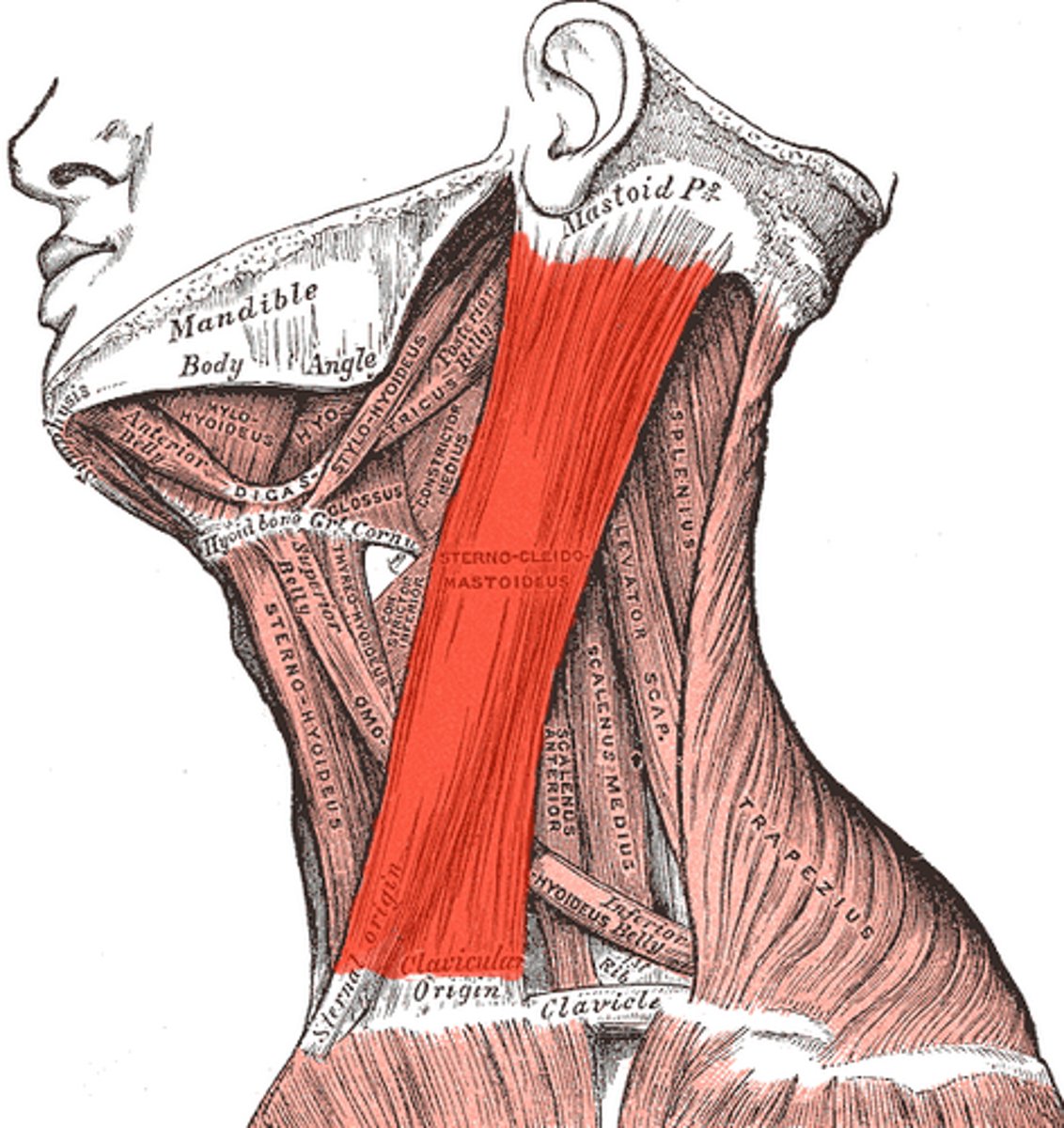
action: rotates head to the opposite side unilaterally, flexes the neck bilaterally, accessory muscle of respiration
innervation: spinal accessory nerve (motor) and C2 and C3 VPR (sensory)
what is the action and innervation of the sternocleidomastoid?
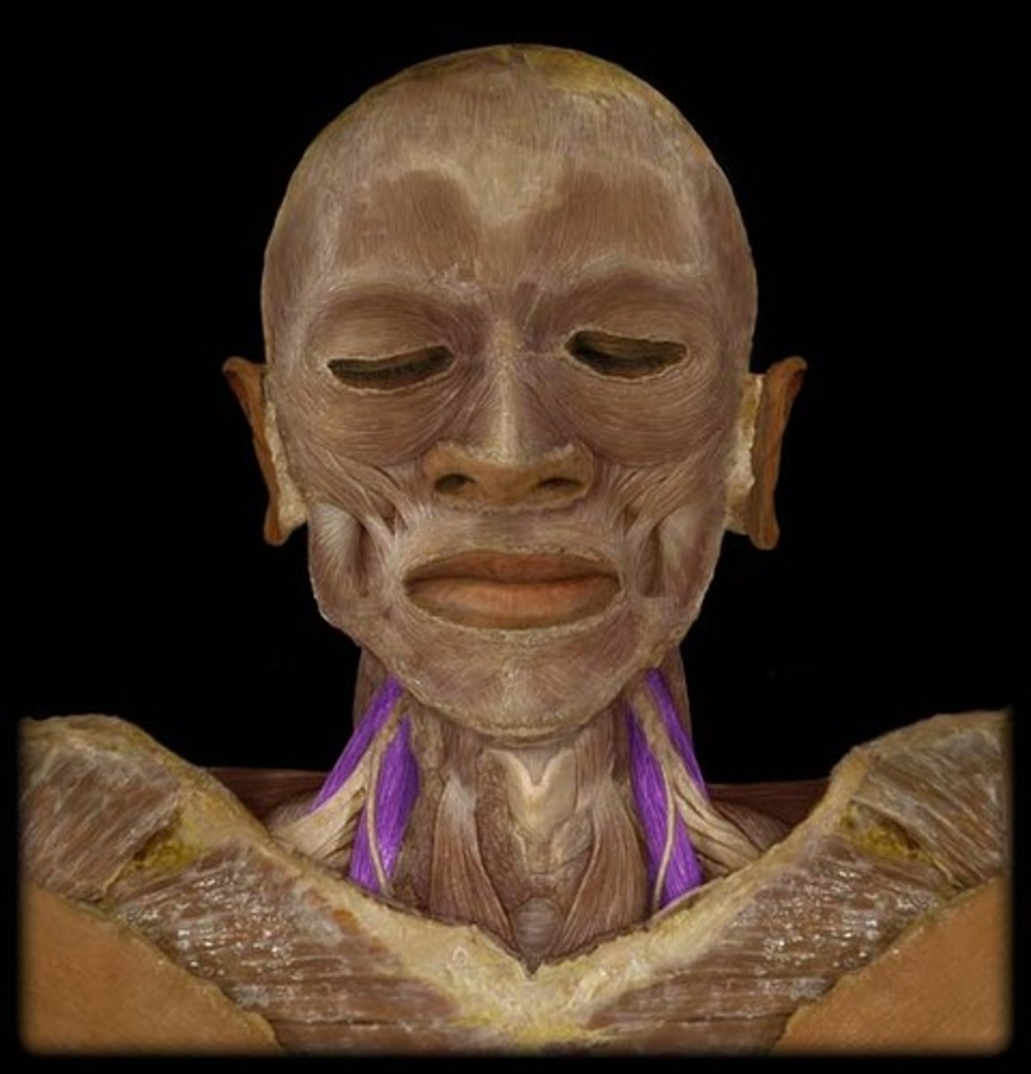
platysma muscle
found in the superficial fascia of the anterolateral neck; runs superficial to the EJV and visible cutaneous nerves; spans fascia over clavicle to inferior mandible
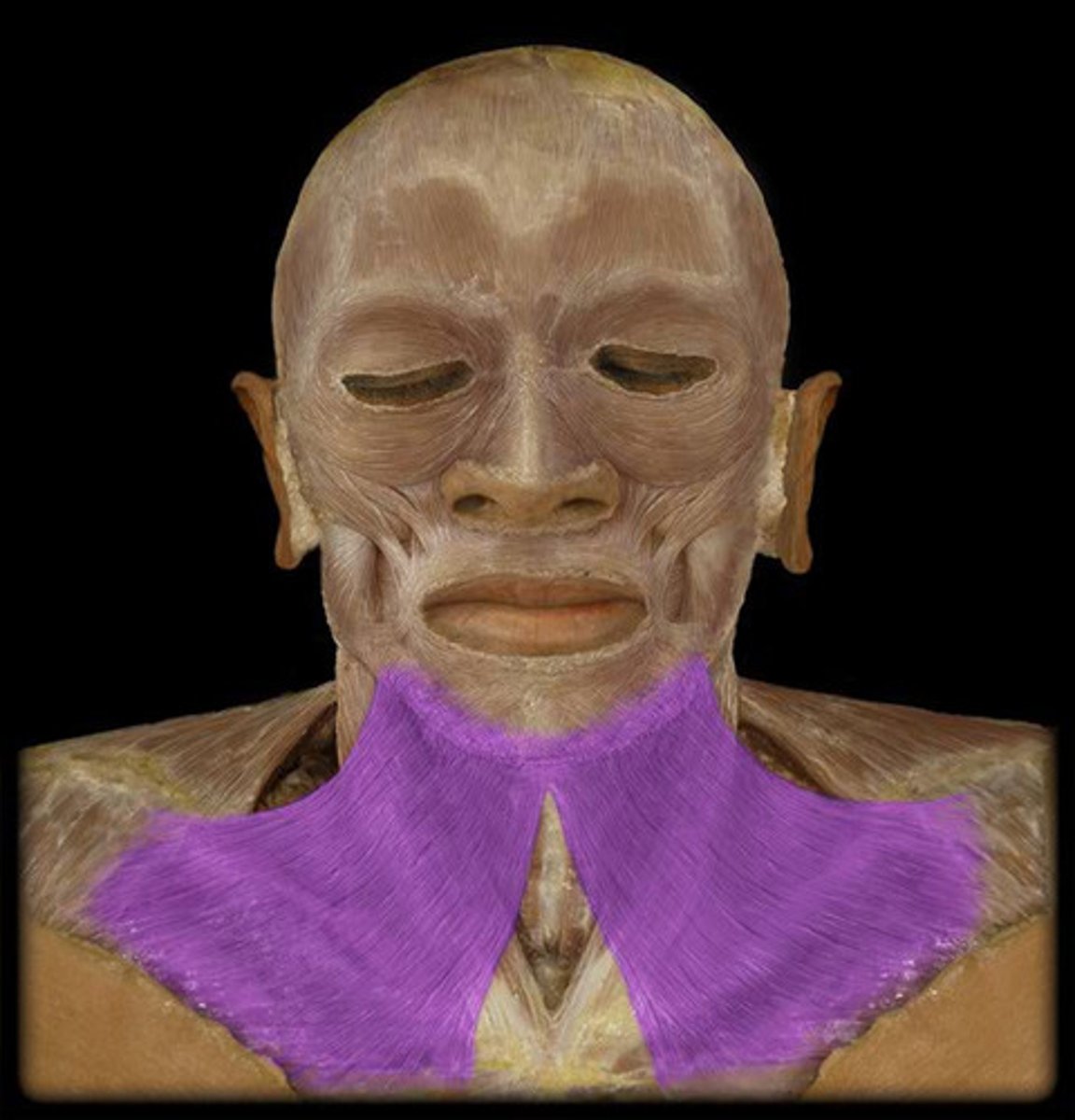
action: tense neck skin and depress angle of the mouth
innervation: cervical branch of facial nerve (CN V||)
what is the action and innervation of the platysma?
origin: upper border of scapula just medial to the scapular notch
insertion: hyoid bone
what is the origin and insertion of the omohyoid muscle?
inferior belly: in the posterior triangle; separates the occipital and omoclavicular/subclavian sub-triangles
superior belly: in the anterior triangle
where are the two bellies of the omohyoid located?
ansa cervicalis (C1-3 VPR)
what is the innervation of the omohyoid muscle?
anterior scalene
what is the key landmark in the posterior triangle?
origin: transverse process of CV3-6
insertion: scalene tubercle of rib 1
what is the origin and insertion of the anterior scalene?
action: flex neck and assist forced inhalation bilaterally; lateral flexion unilaterally
innervation: cervical VPRs
what is the action and innervation of the anterior scalene?
- prevertebral fascia
- subclavian vein
- phrenic nerve
- ascending cervical artery
- transverse cervical artery
- suprascapular artery
- omohyoid muscle
what is superficial and anterior to the anterior scalene muscle?
- subclavian artery
- roots of VPR forming the brachial plexus
what is between the anterior and middle scalene muscles?
origin: transverse processes of CV2-7
insertion: rib 1 posterior to the groove for the subclavian artery
what is the origin and insertion of the middle scalene?
action: flex neck and assist forced inhalation bilaterally; lateral flexion unilaterally
innervation: C3-C7 VPR
what is the action and innervation of the middle scalene?
origin: transverse processes of CV4-6
insertion: second rib
what is the origin and insertion of the posterior scalene?
action: flex neck and assist forced inhalation bilaterally; lateral flexion unilaterally
innervation: C5-C7 VPR branches
what is the action and innervation of the posterior scalene?
- 1st rib
- anterior scalene
- middle scalene
what are the borders of the interscalene triangle?
roots of the brachial plexus and the 3rd part of the subclavian artery
what is in the interscalene triangle?
Erb's point
nerve point on the neck
lesser occipital nerve (C2)
follows posterior border of SCM to reach the mastoid process and behind the eat
great auricular nerve (C2-3)
large; courses vertically on SCM towards the angle of the mandible and parotid region
transverse cervical nerve (C2-3)
crosses SCM horizontally towards the anterior triangle of the neck
suprascapular nerves (C3-4)
crosses the clavicle towards the inferior posterior triangle and upper chest and thorax
dorsal scapular nerve and long thoracic nerve
pierces the lateral side of the middle scalene
inside the carotid sheath, anterior to subclavian artery, and medial to phrenic enrve
where is the vagus nerve located as it passes from the neck to the thorax?
right: right recurrent laryngeal nerve at the right subclavian artery
left: left recurrent laryngeal nerve at the arch of the aorta
what do the right and left vagus nerves give off?
descends on the anterior surface of the anterior scalene (lateral to the ascending cervical artery), enters superior thoracic aperture, courses along pericardium to the diaphragm
what path does the phrenic nerve take as it passes from the neck to the thorax?
- motor innervation to the diaphragm (severance paralyzes the ipsilateral hemidiaphragm)
- sensory innervation from the parietal peritoneum in the right upper quadrant of the abdomen (irritation causes referred pain to the shoulder)
what is the function of the phrenic nerve?
- passes between the anterior and middle scalenes
- ends at the lateral border of the first rib (where it changes to axillary artery)
where are the subclavian arteries located?
anterior scalene
what divides the subclavian artery into three parts?
- vertebral artery: enters the transverse foramen of CV6; ascends to supply the brain and brainstem
- internal thoracic artery: descends posterior to the sternal end of the clavicle into the thorax
- thyrocervical trunk: extends superiorly from the subclavian; splits into the inferior thyroid artery, transverse cervical artery, and suprascapular artery
what branches off the first part of the subclavian artery?
costocervical trunk (from posterior aspect of the vessel; deep to the anterior scalene)
- branches into the deep cervical artery (supplies deep muscles on back of neck) and the highest intercostal artery (supplies the first two intercostal spaces)
what branches off the second part of the subclavian artery?
dorsal scapular artery (weaves between the superior and middle trunks of the brachial plexus)
what branches off the third part of the subclavian artery?
external jugular vein
formed by union of the retromandibular and posterior auricular veins; runs vertically superficial to the SCM; drains into the subclavian vein
internal jugular vein
begins at jugular foramen on skull; runs laterally within the carotid sheath; ends by joining the subclavian vein to form the brachiocephalic vein
subclavian vein
begins at lateral border of first rib; passes anterior to the anterior scalene muscle; joins the internal jugular vein to from the brachiocephalic vein
internal jugular vein and subclavian veins
what makes the venous angle?
right lymphatic duct
drains the R upper limb and shoulder, R upper thorax, and R head/neck
thoracic duct
drains everything that the right lymphatic duct doesn't
trachea
first tracheal cartilage at ~CV7 level; 6-8 tracheal rings palpable in the neck; isthmus of thyroid gland covers rings 2-3
esophagus
begins at the pharyngoesophageal junction at CV6; passes posterior to the trachea
cervical pleura and lung apex
apex of lungs and cervical pleura extend above rib 1 into the posterior triangle; at risk during central line placement or supraclavicular surgery