Week 4: 12-Lead ECG
1/63
There's no tags or description
Looks like no tags are added yet.
Name | Mastery | Learn | Test | Matching | Spaced | Call with Kai |
|---|
No analytics yet
Send a link to your students to track their progress
64 Terms
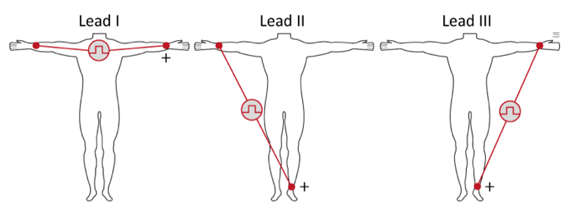
What limb leads view the heart in the frontal plane using bipolar electrodes?
Standard limb leads (I, II, and III)

What mnemonic helps remember limb lead electrode placement?
“Smoke over Fire” and “Snow over Grass.”

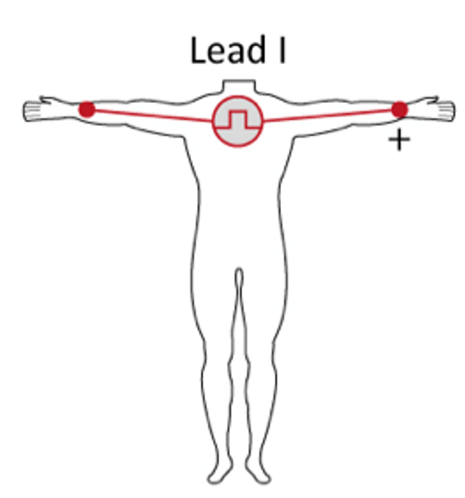
Which limb lead records electrical activity from right arm to left arm?
Lead I
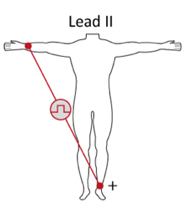
Which limb lead records electrical activity from right arm to left leg?
Lead II

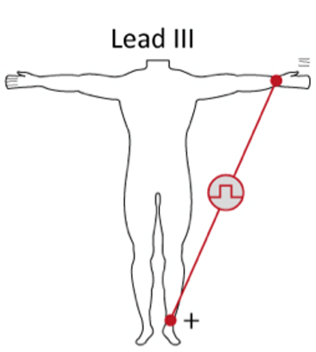
Which limb lead records electrical activity from left arm to left leg?
Lead III
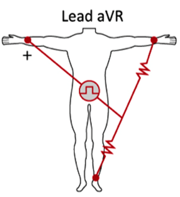
Which augmented lead looks at the heart from the right shoulder?
aVR

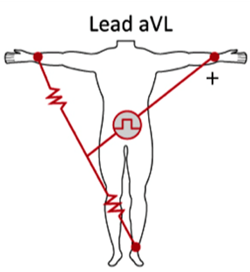
Which augmented lead looks at the high lateral wall of the heart?
aVL
Which augmented lead looks at the inferior wall of the heart?
aVF
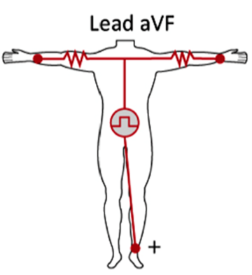
What makes up a unipolar augmented lead?
Unipolar Augmented Lead = 1 Physical Electrode + 1 Virtual Electrode
What structure mathematically creates the virtual negative pole for augmented leads?
Goldberger’s Central Terminal
What does Einthoven’s Law state?
Lead I + Lead III = Lead II.
What happens to R-wave amplitude in Lead II according to Einthoven’s Law?
Lead II R-wave amplitude = Lead I R-wave amplitude + Lead III R-wave amplitude
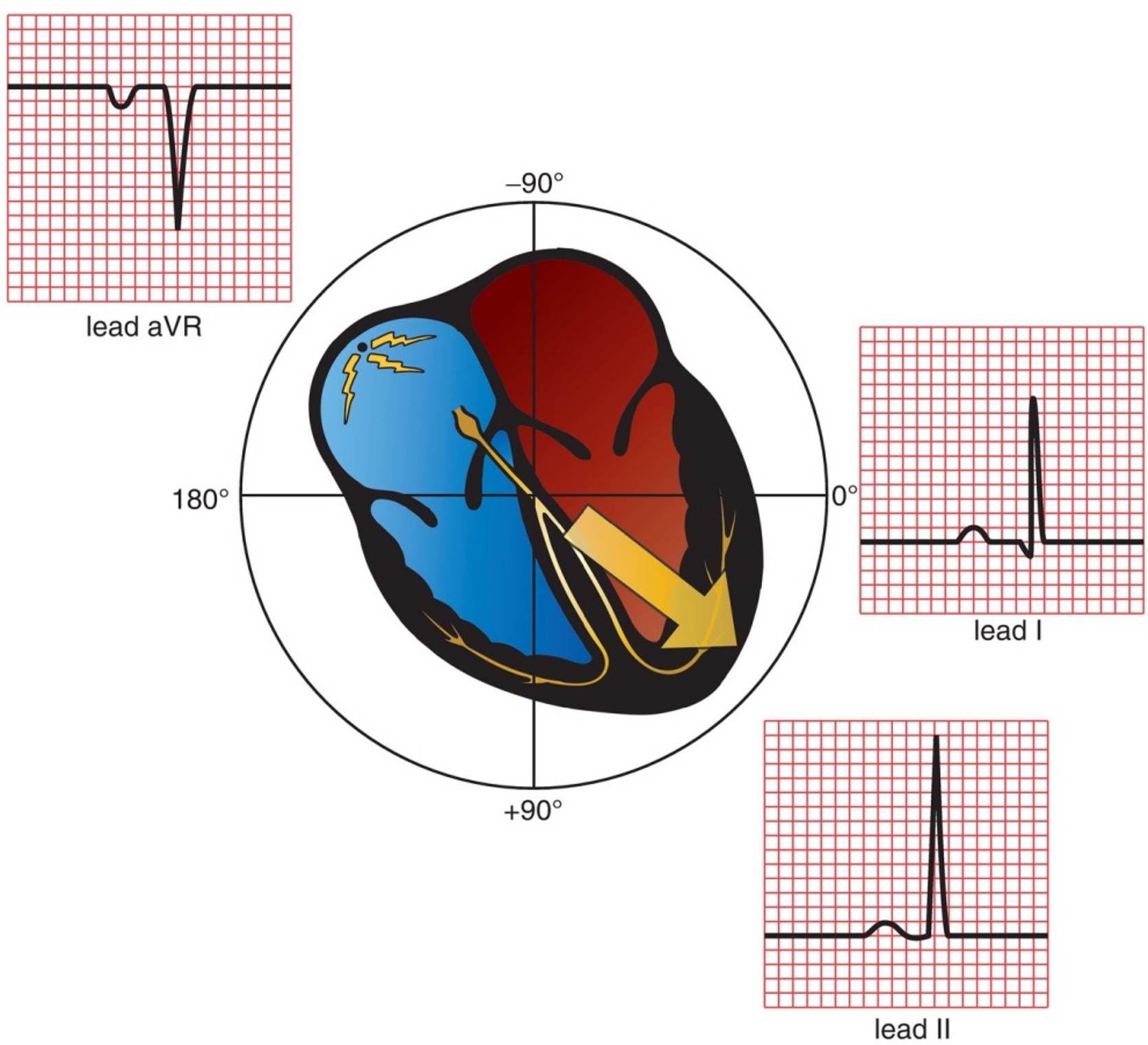
What do negative angles represent in the hexaxial reference system?
Superior leads
What do positive angles represent in the hexaxial reference system?
Inferior leads
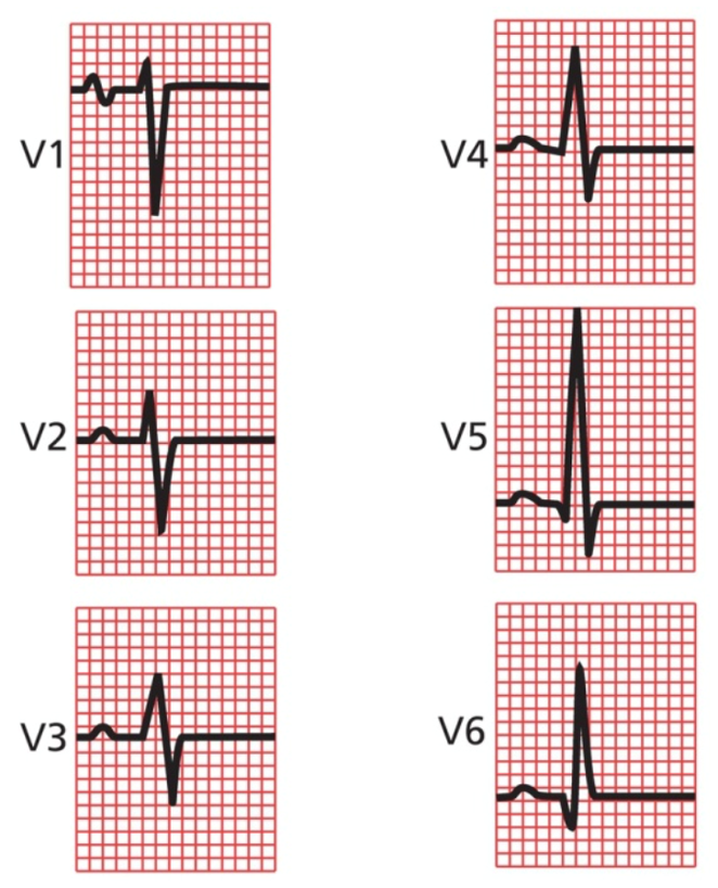
Which leads view the heart in the horizontal plane?
Precordial (V1–V6) leads
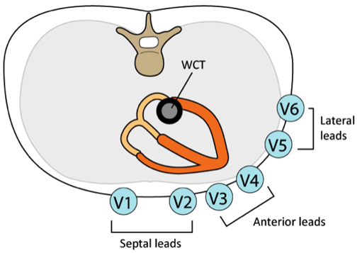
What creates the negative pole for precordial leads?
Wilson’s Central Terminal
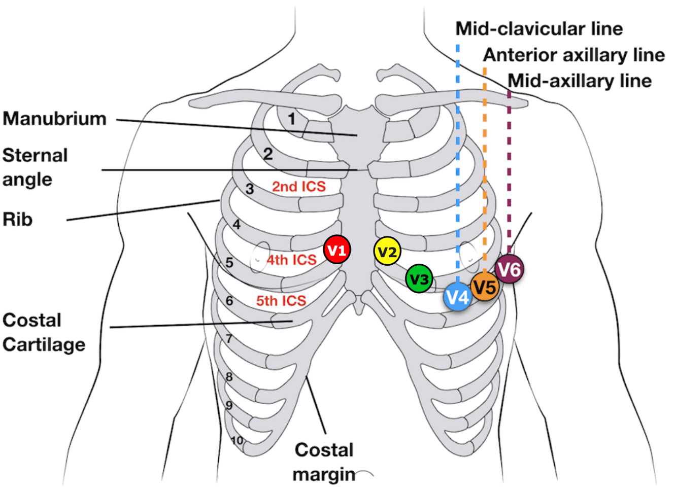
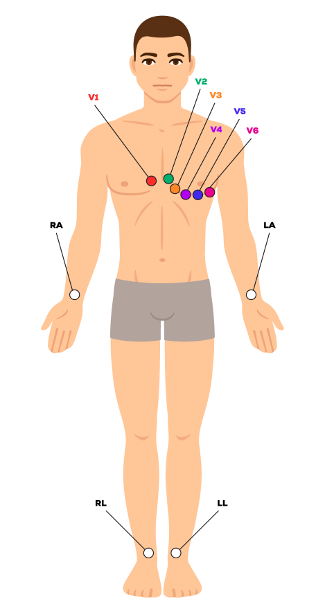
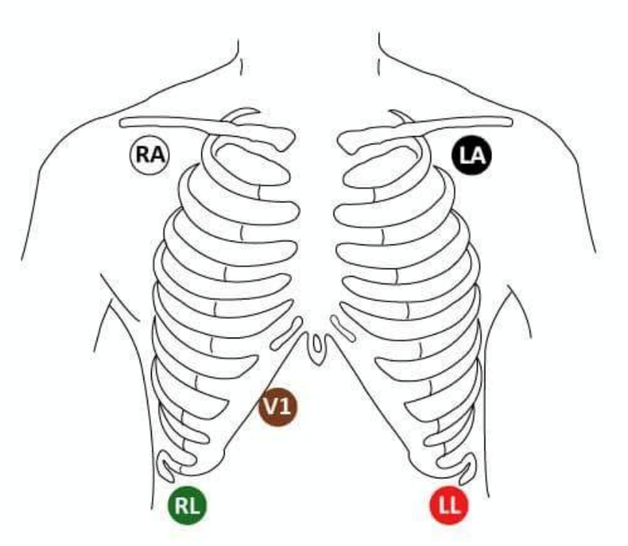
Which chest lead is placed at the 4th intercostal space right sternal border?
V1

Which chest lead is placed at the 4th intercostal space left sternal border?
V2

Which chest lead is placed midway between V2 and V4?
V3

Which chest lead is placed at the 5th intercostal space midclavicular line?
V4

Which chest lead is placed at the anterior axillary line level with V4?
V5

Which chest lead is placed at the midaxillary line level with V4?
V6
Which lead usually has the tallest positive P wave?
Lead II

Which lead usually has the most negative P wave?
Lead aVR

Which limb leads usually show positive P-wave deflection?
I
Which limb lead commonly records a biphasic P wave?
Lead III
Which precordial leads usually show positive P waves?
V5 and V6
Which precordial lead commonly shows a biphasic P wave?
V1
Which chest leads have variable P-wave appearance?
V2–V4
In what direction does septal depolarization normally travel?
Left to right
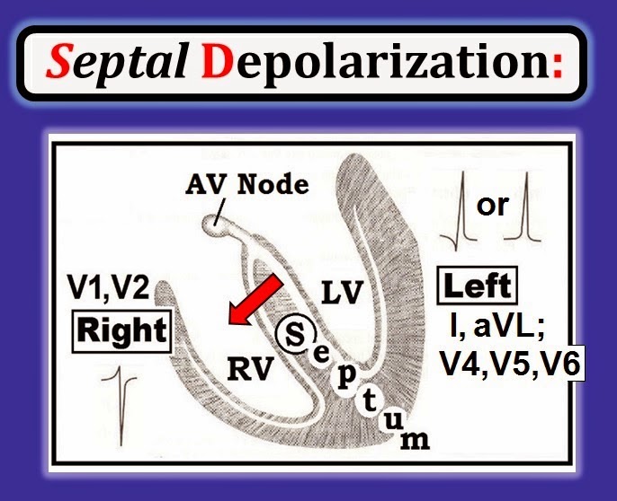
What ECG deflection represents septal depolarization?
Small Q wave
Which leads commonly show normal septal Q waves?
I

What is the normal amplitude limit of septal Q waves?
Usually ≤0.1 mV
Which chamber dominates most of the QRS complex?
Left ventricle
What is the general direction of the mean QRS vector?
Caudad and leftward
Which limb leads usually show large positive R waves?
Left Lateral Leads (I, aVL, V5, V6) and Inferior Leads (II, III, aVF)
Which lead commonly shows a deep negative S wave?
aVR
Which chest lead commonly has a deep S wave?
V1

Which chest leads usually have tall positive R waves?
V5 and V6
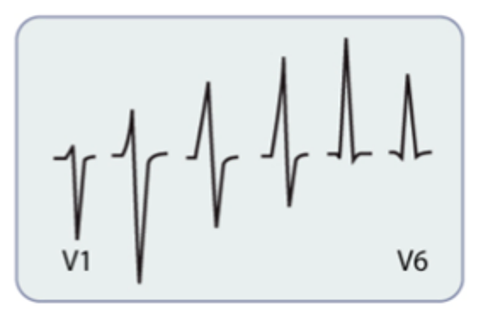
Which chest leads are considered the transition zone?
V3 and V4

What is R-wave progression?
R-wave amplitude increases from V1 to V5
What does the T wave represent?
Ventricular repolarization
Why are T waves usually positive in the same leads as positive R waves?
Repolarization travels opposite depolarization
What is the normal heart rate range in sinus rhythm?
60–100 BPM
What rhythm characteristic defines normal sinus rhythm?
Regular rhythm with a round, consistent P wave before every QRS in a 1:1 ratio.
What is the normal P-wave axis?
0° to +75°
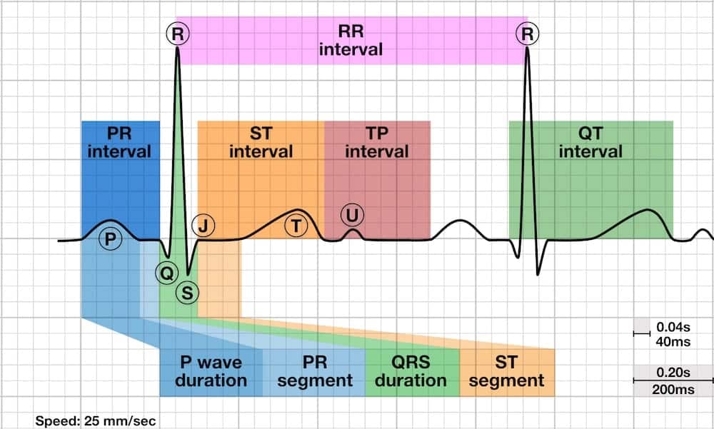
What is the normal PR interval?
120–200 ms
What is the normal QRS duration?
60–100 ms
What is the normal QT interval?
<440 ms
Which leads should normally have a positive QRS complex?
I, II, aVF and V3–V6
Which lead should normally have a negative QRS complex?
aVR

Which leads are considered inferior leads?
II and aVF
Which leads are considered left lateral leads?
I and aVL
Which leads are considered anterior leads?
V2–V4
Which lead is considered the right ventricular lead?
V1
How many skin electrodes are used to create a 12-lead ECG?
10 electrodes

How many total ECG leads are generated from 10 electrodes?
12 leads
Why is standard electrode positioning important?
It allows comparison between ECGs
What color electrode is placed on the right arm in a 5-lead ECG?
White

What color electrode is placed on the left arm in a 5-lead ECG?
Black
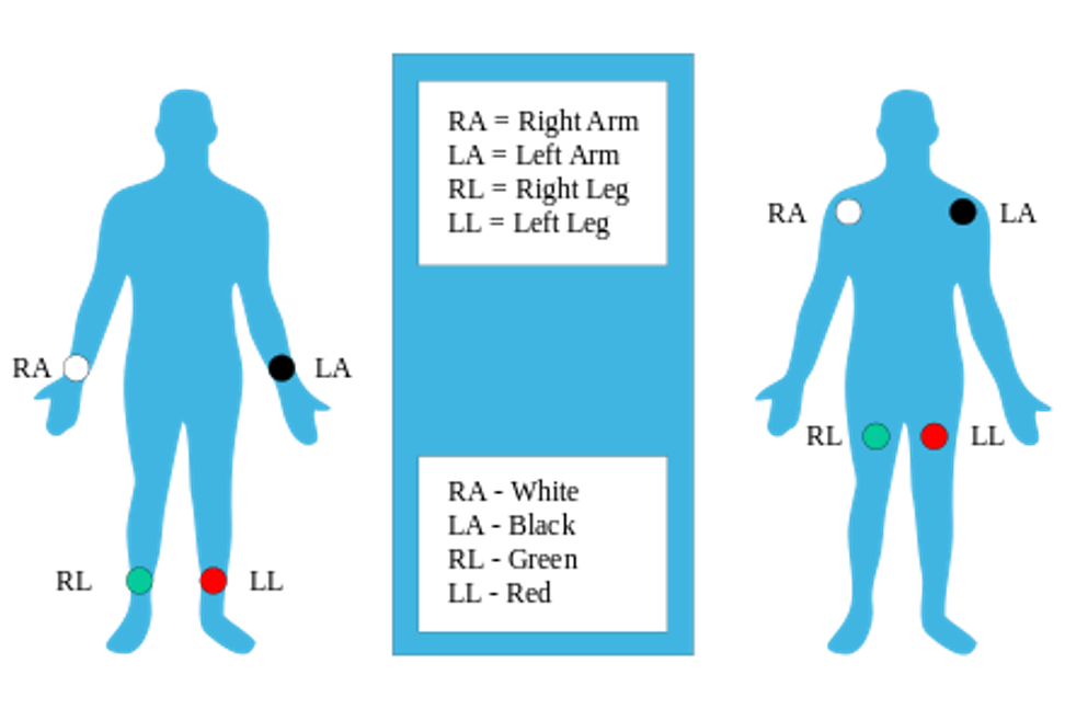
What color electrode is placed on the left leg in a 5-lead ECG?
Red

What color electrode is placed on the right leg in a 5-lead ECG?
Green

What color electrode is typically used for the chest lead in a 5-lead ECG?
Brown
Which lead is most commonly monitored in a standard 3-lead ECG?
Lead II
