CT 1 (parts + important terms)
1/42
There's no tags or description
Looks like no tags are added yet.
Name | Mastery | Learn | Test | Matching | Spaced | Call with Kai |
|---|
No analytics yet
Send a link to your students to track their progress
43 Terms
Gantry Aperture
1

Microphone
2

Sagittal Laser Alignment Light
3

Patient Guide Light
4
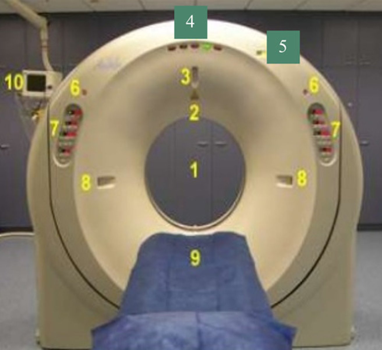
X-Ray Exposure Indicator Light
5

Emergency Stop Buttons
6

Gantry Control Panels
7

External Laser Alignment Lights
8

Patient Couch
9

ECG Gating Monitor
10

X-Ray Tube
1

Filters, Collimator
2

X-ray Tube Heat Exchanger (Oil Cooler)
4

High Voltage Generator (0-75 kV)
5

Direct Drive Gantry Motor
6

Rotation Control Unit
7

Data Acquisition System (DAS)
8
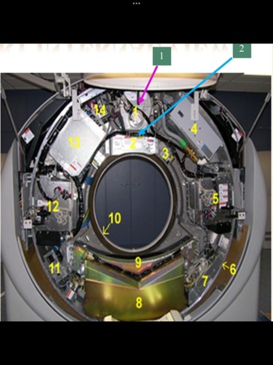
Detectors
9

Slip Rings
10

Detector Temperature Controller
11

High Voltage Generator (75-150 kV)
12

Power Unit (AC to DC)
13

opening of the gantry through which the patient passes during the scan
Aperture
Preset amount of radiopaque contrast medium injected rapidly per IV administration to visualize high-flow vascular structures, usually in conjunction with dynamic scan; most often injected using a pressure injector
Bolus
In multidetector CT, multiple rows of detectors are arranged along the longitudinal (z) axis of the patient. Each detector row consists of numerous elements
Channel
Arbitrary number assigned by computer to indicate relative density of a given tissue; CT number varies proportionately with tissue density
CT number
Image distortion caused by combination of table indexing and respiration; table moves in specified increments, but patient movement during respiration may cause anatomy to be scanned more than once or not at all.
Image misregistration
Spatial resolution of a voxel in which all three axes of the volume element are equal. Slice thickness is equal to pixel size
Isotropic spatial resolution
assignment of appropriate gray level to each pixel in an image
Mapping
Instructions for CT examination specifying slice thickness, table incre-ments, contrast administration, scan diameter, and any other requirements specified by the radiologist.
Protocol
Ability to process or reconstruct incoming data in milliseconds.
Real time
Process of creating a digital image from raw data.
Reconstruction
Process of changing the shading of a three-dimensional image; commonly used to increase depth perception of an image.
Rendering
Actual rotation of x-ray tube around the patient; used as a generic reference to one slice or an entire examination.
Scan
Also referred to as the zoom or focal plane of a CT scan; predetermined by the radiographer to include the anatomic area of interest; determines FOV.
Scan diameter
Amount of time used to scan an entire volume during a single spiral scan.
Scan duration
X-ray exposure time in seconds.
Scan time
Method of cropping or editing target objects from image data.
Segmentation
One scan through a selected body part; also referred to as a cut; can vary from 0.35 mm to 1 cm, depending on the exam
Slice
low-voltage electrical contacts within the gantry designed to allow continuous rotation of an x-ray tube without the use of cables connecting internal and extenders component
Slip ring
Individual pixel with the associated volume of tissue based on the slice thickness.
Voxel
controls the overall gray level and affects image contrast
Window width
controls subtle gray images within a certain width range and ultimately affects the brightness and overall density of an image
Window level