Chapter 10
1/169
Earn XP
Description and Tags
digestion
Name | Mastery | Learn | Test | Matching | Spaced | Call with Kai |
|---|
No analytics yet
Send a link to your students to track their progress
170 Terms
The gastrointestinal (GI) tract consists of a tube that extends from mouth
to anus
The contents of this tube are surprisingly not part of our bodies until they are
digested and absorbed into our blood stream to be used for cellular metabolism and growth
Mechanical digestion begins in the
mouth (buccal cavity) by mastication (chewing)
Mechanical digestion does what
This increases surface area to volume ratio and moistens food with secretions from salivary glands
Once swallowing is initiated, one direction movement is accomplished by
rhythmic, muscular contractions (peristalsis) of the muscularis externa
Chemical digestion is accomplished by
enzymes from the mouth, stomach, and most importantly, pancreatic secretions into the small intestines.
Our digestive tract breaks down ingested food to
macromolecules such as glucose, amino acids, fatty acids, and nucleic acids using enzymes during the process of digestion.
Absorption of water and nutrients occurs through the
intestinal lining
The small intestine absorbs most
nutrients, vitamins, and water
The large intestine hosts a plethora of
microbes, completes water reabsorption, and compacts feces
The entire length of the gi tract is made of four layers:
the mucosa
submucosa
muscularis externa
and serosa or adventitia
what is the serosa
outermost thin layer of the last portion of esophagus to the sigmoid colon
what is the adventitia
outermost layer of the pharynx and most of esophagus and rectum
Both the serosa and adventitia serve to anchor the
tube to surrounding tissue
Regional specializations occur in each major division of the
GI tract
Modifications of the mucosa and submucosa layers dictate the type of
secretion introduced into the lumen of the tube
the secretions from the entrire GI tract averages about
7–8 liters/day
This large amount of body water must be reabsorbed, mainly by the intestines or dehydration can occur.
The digestive tract, or alimentary canal, consists of the
esophagus, stomach, small intestines, and large intestine
Each region contains specialized glands and modifications.
The esophagus secretes
mucous to reduce friction as food is swallowed.
what is mucous
esophageal glands in the submucosa
The stomach produces
hydrochloric acid (HCl) and pepsinogen from mucosal gastric glands
The small intestine contains
villi and microvilli
what is modifications of the mucosa
microvilli
The large intestine possesses
crypts which aid in water reabsorption and mucous secretion
Peristalsis pushes food from mouth
to anus along the tube
what is Peristalsis
muscular wave of contraction of the muscularis externa
As food fills the stomach (now called chyme), it
stretches
The gastric rugae (wrinkles) allow this to occur
The muscular wall contractions mix the ingested contents with
secretions from mucous cells (mucous)
parietal cells (HCl)
chief cells
enteroendocrine hormones that regulate digestion.
what is chief cells
pepsinogen, an inactive form of the protein digesting enzyme, pepsin
HCl (hydrochloric acid) is a strong acid that helps kill
ingested pathogens and activates pepsinogen into pepsin.
The duodenum is the first segment of the
small intestines and is about 25 centimeters or 10 inches in length
As chyme exits the pylorus of the stomach, in small amounts controlled by the pyloric sphincter into the duodenum, the acidic contents and stretching of the duodenal wall trigger the release of
enzymes, sodium bicarbonate, and zymogens from the pancreas
Bile salts from the gallbladder help emulsify
fats
what does emulsify mean
break large lipids into tiny, suspended droplets
Alkaline mucus secretions of duodenal glands and brush border enzyme activity of the duodenal epithelium help neutralize
stomach acid and activate pancreatic enzymes
The circular folds (plicae circulares) help mix the chyme with
enzymes, mucus, and bile salts.
All enzymes of digestion are produced by the
pancreas
Starch, fat, protein, DNA, and RNA are all broken down to the major
macromolecules
glucose, fatty acids, amino acids, and nucleotides
The pancreas produces and secretes —— into the duodenum
enzymes, zymogens, and sodium bicarbonate
what us zymogens
inactive form of protein digesting enzymes
what does sodium bicarbonate do
buffers the acidic chyme from the stomach
The liver produces thousands of unique
proteins for various of functions
Bile formation has the only role in
digestion
Bile is composed of
bile salts, minerals, and pigments.
Pigments are produced by the breakdown of the heme portion of
hemoglobin into bilirubin
Only bile salts contribute to digestion by emulsifying and preparing
fats for absorption
Bile salts are derived by the addition of
glycine or taurine to cholesterol
This creates ampipathic molecules that separate fat globules into lipid droplets (emulsify)
Bile salts then combine with fatty acids and monoglycerides and form
tiny micelles, about 4–7 nanometers in diameter
can be absorbed into columnar epithelial cells of the small intestines.
The jejunum is where most
chemical digestion and absorption of nutrients takes place
facts about the jejunum
It’s about a meter and a half long and is highly vascula
Pancreatic enzymes continue to actively digest introduced
chyme
villi and microvilli greatly increase the surface area of the
jejunum’s epithelium
jejunum’s epithelium is where
where membrane transport pumps move nutrients into the circulatory system or lymphatic (fats) system
About the last half of the small intestines is the
ileum
More digestion and absorption of nutrients takes place here
The ileum transforms in structure as it approaches the
cecum of the large intestine
Clusters of lymphatic tissue (Peyer’s patches) become more
prevalent, housing numerous lymphocytes to resist infection.
Villi and microvilli greatly increase the surface area of the small intestines about
200 times
Without these structures, our intestines would need to be about 1,200 feet long to provide the same surface area
All nutrient absorption occurs through the
simple columnar epithelial cells of the mucosa
The large intestine is about
1.5 meters long and extends from the cecum to the rectum
The mucosa of the large intestine is
simple columnar epithelium that’s specialized for absorption
Motility is slower in the
large intestine
allowing most of the water of chyme that enters to be reabsorbed
motility is important in preventing dehydration, which is a serious concern during extended bouts of
diarrhea
The colon is home to about 800 species of
bacteria that number in the billions
the bacteria in our colon do what
They further digest cellulose (we do not make enzymes that can break it down) and release glucose.
some bacteria produce
sulphur containing gas that gives rise to the foul odor of flatus
About 60% of the dry mass of feces is
bacteria
The epithelium makes a transition from simple columnar in the rectum to
stratified squamous in the anal canal.
Splenic flexure
the spleen can be seen through the colon wall at the sharp bend between the transverse and the descending colon
Cecum
beginning of the large intestine
Appendiceal orifice
opening into the appendix
Terminal ileum
last region of the small intestines
Ileo-cecal valve
prevents material in the colon from traveling back into the small intestine
Diverticulum in the ascending colon
diverticula are pouches that form in the mucosa and submucosa causing a bulge
Rectum
about 12 cm long
As material fills, the rectal walls expand and stimulate stretch receptors involved in defecation.
Parotid salivary gland
produces the majority of saliva, which helps moisten masticated food (bolus) through the esophagus
Wisdom teeth (3rd molars)
sometimes do not descend or have to be pulled to make room

a.
Anal canal

b
Mid-descending colon

c.
Splenic flexure
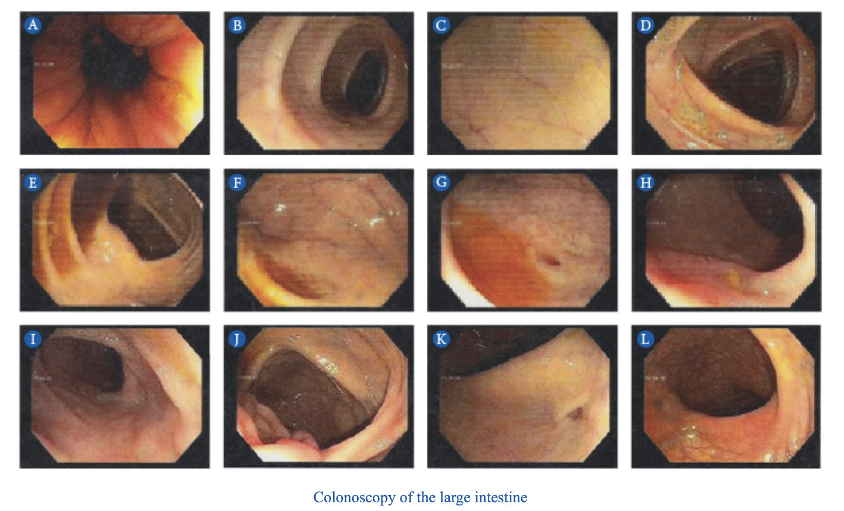
d
Mid-transverse colon

e
e. Cecum

f
f. Appendiceal orifice

g
g. Appendiceal orifice

h
h. Terminal ileum

i
i. Terminal ileum

j
j. Ileo-cecal valve

k.
Diverticulum in the ascending colon

L
L. Rectum

a
Ileum

b
Ileocecal valve

c
Cecum

d
Appendix

e
Ascending colon

f
transverse colon

g
Descending colon

h
Sigmoid colon

i
rectum
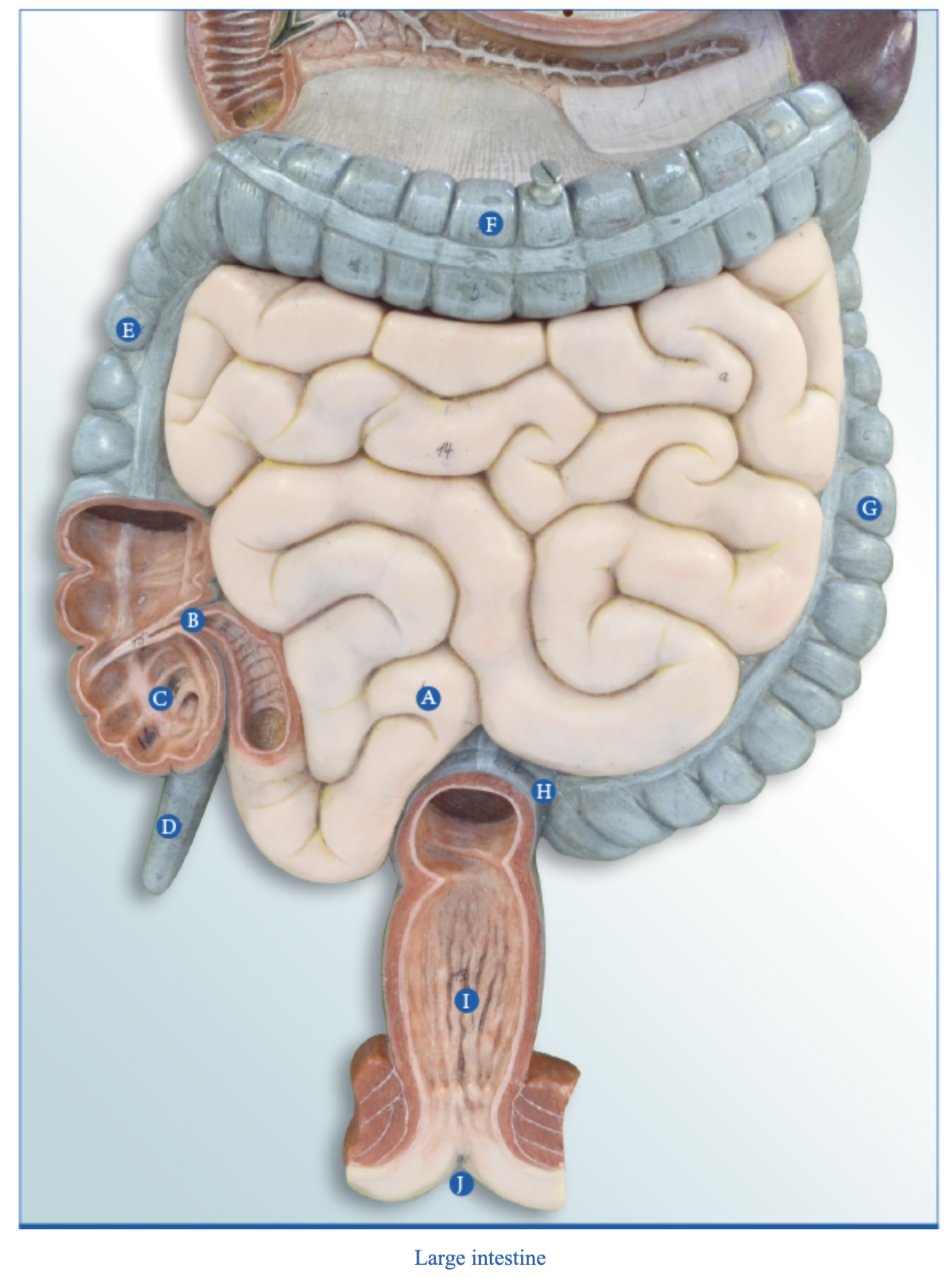
j
anus

a
Villus
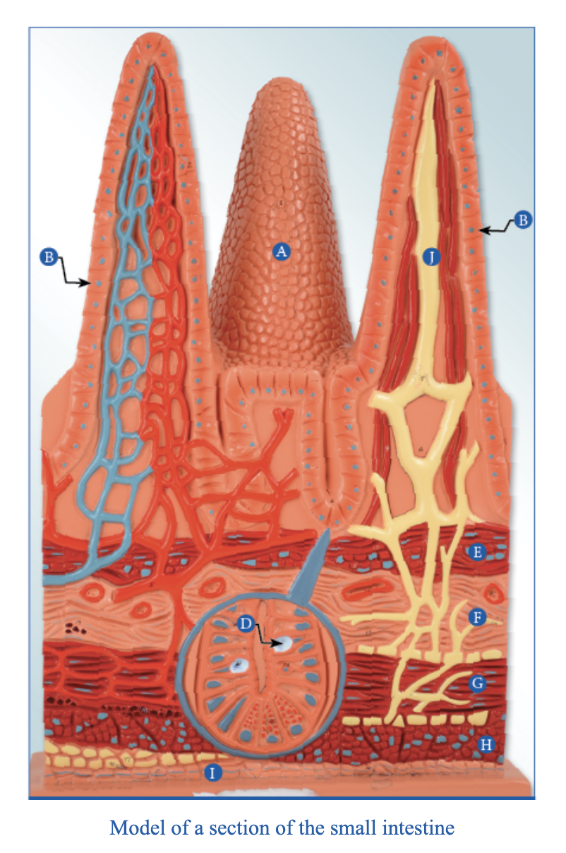
b
Simple columnar epithelial cells