Cytoplasmic membrane part 2
 Call Kai
 Learn
Learn Practice Test
 Spaced Repetition
 Match
Match Flashcards
 Knowt Play
1/160

Earn XP
Description and Tags
Cell Biology
Name | Mastery | Learn | Test | Matching | Spaced | Call with Kai |
|---|
No analytics yet
Send a link to your students to track their progress
161 Terms
TGN
(trans-Golgi network) the Major Processing Center where Proteins Destined for Different Sites are Sorted into Different Membrane-Bound Carriers
The best understood post-Golgi pathway involves
Lysosomal enzymes
Where are Lysosomal enzymes synthesized?
they are Synthesized in the RER and Carried to the Golgi, Where they are Recognized Due to Signal Patches (Signals that are Part of their 3D structure)
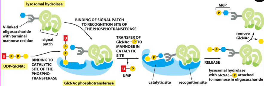
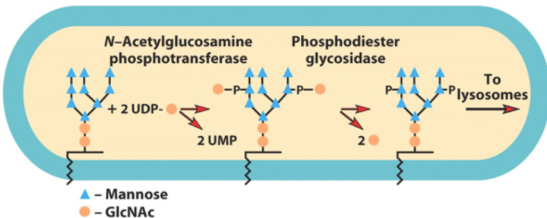
How are lysosomal enzymes recognized?
by Enzymes that Catalyze the 2-Step Addition of a Phosphate Group to Certain Mannose Residues that Act as a Recognition Signal
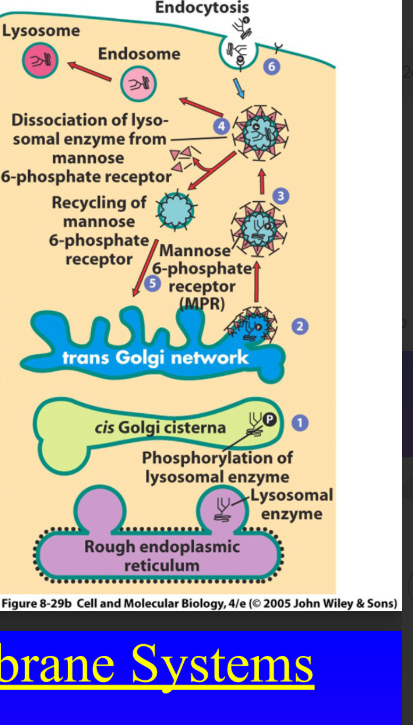
Lysosomal Enzymes Carrying Mannose-6-Phosphate Signal are recognized by what?
they are Recognized and Captured by Mannose-6-Phosphate Receptors, that are Integral Membrane Proteins of the TGN
How are lysosomal enzymes transported from the TGN?
in Clathrin-coated vesicles who has a Honeycomb-Like Lattice of the Protein Clathrin
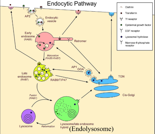
What does clathrin do for adaptor proteins?
Forms a Scaffold for an Inner Shell of Adaptor Proteins that Cover the Surface of the Vesicle that Faces the Cytosol
What proteins escort lysosomal enzymes from the TGN
adaptor proteins like GGAs or AP complexes
AP-1 and GGAs
are Associated with TGN Vesicles Moving to Early, Late or Recycling Endosomes
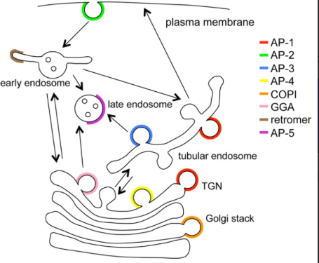
AP-2
is Involved with the Endocytosis of Vesicles from the Plasma Membrane to Early Endosomes
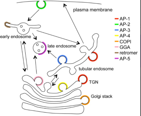
AP-3
is Involved with the Movement of Vesicles from Recycling (Tubular) Endosomes to Late Endosomes
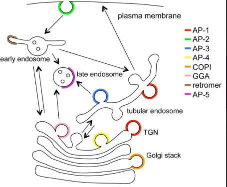
AP-4
is Involved with the Movement of Vesicles Between the TGN, Early, Late and Recycling Endosomes
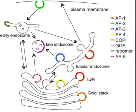
AP-5
is Involved with the Movement of Vesicles Between Late Endosomes and Lysosomes
retromer coat complexes
are Involved in Vesicles Returning Mannose-6-Phosphate Receptors from Early or Recycling Endosomes to the TGN
TIP47 (perilipin)
is Involved in Vesicles Returning Mannose-6-Phosphate Receptors from Late Endosomes to the TGN
Which AP complexes can associate with Clathrin?
The AP complexes that can associate with Clathrin include AP-1, AP-2, and AP-3, AP-4
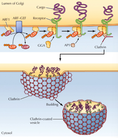
Clathrin-Coated Vesicles have Adaptor Proteins Like GGA, that are Recruited to the Membrane by
ARF1-GTP (GGA can then recruit AP1)
Adapter proteins bind to
the sorting signal in the membrane proteins such as the mannose-6-phosphate receptor, helping to recruit clathrin and other coat proteins for vesicle formation.
what are mannose-6-phosphate receptors bound to?
soluble lysosomal enzymes in the vesicle lumen
the G-protein ARF1 Effectively Allows
the Binding of Coat Proteins at the Site where ARF1 is Itself
Bound to the Membrane
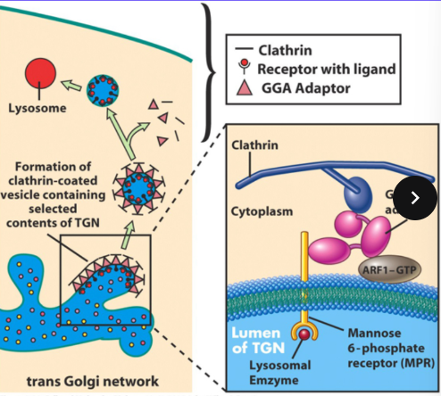
Once the Vesicle Buds from the TGN
ARF1’s GTP is Hydrolyzed to GDP, Releasing the Coat, the Clathrin is Shed into the Cytoplasm Again The Uncoated Vesicle then Fuses with Early, Late or Recycling Endosomes
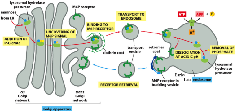
What happens in endosomes with lower pH, particularly late endosomes?
the Mannose-6-Phosphate Receptors Release the Bound Soluble Lysosomal Enzymes who Carry on their Route to the Lysosome, or the Late Endosome Fuses with a Lysosome; The Mannose-6 Phosphate Receptors are Returned to the TGN in Retromer or TIP47 Vesicles
What can mannose-6-phosphate receptors do for the plasma membrane?
they can be sent there to retrieve certain secreted enzymes from outside the cell using endocytosis
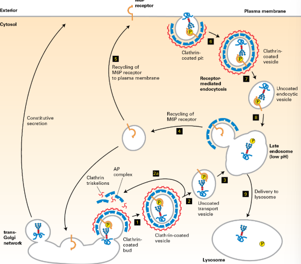
Secretory and Membrane Proteins Destined for the Plasma Membrane Also Leave the TGN in
vesicles or secretory granules
why do secretory or membrane proteins destined for the plasma membrane go through the TGN
Here the Proteins Concerned are Thought to Form Aggregates which are then Partitioned into Particular Membrane Bodies as the TGN Fragments into Vesicles/Secretory Granules
The Delivery of Integral Proteins to the Plasma Membrane Occurs Due to
Sorting Signals Present in their Cytoplasmic Tails
The Sorting Signals for Each Membrane Domain are Different and Sorting Occurs in the
TGN or recycled endosomes
What makes basolateral proteins targetable?
Short Hydrophobic Amino Acid Sequences Such as Di-Leucine (LL) or Tyrosine Containing Motifs
what makes apical proteins targetable?
although not fully understood but involves carbohydrate modifications rather than amino acid sequences
a known apical membrane protein sorting mechanism
Transcytosis, Where Proteins are Delivered Initially to the Basolateral Membrane Before Being Endocytosed
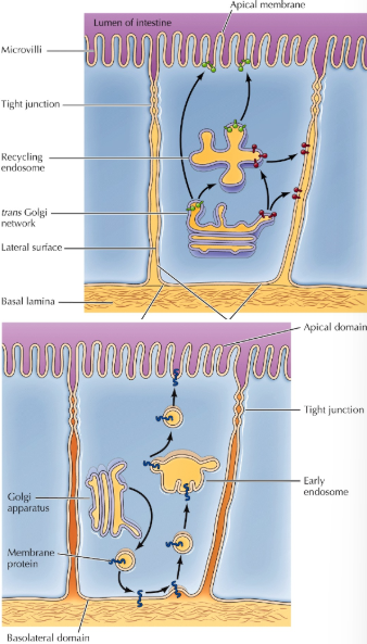
endocytosed proteins are re-sorted via
Early Endosomes and Sent to the Apical Membrane
Plasma Membrane Proteins in Non-Polarized Cells Do Not Need
Any Kind of Special Sorting Machinery and Can Just be Carried to the Surface in Vesicles of the Constitutive Secretory Pathway
selective fusion
One Factor that Ensures a Directed Flow Through the Various Endomembrane Compartments Starting at the ER
parts of the vesicle fusion process
1) Movement of Vesicles Towards the Target Compartment For Fusion is Mediated by Microtubules
2) Vesicles are Often Tethered Once they Reach the Target Compartment by Extended Fibrous Proteins
3) At Some Point During Fusion the Membranes Come Close to Each Other
4) When Artificial Liposomes Containing v- or t-SNAREs are Mixed, the 2 Types of Vesicles Fuse Together, but Not Vesicles of the Same Kind
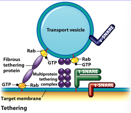
compatibility between the vesicle and the target compartment is determined by members of the
Rab G-Protein Family
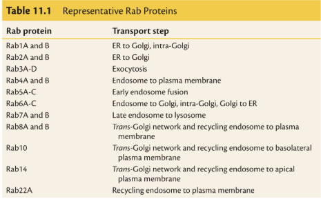
Rabs associate with membranes by a
lipid anchor
When GTP is Bound to the Rab G-Proteins, they are Thought to
Recruit Rab-Effector Tethering Proteins to the Membrane Surfaces
Griscelli Syndrome
Patients Exhibit Partial Albinism and are Immuno-deficient, is Caused by Mutations in Rab27a, Rab27a Plays a Role in Transport of Pigment Containing Vesicles in Skin and Hair and in Exocytosis in T Lymphocytes
At Some Point During Fusion the Membranes Come Close to Each Other due to
Integral Proteins Called SNAREs in the 2 Membranes
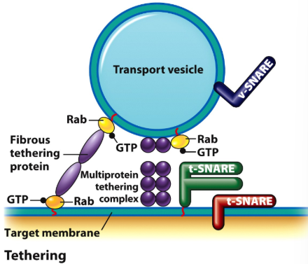
SNAREs Vary in Structure and Size, But All Contain a Segment in their Cytosolic Domain Called
a SNARE Motif
v-SNAREs
Become Incorporated into the Vesicle During Budding
t-SNAREs
Located in the Target Membrane Compartment
The Best Studied SNAREs are
those Involved in Docking Synaptic Vesicles During Nerve Cell Neurotransmitter Release
The Plasma Membrane of the Nerve Cell Contains 2 t-SNAREs, Called
Syntaxin and SNAP-25
the Synaptic Vesicle Membranes Contain a Single v-SNARE
a Member of the Vesicle Membrane Associated Protein (VAMP) Family, Called Synaptobrevin
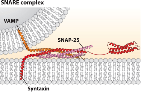
the Membranes Approach One Another, the SNARE Motifs of Opposing v- and t-SNAREs form a
4-Stranded Bundle, Each Bundle Consists of 4-Alpha Helices, 2 from Snap-25 and 1 Each from Syntaxin and Synaptobrevin, Together these Helices Form the Coiled Coil that Pulls the 2 Membranes into Close Proximity
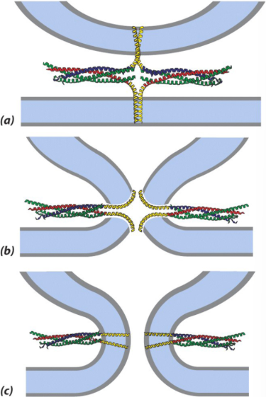
The 4 SNAREs in the Bundle in Nerve Cells are Locked in an Inactive Conformation by Another Bound Protein Called
Complexin
Vesicles with SNAREs Inactivated by Complexin, are
Docked at the Membrane Ready to Discharge their Contents Virtually Instantly
The Activating Signal which Causes Vesicle Fusion
is a Rise in Intracellular Ca2+ Ion Concentration
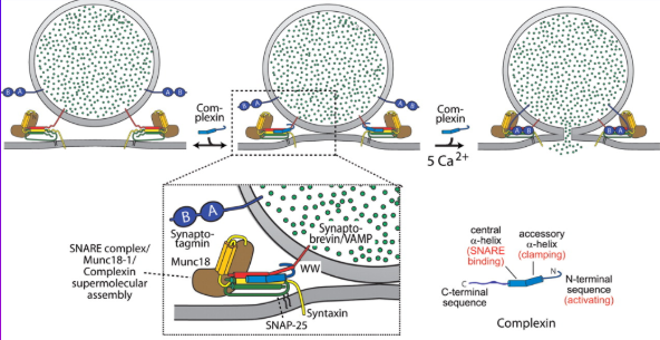
Nerve Cells, Vesicle Fusion is Regulated by the Calcium Binding Protein,
Synaptotagmin Present in the Synaptic Vesicle Membrane, After a Rise in Intracellular Calcium, Synaptotagmin Displaces Complexin, Which Releases the Inhibition of the SNARE Complex, Causing Vesicle Fusion and Neurotransmitter Release
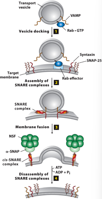
Dissociation of the 4-Stranded SNARE Bundles is Achieved by a
Doughnut-Shaped Cytosolic Protein Complex, Containing NSF and α-SNAP Proteins, these Attach to the Bundle and Twist it Apart Using Energy From ATP
exocytosis
Fusion of a Secretory Vesicle or Granule with the Plasma Membrane
exocysts
Protein complexes that mediate exocytosis by guiding vesicles to the plasma membrane for fusion.
exocysts consist of
8 Proteins (Sec3, 5, 6, 8, 10, 15 and Exo70 and 84) Plus a Rab Protein in the Vesicle, Sec3 and Exo 70 are Located on the Plasma Membrane, the Rest are on the Docking Vesicle
a typical lysosome contains
50 Hydrolytic Enzymes Produced in the ER and Shipped to Lysosomes in Vesicles
Lysosomal Enzymes All Have an Optimal Activity at an Acid pH and are thus
Acid Hydrolases
The Internal Lysosomal pH is Maintained by
a Membrane-Bound V-Type, H+-ATPase Pump
Mutations in the Genes that Encode Lysosomal Enzymes are Responsible for 40 Different Genetic Diseases Called
Lysosomal Storage Diseases that Affect 1 in 5000 Births,
Lysosomal Storage Diseases
These Lead to the Accumulation of Non-Degraded Products that Leads to a Build up in the Size and Number of Lysosomes
Gaucher’s Disease
a Mutation in the Gene for an Enzyme Required for the Breakdown of Glycolipids
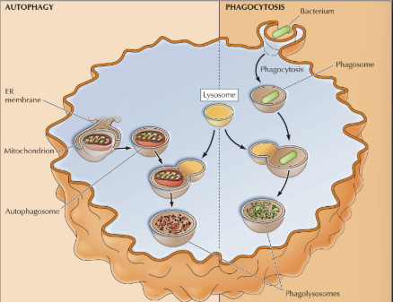
phagolysosomes
are formed by the fusion of phagosomes containing engulfed material with lysosomes, allowing for the degradation of the contents,
envelopment of pseudopodia surrounding microbes involves
Phosphorylation of Membrane Phospholipids PI(4,5)P2 to Form PI(3,4,5)P3
Once Engulfed, Microbes May be Killed by
Lysosomal Enzymes or Oxygen Free Radicals Generated in the Lumen of the Phagosome
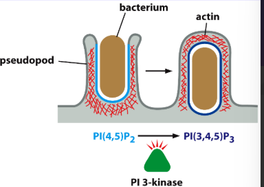
Engulfment of Materials Involves the Activities of
Actin Microfilaments Underneath the Plasma Membrane
Mycobacterium tuberculosis is Taken into Macrophages by Phagocytosis,
but the Phagosomes Fail to Fuse with a Lysosome, The Bacteria Inhibits Membrane Fusion and Instead Multiplies Inside the Cell
Q Fever, Coxiella burnetii, Becomes Enclosed in the Phagosome,
but Neither the Acidic Environment or the Lysosomal Enzymes Can Destroy it
Listeria monocytogenes, a Bacterium that Causes Meningitis,
Produces Proteins that Destroys the Lysosomal Membrane, This Allows the Listeria Bacterium to Escape into the Cell Cytoplasm
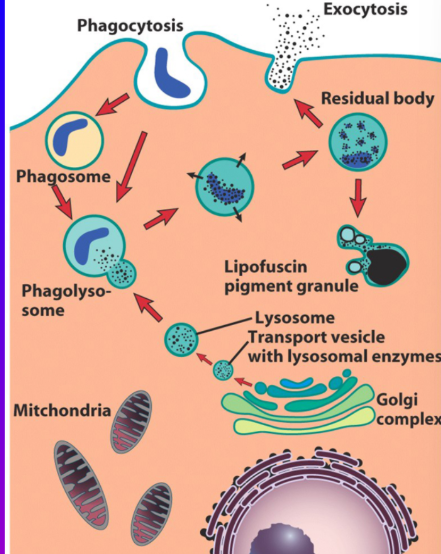
residual body or lipofusin pigment granule
phagolysosomes that contain undigested material leftover from phagocytosis. These structures can accumulate in cells and indicate previous immune responses.
autophagy
is a cellular process that degrades and recycles cellular components through the formation of autophagosomes that fuse with lysosomes.
Autophagic Vacuole
A double-membrane structure in cells formed during autophagy that encloses cellular components targeted for degradation and recycling.
autophagolysosome
organelles like mitochondria are initially surrounded by the double membrane part of the ER forming the autophagic vacuole, the ER then fuses with a lysosome to form this
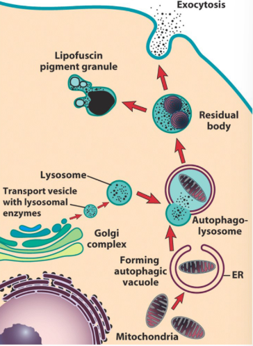
Once the Digestive Process of the Autophagolysosome is Completed,
the Organelle Becomes a Residual Body
tonoplast
a membrane-bound structure in plant cells that encloses the central vacuole. It regulates the movement of ions and molecules in and out of the vacuole and helps maintain turgor pressure.
Vacuoles have Homologues of
Lysosomal Acid Hydrolase Enzymes
in vacuoles, internal pH is kept low by
Tonoplast V-Type H+-ATPases Pumping Protons into the Vacuole
Where are vacuole proteins made?
Vacuole proteins are synthesized in the endoplasmic reticulum (ER) and processed in the Golgi apparatus before being transported to the vacuole.
Proteins are Targeted to Vacuoles By
Short Peptide Signals Rather Than Carbohydrates (Mannose-6-Phosphate) as in Lysosomes
endocytosis
is where the Cell Internalizes Membrane Receptors and Their Bound Ligands
phagocytosis
Involves the Uptake of Particles from Extracellular Fluid
the endocytic pathway starts with
the Invagination of the Plasma Membrane to Form Cytoplasmic Vesicles that are Transported into the Cell
Bulk-Phase Endocytosis or Pinocytosis
is the Non-Specific Uptake of Extracellular Fluid, any molecules in the enclosed fluid enter the cell
Pinocytosis Also Removes Plasma Membrane and Functions Primarily to
Recycle Membrane Between the Cell Surface and the Interior
Receptor-Mediated Endocytosis (RME)
Absorbs Specific Macromolecules (Ligands) into the Cell, as they are Associated with Receptors on the Cell Surface
Clathrin-Coated Pits
are specialized structures in the plasma membrane that facilitate receptor-mediated endocytosis by assisting in the internalization of specific ligands.
Each Clathrin Molecule Consists of 3 Heavy Chains and 3 Light Chains Joined Together to Form a
triskelion
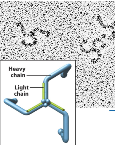
The Clathrin Triskelion is a Very Adaptable Structure for
Forming Polyhedral Shapes, as the clathrin curves it restructures and some of the hexagons become pentagonal
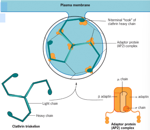
Like Clathrin Coated Vesicles Budding From the TGN, Endocytic Vesicles Also have
AP-2 Adaptor Complexes Between the Clathrin and the Vesicle Membrane
AP-2 Adaptors are a Complex of Multiple Subunits,
Consisting of α, β, σ, μ
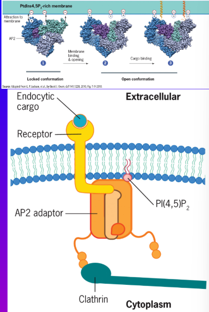
Membrane Phosphotidyl-inositide PI(4,5)P2
recruits AP2 to the membrane and Also Changes AP2’s Conformation to Activate (Open) it so that the μ Chain Engages the Cytoplasmic Tails of Specific Plasma Membrane Receptors Leading to their Concentration (and their Bound Ligands) in the Vesicle, the β Adaptin Subunit Binds and Recruits the Clathrin Molecules Overlying the Adaptors
how many different kinds of PI are ther?
There are 7 Different Kinds of PI, [PI(3)P, PI(4)P, PI(5)P,
PI(3,4)P2, PI(4,5)P2, PI(3,5)P2 and PI(3,4,5)P3] Which have Phosphates Attached at Different Positions
![<p>There are 7 Different Kinds of PI, [PI(3)P, PI(4)P, PI(5)P,</p><p>PI(3,4)P2, PI(4,5)P2, PI(3,5)P2 and PI(3,4,5)P3] Which have Phosphates Attached at Different Positions</p>](https://assets.knowt.com/user-attachments/b4134284-6683-4a8d-b9fa-53f50cb2be80.png)
In Endocytosis, AP2 (and Dynamin) Recruitment to the Membrane is Facilitated by
PI(4,5)P2
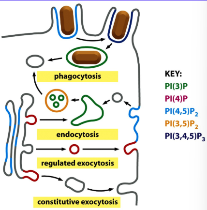
PI(3)P is
Localized to Early Endosomes and Late Endosome Intraluminal Vesicle Membranes
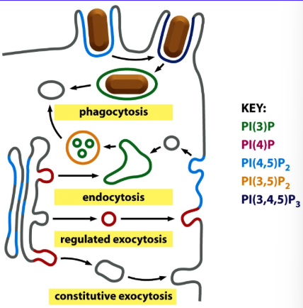
PI(4)P is
localized to the TGN, Secretory Granules, and Synaptic Vesicles
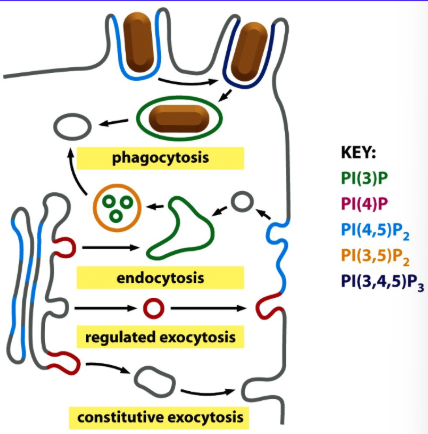
PI(3,5)P2 is
Localized to Late Endosome Boundary Membranes
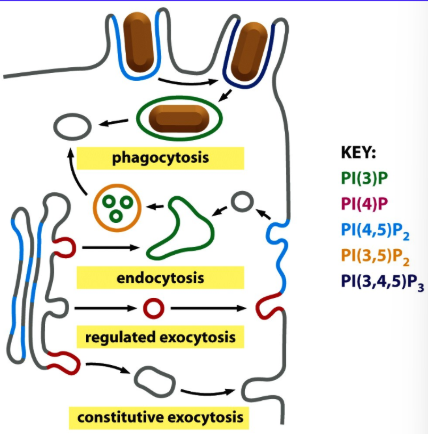
PI(3,4,5)P3 is
Involved in the Later Stages of Phagocytosis
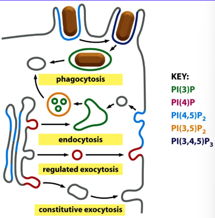
dynamin
is a G-Protein Required for the Release of Clathrin-Coated Vesicles from the Plasma Membrane
Dynamin Self-Assembles into
a Helical Collar Around the Neck of the Invaginated Coated Pit, Just Before it Pinches Off From the Membrane
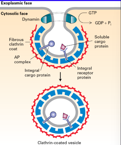
Hydrolysis of GTP Bound to Dynamin
Induces a Conformational Change that Severs the Vesicle from the Membrane either directly or through the action of another protein
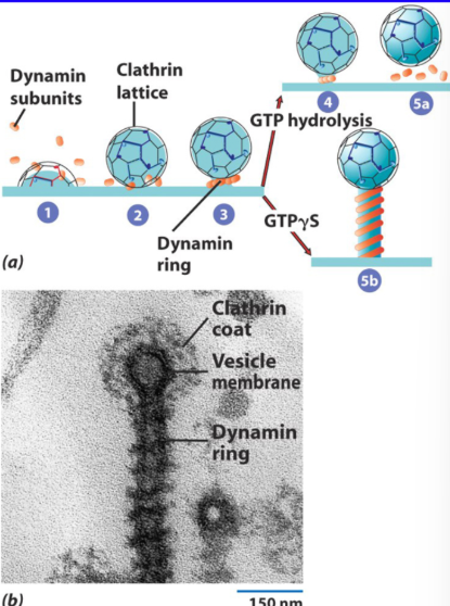
If a Non-Hydrolyzable Analogue of GTP (GTPγS) is Bound to Dynamin,
It Continues to Build the Helical Ring of Dynamin Subunits Without Severing Off the Vesicle
the two types of receptors that utilize the endocytic pathway
Housekeeping Receptors that Bring Nutrients into the Cell and Signaling Receptors