the liver
1/72
There's no tags or description
Looks like no tags are added yet.
Name | Mastery | Learn | Test | Matching | Spaced | Call with Kai |
|---|
No analytics yet
Send a link to your students to track their progress
73 Terms
where is the liver located?
in the right hypochondrium, epigastric and extends to the left hypochondrium
what 2 surfaces can the liver be split into?
- diaphragmatic surface
- visceral surface
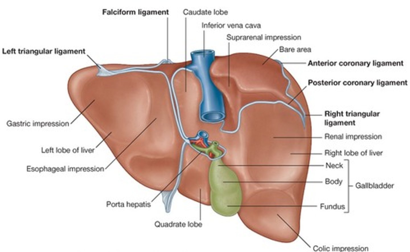
what is the diaphragmatic surface?
anterosuperior aspect of the liver that fits under the curvature of the diaphragm
what is the 'bare area' of the liver?
the posterior portion of the diaphragmatic surface that is not covered by the peritoneum
what else demarcates the bare area of the liver?
gap between the posterior and anterior folds of the coronary ligament
what is the visceral surface?
the posteroinferior surface
what is the visceral surface in contact with? (8)
- right kidney
- right adrenal gland
- right colic flexure
- transverse colon
- first part of duodenum
- gallbladder
- oesophagus
- stomach
is the visceral surface of the liver intraperitoneal?
yes it is covered in peritoneum except from the fossa of the gallbladder and the porta hepatis
how are the ligaments of the liver formed and what do they do?
- formed by a double layer of peritoneum
- they attach the liver to the surrounding organs
what are the ligaments of the liver? (5)
- falciform ligament
- anterior and posterior layers of the coronary ligament
- left and right triangular ligaments
- lesser omentum (hepatogastric and hepatoduodenal ligaments)
- round ligament/ligamentum teres
what is the falciform ligament? (2)
- attaches the anterior surface to the anterior abdominal wall
- forms a natural division between the left and right lobes of the liver
what does the free edge of the falciform ligament contain?
the ligamentum teres - the remnant of the umbilical vein
what are the anterior and posterior layers/folds of the coronary ligament?
they attach the superior surface of the liver to the diaphragm
what are the right and left triangular ligaments?
where the anterior and posterior coronary ligaments join
what is the lesser omentum in correlation to the liver?
attaches the liver to the lesser curvature of the stomach
- it consists of:
- the hepatoduodenal ligament (which extends from the duodenum to the liver and surrounds the portal triad)
- the hepatogastric ligament (from stomach to liver)
what ligaments does the lesser omentum contain in correlation to the liver? (2)
- the hepatoduodenal ligament
- the hepatogastric ligament
what are the hepatic recesses?
- subphrenic spaces
- subhepatic space
- hepatorenal recess (Morison's pouch)
where are the subphrenic spaces? (2)
- located between the diaphragm and the anterior and superior aspects of the liver
- divided into right and left spaces by the falciform ligament
where is the subhepatic space?
between the inferior surface of the liver and the transverse colon
where is the hepatorenal recess/Morison's pouch?
space between the visceral surface of the liver and the right kidney
what is important about Morison's pouch/heptaorenal recess?
deepest part of the peritoneal cavity when supine
are the peritoneal spaces always there?
no, they are all only potential spaces containing just enough peritoneal fluid to lubricate adjacent peritoneal membranes
what is the liver covered by?
a fibrous layer called Glisson's capsule
what are the 4 lobes of the liver?
- right
- left
- caudate
- quadrate
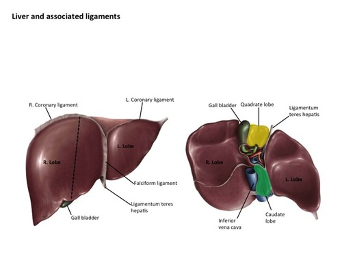
where is the caudate lobe? (2)
- upper aspect of visceral surface
- lies between inferior vena cava and a fossa produced by ligamentum venosum
where is the quadrate lobe? (2)
- lower aspect of visceral surface
- lies between gallbladder and a fossa produced by ligamentum teres
which out of the right or left lobe is larger?
right lobe
how many segments of the liver are there?
8
what is the segment classification called?
Coinaud classification
what is the significance of the 8 segments of the liver?
they are all have an independent function system
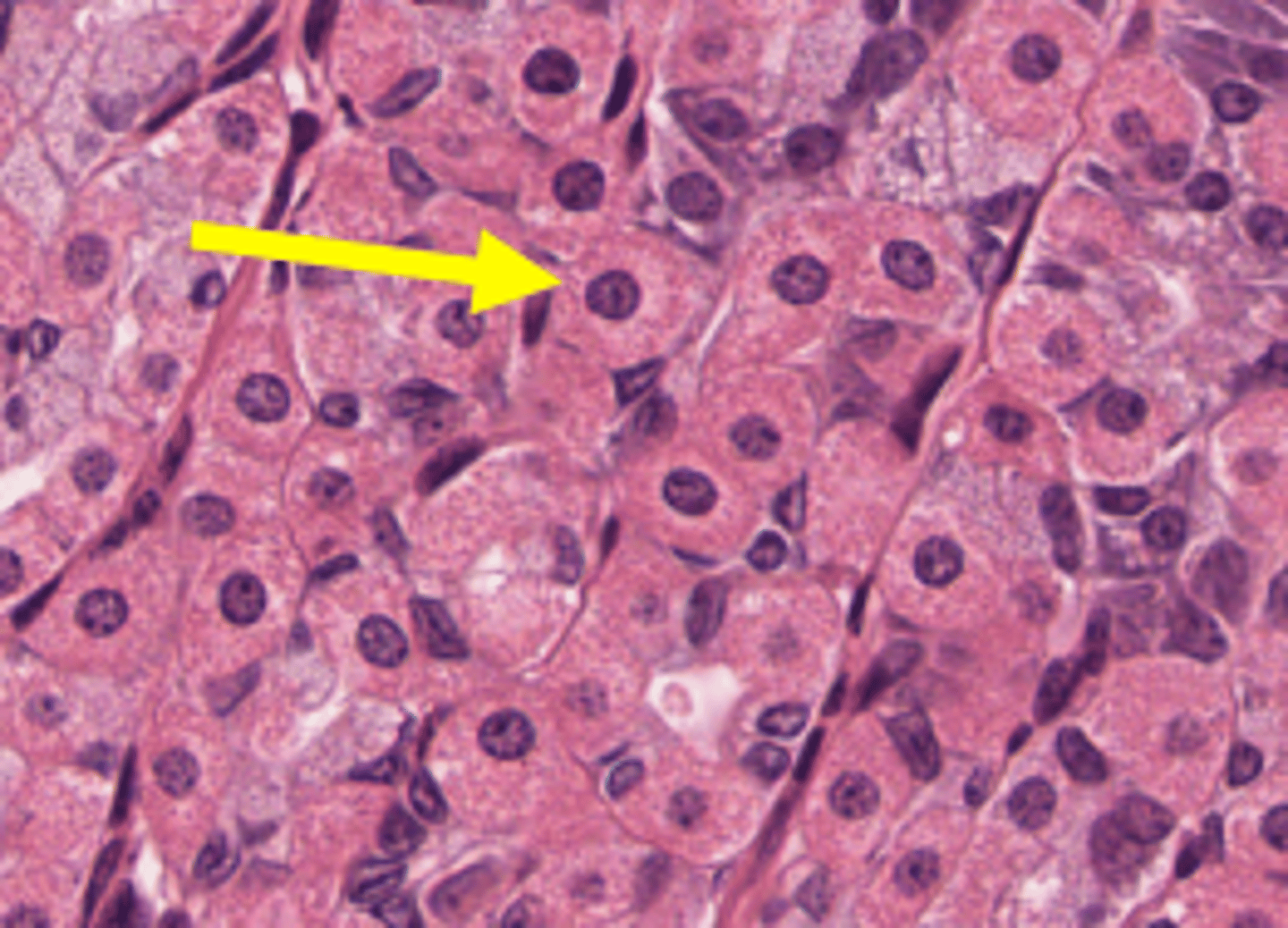
what is the main cell type in the liver?
hepatocytes
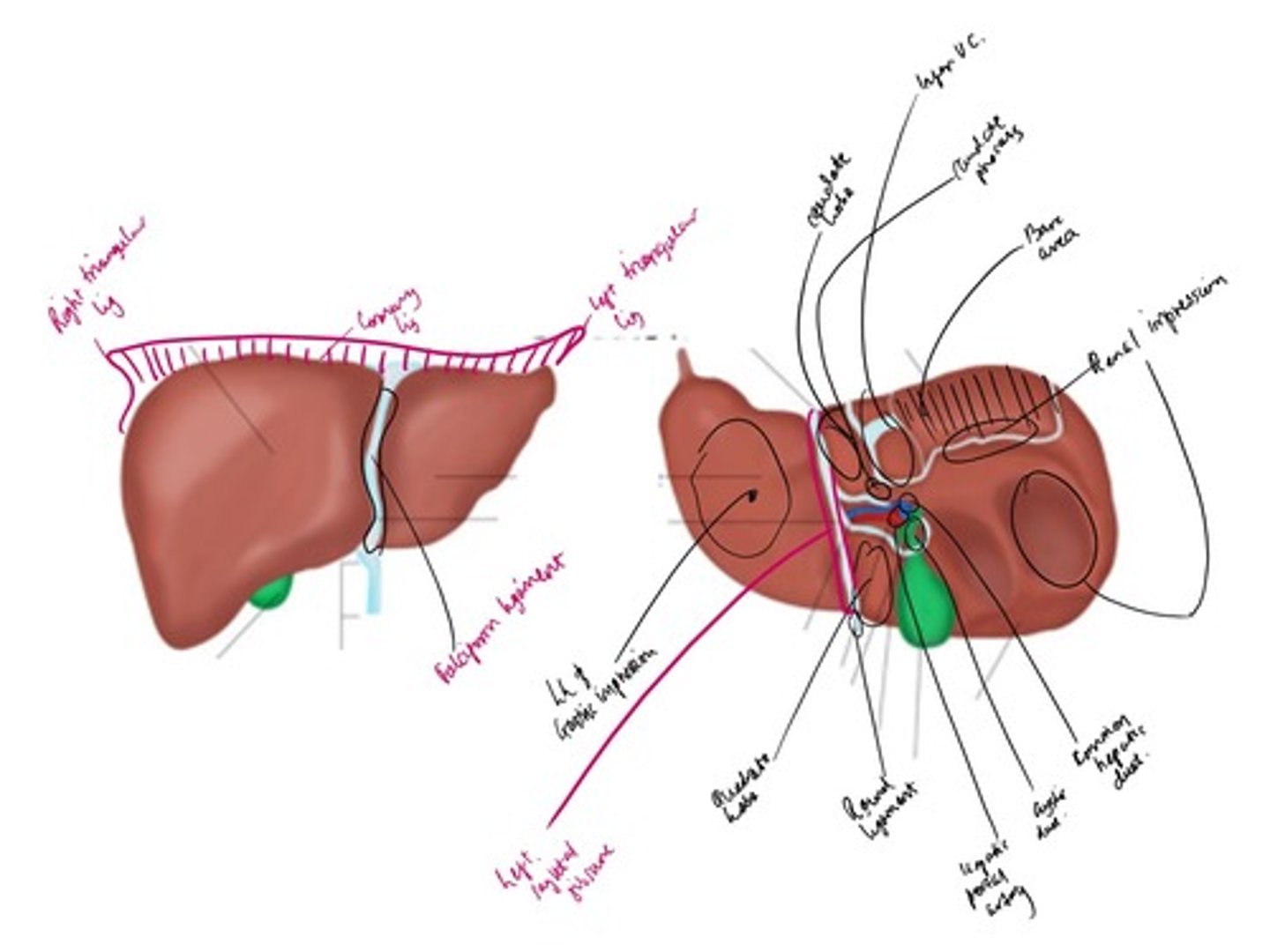
draw a labelled diagram of the 8 segments of the liver
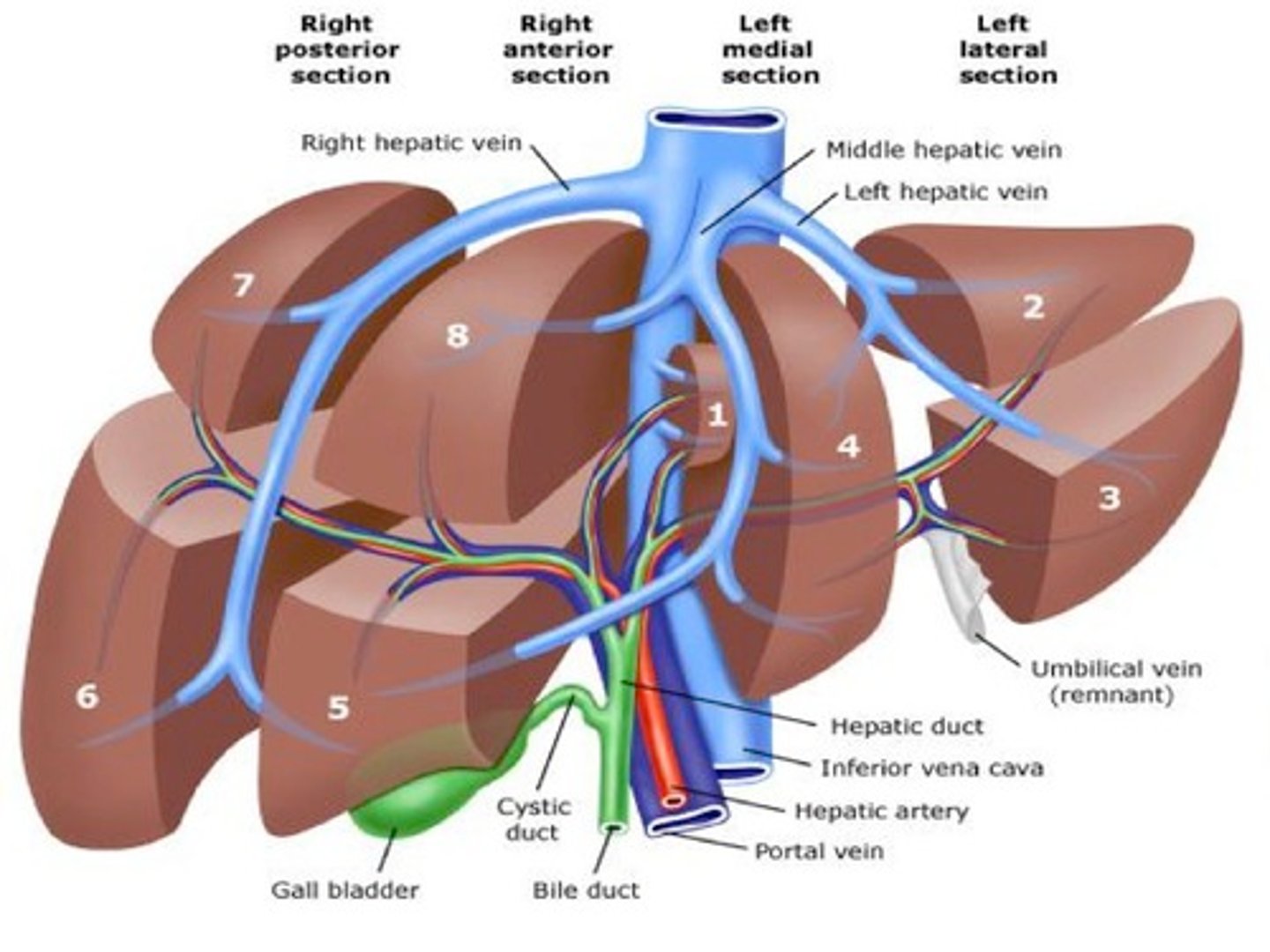
what does each independent segment have?
its own:
- bile duct
- hepatic artery
- portal vein
draw a labelled diagram of the lobes of the liver from the posterior view
draw a labelled diagram of the lobes of the liver from the anterior view
what happens if one segment is damaged?
has no effect on the other segments
where and what is the porta hepatis?
- it lies between the caudate and quadrate lobe
- it transmits all the vessels, nerves and ducts entering or leaving the liver - apart from the hepatic veins
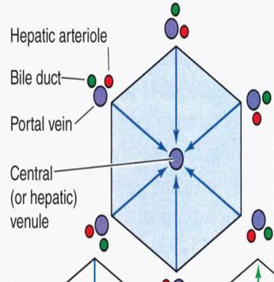
what is liver arranged into microscopically?
hexagonal lobules
what is the gap between the sinusoids and hepatocytes called?
space of Disse
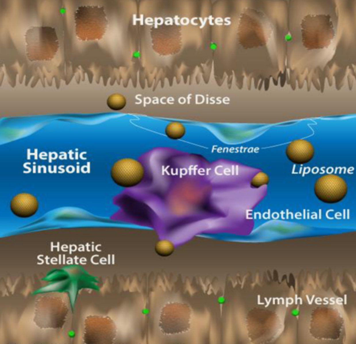
what are 3 other cell types present in the liver?
- Kupffer cells
- endothelial cells
- stellate cells
where are stellate cells found?
space of Disse
what is the function of stellate cells? (2)
- damage response as it produces type I collagen
- vitamin A storage
what is the function of Kupffer cells?
specialised immune cells - macrophages
where are endothelial and Kupffer cells found?
on endothelium of sinusoids
what are the hepatocytes arranged into?
arranged in laminae
what is at the centre of each lobule?
a central vein
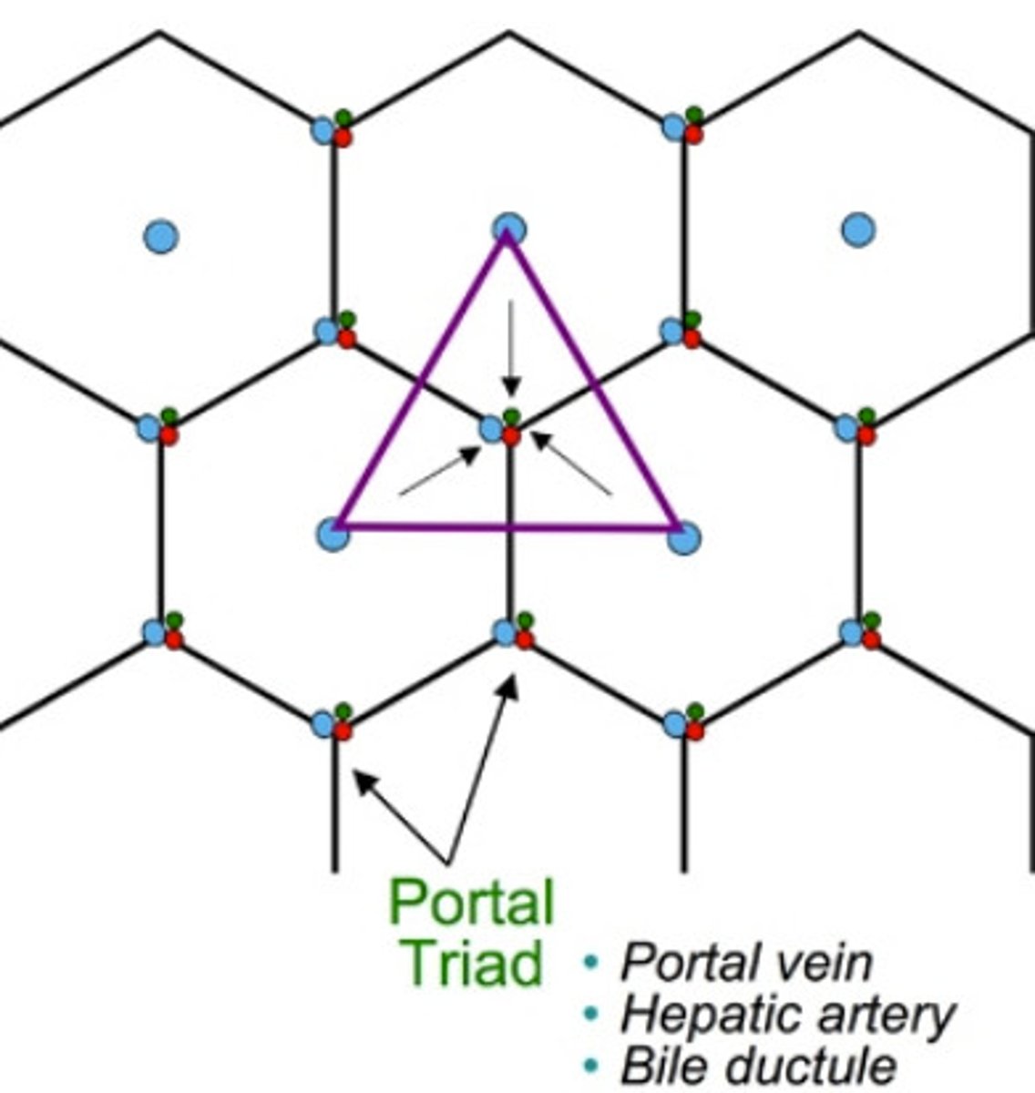
what is at the periphery of each lobule?
the portal triad
what does the portal triad consist of?
arteriole - a branch of the hepatic artery entering the liver
venule - a branch of the hepatic portal vein entering the liver
bile duct - branch of the bile duct leaving the liver
- also contains lymphatic vessels and vagus nerve fibres
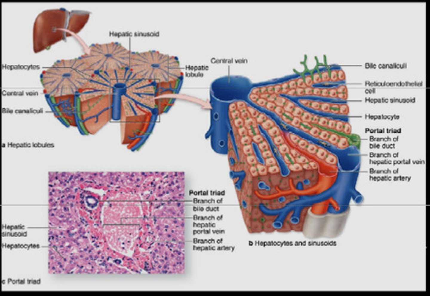
how does oxygenated blood reach the hepatocytes?
capillaries called sinusoids
where do the sinusoids travel from and to?
from hepatic arteries and potal veins in the portal triad into the central vein
how does bile get taken from hepatocytes?
through bile canniculi
which direction do bile canniculi run?
in the opposite direction to sinusoids, away from central vein
what do the nuclei of hepatocytes stain strongly with?
hematoxylin (pink)
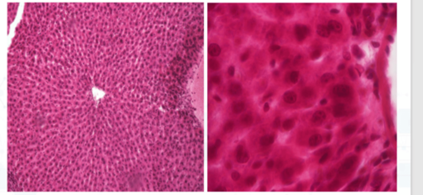
what do do the mitochondria stain strongly with?
eosin (purple)
what are the 3 lobule classifications?
- classic lobule
- portal lobule
- acinar lobule
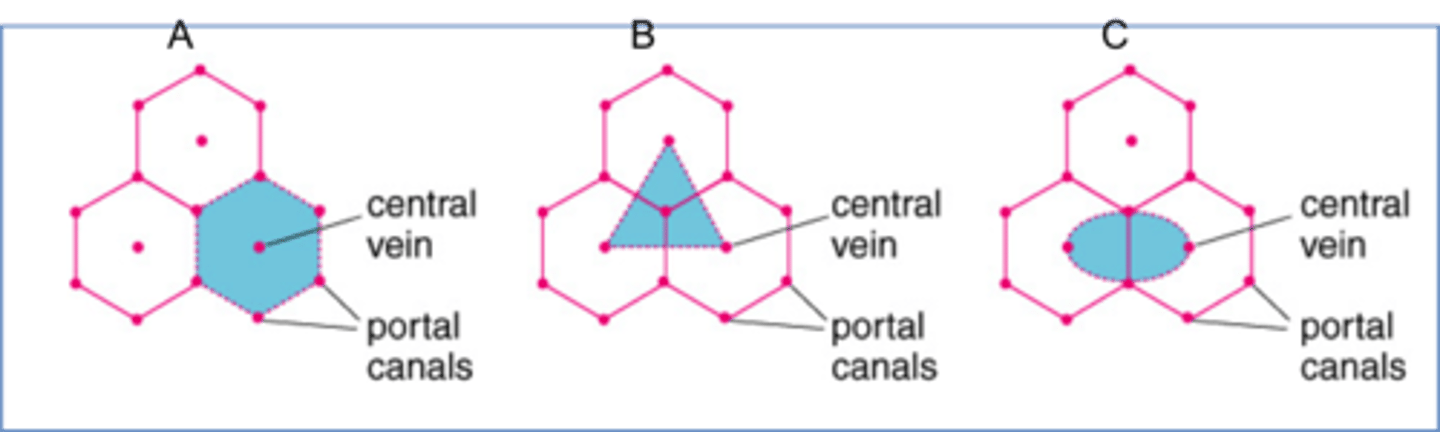
what are the features of the classic lobule? (2)
- central vein in the centre of each lobule
- blood drains from portal triad to central vein
what are the features of the portal lobule? (2)
- bile ducts (portal triad) are at the centre of each lobule
- bile moves in direction from central vein to portal triad
what are the features of the acinar lobule? (2)
- the hepatic arteries (portal triad) are at the centre of the lobules
- preferred structural and functional lobule
what are the 3 zones of the acinar lobule?
1) periportal
2) midlobular
3) perivenous/central
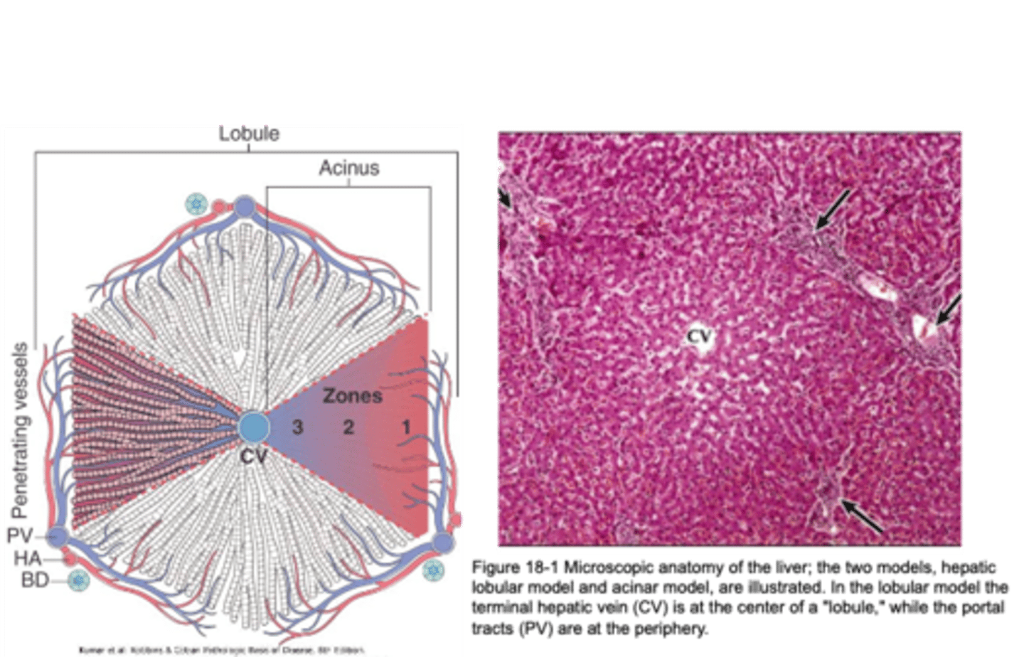
which is the most susceptible zone to toxic damage and why?
zone 1 - due to close proximity to portal vein
which is the most susceptible zone to hypoxia and why?
zone 3 - due to distance from portal triad
what supplies the blood to the liver?
- hepatic artery from the coeliac trunk - (25%) supplies the non-parenchymal structures of the liver
- hepatic portal vein (75%) supplies the liver parenchyma with partially deoxygenated blood
what is the purpose of carrying partially deoxygenated blood to the liver?
so it can detoxify any absorbed nutrients from the GI
what is the venous drainage of the liver? (3)
hepatic veins
- right
- left
- middle
which segments does the right hepatic vein drain?
- 6,7 mainly
- 5,8 also
which segments does the middle hepatic vein drain?
4,5,8
which segments does the left hepatic vein drain?
2,3
which vein drains segment 1?
directly from inferior vena cava
what is the route of the veins from the liver? (4)
central vein - collecting veins - multiple hepatic veins - inferior vena cava
what is the innervation of the liver?
by the hepatic plexus
what is the parasympathetic nervous supply?
vagus nerve
what is the sympathetic nervous supply?
coeliac plexus
where do the nerves enter the liver?
porta hepatis