Orbit/Eye anatomy
1/34
There's no tags or description
Looks like no tags are added yet.
Name | Mastery | Learn | Test | Matching | Spaced | Call with Kai |
|---|
No analytics yet
Send a link to your students to track their progress
35 Terms
Pneumonic: Five Eager Ladies Make Zebras Smile Pretty
Frontal, ethmoid, lacrimal, maxilla, zygomatic, sphenoid, and palatine
What bones make up the roof of the orbit? What important structure is here?
Frontal and lesser wing of sphenoid, the lacrimal fossa is located here
What bones make up the floor of the orbit? What is the clinical significance?
The maxilla, zygomatic, and palatine, inferior recuts entrapment usually occurs due to an injury in this area
What bones make up the medial wall of the orbit? What is to be noted about this wall?
Frontal, ethnoid, lacrimal, and sphenoid. This is the thinnest wall.
What bones make up the lateral wall of the orbit? What is importantly to remember about this wall.
The greater wing of the sphenoid and zygomatic, this is the strongest wall

What wall is crossed out in red, what bones make it?
This is the lateral wall created by the greater wall of the sphenoid bone and the zygomatic, strongest wall

What wall is crossed out in yellow, what bones make it? Any clinical significance?
This is the superior wall aka the roof of the orbit, created by the frontal and lesser wing of sphenoid. This is where the lacrimal fossa is located.

What wall is crossed out in blue, what bones make it? Any clinical significance?
This is the inferior wall aka the floor of the orbit, created by the maxilla, zygomatic, palatine. When this is blownout out, inferior rectors entrapment occurs.
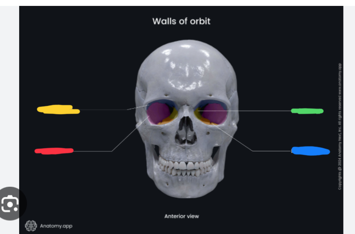
What wall is crossed out in green, what bones make it? Any clinical significance?
This is the medial wall created by the frontal, ethnoid, sphenoids and lacrimal bones. This is the thinnest wall and ethnoid sinusitis can spread here.
What are the main 5 orbita foramina?
Optic canal, superior orbital fissure, inferior orbital fissure, surpaorbitsl fissur, Supra orbital Foramen, and infra orbital Foramen
what are the contents of the optic canal?
CN 2 and opthalmic artery
What are the contents of the superior orbital fissure?
CN 3, 4, CN 5 V 1, and CN 6
What are the contents of the inferior orbital fissure?
Infraorbital artery and CN 5 V2 nerve
What are the contents of the supraorbital foramen?
Supraorbital nerve and vessels of V1
What are the contents of the infraorbital foramen?
Infraorbitla nevre and the vessels of V2

What structure is highlighted in green? What would a lesion to this area cause?
This is the optic canal. Lesions would cause Vision loss to the entire eye, possible afferent pupil defect
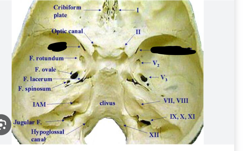
What figure is crossed out in black? What would a lesion to this area cause?
SOF, lesion to the area would cause paralysis to all ocular muscles and V1 sensory loss aka SOF syndrome
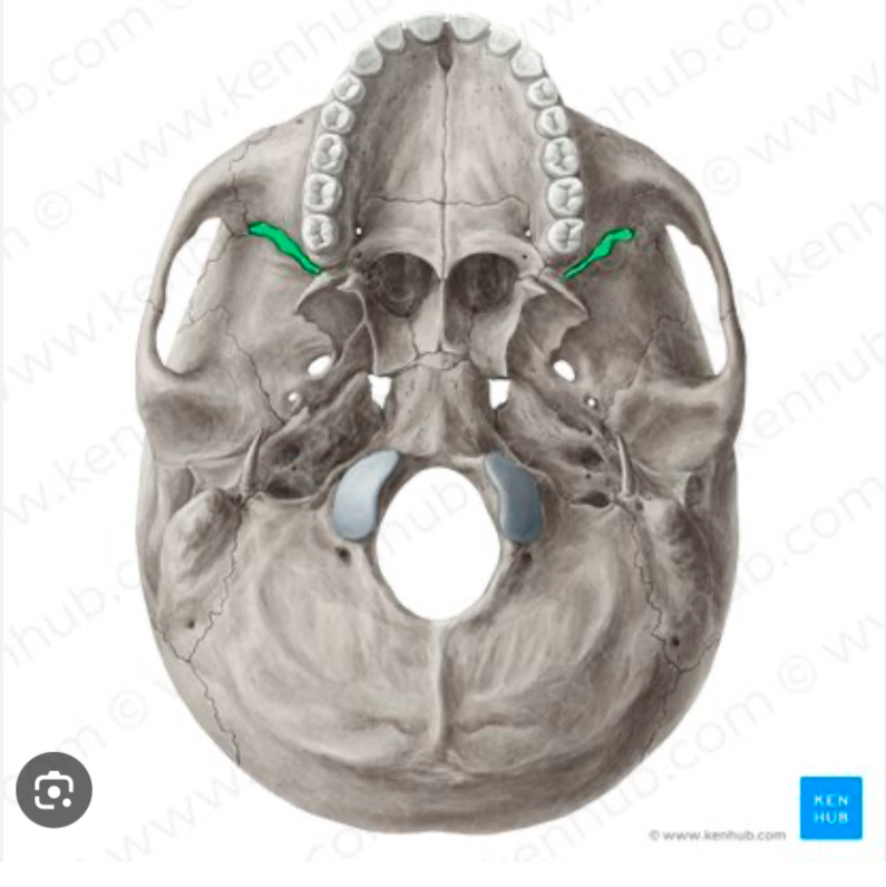
What figure is highlighted in green and what would a lesion to this area cause?
Inferior orbital fissure, lesion would cause upper lip and cheek numbness
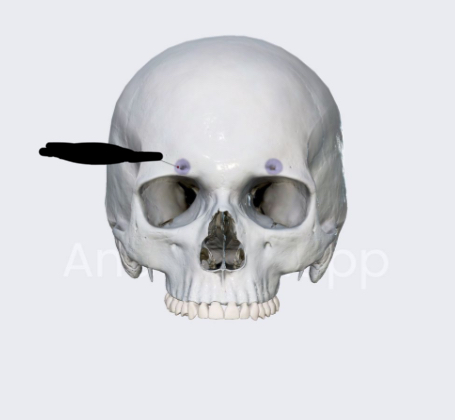
What figure is highlighted in purple? Lesion to this would cause?
Forehead numbness, this is the supraorbital foramen
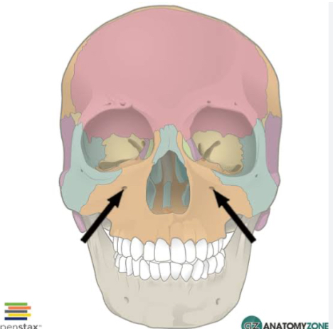
What figure are the black arrows pointing too? What would a lesion to this cause?
Upper teeth numbness most notably but also upper lip/cheek numbness, this is the infraorbital foramen
What is orbital apex syndrome?
Basically SOF syndrome (CN 3,4,6, and V1) which would cause motor and sensory loss around the eye but also involves vision loss due to optic canal lesion (CN2)
What are the 3 layers of the eye from outer to inner?
Fibrous, vascular, and neural
What sutrcutre are in the fibrous layer of the eye?
Sclera (5/6) and cornea (1/6)
What structures are in the vascular layer of the eye?
The choroid, ciliary body, and iris
What structure are in the neural layer of the eye?
Retina

What structure is crossed out in blue? Function and parts?
The fibrous layer which has both the sclera and the cornea. It provides structural support and shape to the eye

What structure is crossed out in red? Function and parts?
The vascular layer aka middle layer which involved the choroid, cillary body, and iris. Iris is colored, choroid provides nourishment, and the cilliary body creates aqueous humor.
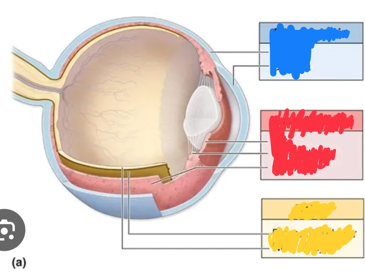
What structure is crossed out in yellow? Function and parts?
This is the neural layer of the eye aka the innermost layer which involves only the retina. The job of this layer is to transduce light into electrical signs.
What is the aqueous humor? Created by?
This is a clear liquid made by the cilliary process in order to nourish the eye and keep it inflated.
What is the flow of the aqueous humor flow?
Cilliary process then Postieor chamber, goes though pupil, anterior chamber, then drains via canal of schlemm at iridocorneal angle
What would blockage of drainage at the iridocorneal angle cause?
Build up of infra ocular process aka glaucoma. It can be open angle aka gradual or closed angle which is fast, painful, and emergent
What are the 3 structures involved in pupil control
The sphincter pupilae, dilator pupilae, and ciliary muscle
What does the sphincter do and which ANS system is it controlled by via which nevre?
It constrict the pupil using the parasympathetic nervous system via CN 3 THIS HELPS WITH NEAR VISION
What does the dilator do and which ANS system is it controlled by via which nevre?
Dilates the pupil using the sympathetic nervous system via the superior cervical ganglion THIS HELPS WITH FAR VISION
What does the ciliary muscle do and which ANS system is it controlled by via which nevre?
This muscle helps the lens thicken which is for near vision. This is for near vision CN 3