Muscular System
1/54
There's no tags or description
Looks like no tags are added yet.
Name | Mastery | Learn | Test | Matching | Spaced | Call with Kai |
|---|
No analytics yet
Send a link to your students to track their progress
55 Terms
6 Functions of Skeletal Muscles
Moves bones
Posture
Support soft tissue
Sphincter protection
Heat
Store nutrients
Nutrients
Glycogen and proteins
4 Components
Muscle tissue
Connective tissue
Nerves
Blood vessels
Muscles Organs
They contain multiple tissue types (muscle, connective, nervous, vascular)
Nerves
Control voluntary muscle contraction
Control System
CNS
Extensive Blood Supply
Delivers oxygen
Deliver nutrients
Remove waste products
Oxygen
Needed for ATP production
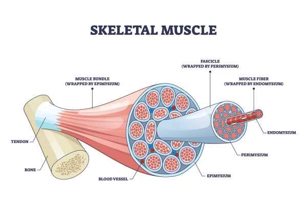
3 Connective Tissue Layers
Endomysium
Perimysium
Epimysium
Smallest → Largest
Endomysium → Perimysium → Epimysium

Endomysium
Surrounds individual muscle fibers

Inside Endomysium
Capillaries
Nerve fibers
Myosatellite cells
Myosatellite Cells
Stem cells that repair muscle damage
Endomysium Importance
Supports and nourishes each muscle fiber
Perimysium
Surrounds fascicles
Inside Perimysium
Blood vessels and nerves supplying fascicle
Fascicle
Perimysium
Bundle of muscle fibers
Epimysium
Surrounds entire muscle

Epimysium Connects to
Deep fascia
Epimysium Function
Separates muscle from surrounding tissue
Muscle Ends
Endomysium, perimysium, and epimysium merge together
Attachment to Bone
Occurs when connective tissue merges
Tendon
Rope-like bundle
Aponeurosis
Flat sheet
Muscle Fiber Structure
Muscle fiber → Fascicle → Whole muscle
Muscle Fiber
Endomysium
Whole Muscle
Epimysium
Connective Tissue Importance
Provides structure
Carries blood vessels/nerves
Forms attachments to bone
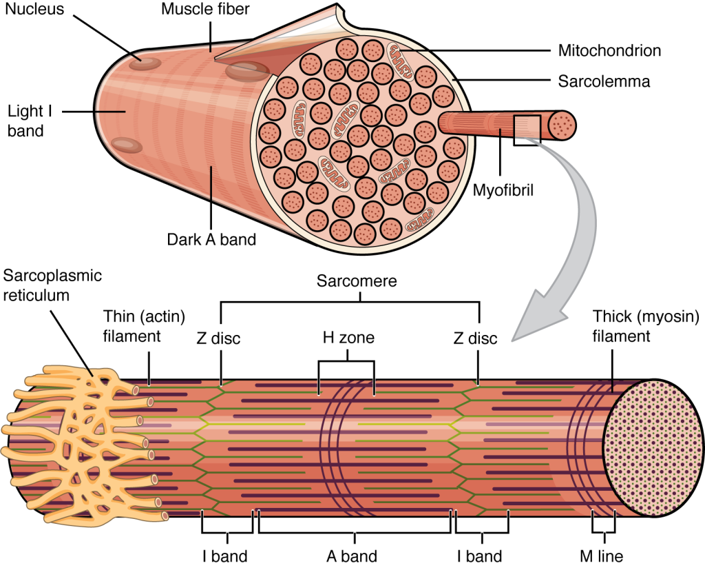
Transverse Tubules
Transmit action potential through cell
Allow entire muscle fiber to contract simultaneously
Have same properties as sarcolemma
Sarcoplasmic Reticulum
Membranous structure surrounding each myofibril
Helps transmit action potential to myofibril
Similar in structure to smooth ER
Forms chambers (terminal cisternae) attached to T tubules
CIsternae
Concentrate Ca2+ via ion pumps
Release Ca2+ into sarcomeres to begin muscle contraction
Triad
Triad
Formed by 1 T tubule and 2 terminal cisternae
Myofibrils
Lengthwise subdivisions within muscle fiber
Made up of protein filaments
Myofilaments
Responsible for muscle contraction
Thin Filaments
Made of actin
Thick Filaments
Made of myosin
Sarcomeres
Contractile units of muscle
Structural units of myofibrils
M Line
Center of A band
At midline of sarcomere

H Zone
Area around M line
Has thick filaments but no thin filaments
Zone of Overlap
Densest, darkest area on a light micrograph
Where thick and thin filaments overlap

I Band
Z lines
Titin
A Band
M line
H Zone
Zone of overlap
Z Lines
Centers of I bands
At 2 ends of sarcomere
Titin
Strands of protein
Stabilize filaments

Sarcomere Function
T Tubules encircle sarcomere near zones of overlap
Ca2+ released by SR causes thin and thick filaments to interact
4 Thin Filament Proteins
F actin
Nebulin
Tropomyosin
Troponin
F actin
2 twisted rows of globular G actin
Active sites on G actin strands bind to myosin
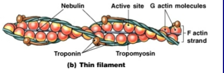
Nebulin
Holds F actin strands together

Tropomyosin
Double strand
Prevents actin-myosin ineraction

Troponin
Globular protein with 3 subunits
Binds tropomyosin to G actin
Controlled by Ca2+

Action Potential Travels
Across sarcolemma
Action Potential Enters
Through T tubules until it reaches triad
Action Potential in Triad
Binds to troponin → change shape
Change in shape
Moves to tropomyosin
Moves to Tropomyosin
Myosin binds to actin