Integumentary System & Skin
1/46
There's no tags or description
Looks like no tags are added yet.
Name | Mastery | Learn | Test | Matching | Spaced | Call with Kai |
|---|
No analytics yet
Send a link to your students to track their progress
47 Terms
What does the integumentary system consist of?
Skin, hair, nails, sweat & sebaceous glands, & sensory receptors
State 6 functions of the integumentary system
Regulation of body temperature
Blood storage
Protection from external environment
Excretion & absorption of substances
Vitamin D synthesis (in the skin)
Detection of cutaneous sensations
What are the two layers of skin?
Epidermis: outermost, thinner layer composed of epithelial cells
Dermis: deeper, thicker layer composed of dense connective tissue
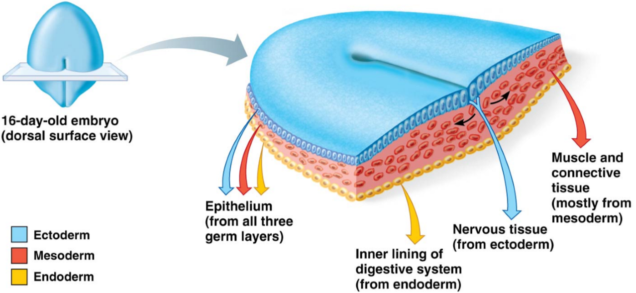
Are the epidermis & dermis vascularised? Explain
Only the dermis is vascularised; the epidermis is not
The dermis & hypodermis develop from the mesoderm along with blood vessels, but the epidermis is derived from ectoderm
The epidermis receives its nutrients via diffusion through interstitial fluid from vessels in the dermis
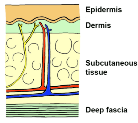
What is the hypodermis (superficial fascia)?
Subcutaneous tissue deep to the skin
Does the epidermis have a high or low degree of functional capacity?
Low degree of functional capacity
The epidermis does have pores for sweat glands, however;
Many functional organs for sweat glands are located in the dermal layer, not the epidermal layer
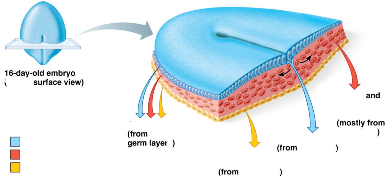
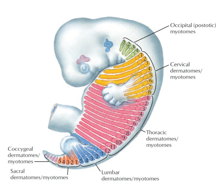
Label the following diagram of an embryo in relation to the integumentary system
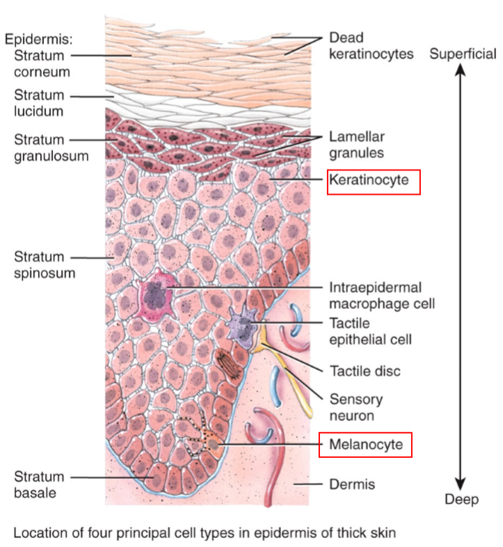
List the 5 layers of epidermis from superficial → deep
Stratum corneum (h0rny layer) - thickest of epidermal layers, comprised of dead cells
Stratum lucidum (clear layer)
Stratum granulosum (granular layer) - 1-5 layers of flattened cells
Stratum spinosum (prickly layer) - same thickness as stratum granulosum, several layers of keratinocytes unified by desmosomes
Stratum basale (basal layer) - 1 layer of newly formed stem cells which push their way upwards superiorly
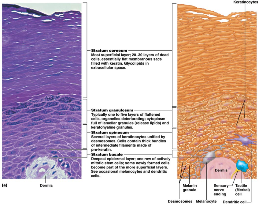
Where are melanocytes produced & how do they contribute to skin pigmentation?
Produced in the stratum basale (deepest layer) of the epidermis
Produce melanin pigment → transferred to overlying keratinocytes
Skin pigmentation depends on the amount of pigment produced by melanocytes, not the number of melanocytes (same for all people
How does the structure of skin vary over the body?
Variability in the epidermal thickness
Dermis is mainly static
Basal layer of epidermal tissue has cells which push their way upward → epidermis is a changing tissue
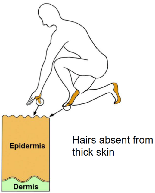
What are the 2 types of epidermis, in terms of epidermal thickness?
Thin skin (hairy): absent stratum lucidum (2nd most superficial layer) + other strata are thinner
Thick skin (hairless): covers area subject to abrasions + thicker stratum corneum (most superficial layer) in response to greater mechanical stress
Describe the physical & mechanical properties of the dermis
Physical properties:
Composed of dense irregular connective tissue (collagen + elastic fibres)
Much thicker than epidermis
Rich supply of nerve fibres, blood vessels & lymphatic vessels
Mechanical properties:
High tensile strength
Easily stretches & recoils
State the 2 layers of the dermis
Papillary
Reticular
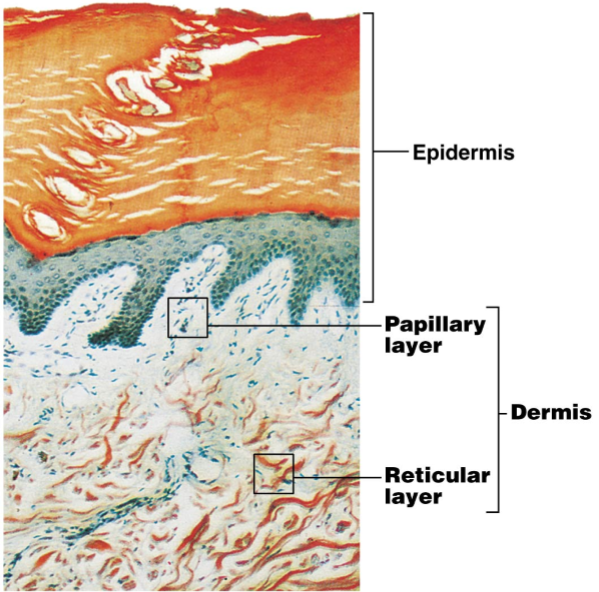
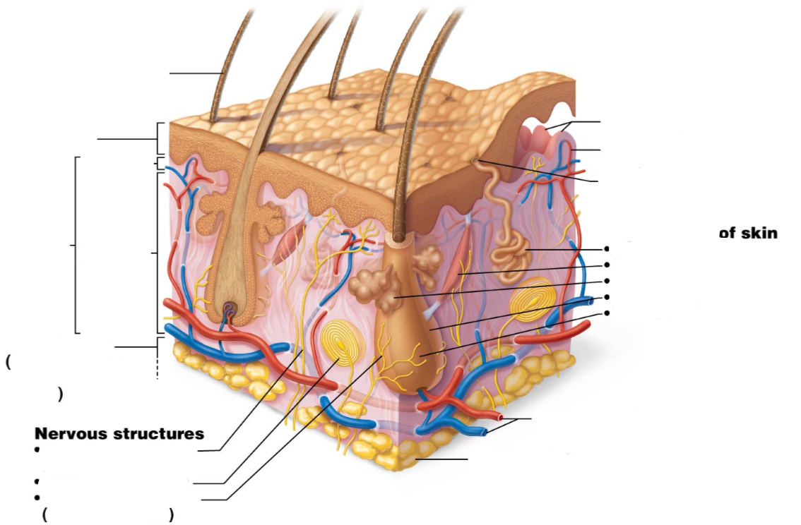
Label the following diagram
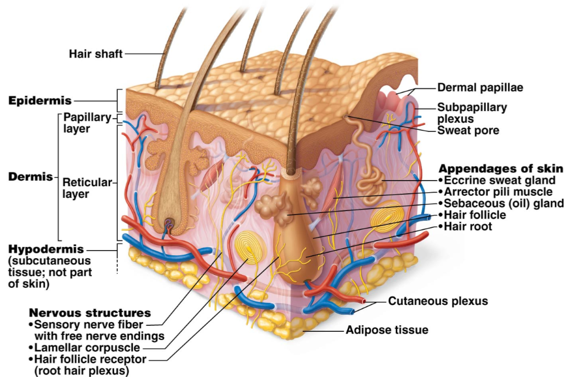
Describe the papillary layer of the dermis
More superficial
Makes up ~ 1/5 of the thickness of dermis
Contains blood vessels (capillaries) & sensory receptors
Forms dermal ridges in regions of thick skin (→ fingerprints & footprints)
In place by the 4th month of embryonic development
Describe the reticular layer of the dermis
Deepest layer of dermis, attached to the subcutaneous tissue/hypodermis/superficial fascia
Majority of the thickness of the dermis
Consists of collagen fibres running in specific planes → cleavage/tension lines
Contains blood vessels, nerves, hair follicles, sebaceous glands & sweat glands
How is blood transmitted to the skin?
Via subcutaneous tissue; projections of blood vessels are sent into the dermis
What are cleavage/tension lines?
Separations/less dense regions of collagen fibre bundles in the reticular dermis due to natural tension in the region
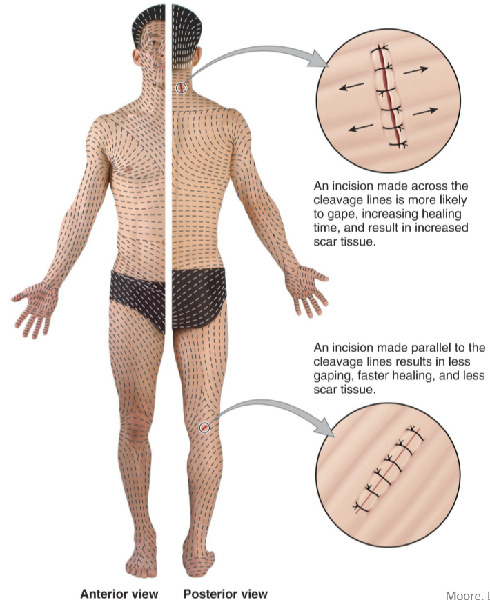
Describe the appearance of tension lines in the skin of the:
Head & limbs
Neck & trunk
Head & limbs - longitudinal tension lines
Neck & trunk - circular tension lines
How are tension lines clinically significant for surgeons?
Incisions parallel to tension lines gape less → reduce scarring
Incisions perpendicular to tension lines → more gaping → more bleeding + ^ scarring → harder to heal
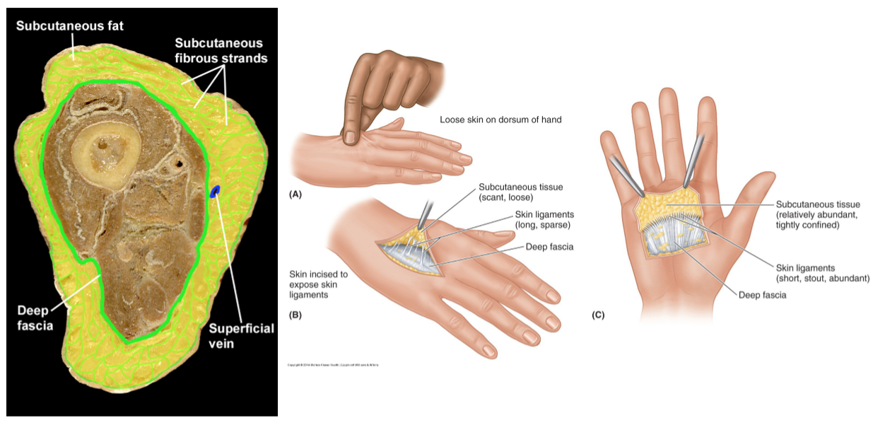
What are septa?
Fibrous strands which bind subcutaneous tissue to underlying dense connective tissue, especially at the palms, soles & scalp
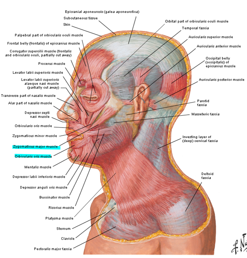
What regions of the body contain muscle in their subcutaneous tissue?
Face
Neck
Palm
Scrotum
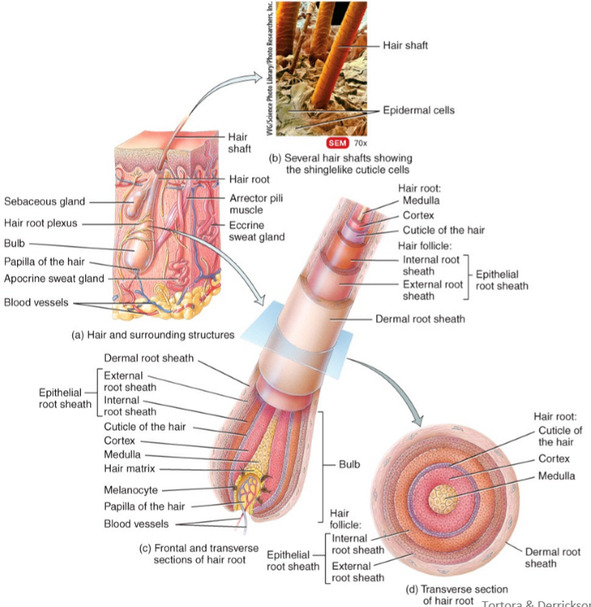
Describe the structure of hair
Dead, keratinised cells organised into 3 concentric layers, bound together by extracellular proteins
Medulla (deepest): 2-3 rows of irregularly shaped cells
Middle cortex: major part of shaft of hair, consisting of elongated cells
Cuticle (most superficial): just deep to the epithelial root shaft, consisting of a single layer of thin, flat cells
Hair follicle/root sheath: a continuation of the epidermis, surrounding the 3 layers of keratinized cells
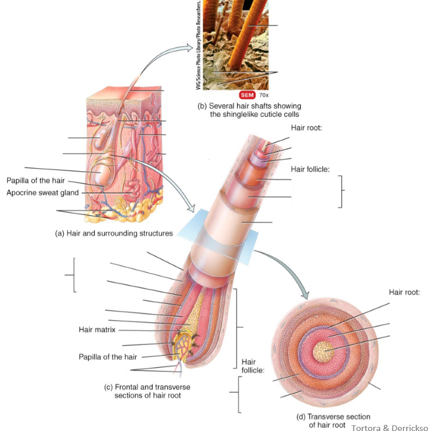
Label the following diagram
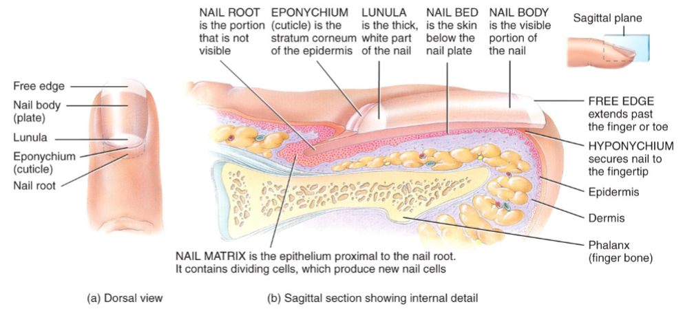
What are nails made of?
Tightly packed, hard, dead keratinized epidermal cells
Nail root + lunule + nail plate
What is the nail bed?
Skin beneath the nail plate
What is the subungual dermis?
Thick, highly vascular underlay of the nail bed
What is the lunule?
The half-moon crescent @ the inferior aspect of the nail body
What is the free-edge of a nail?
The other white edge at the surface (the ‘fingernail’)
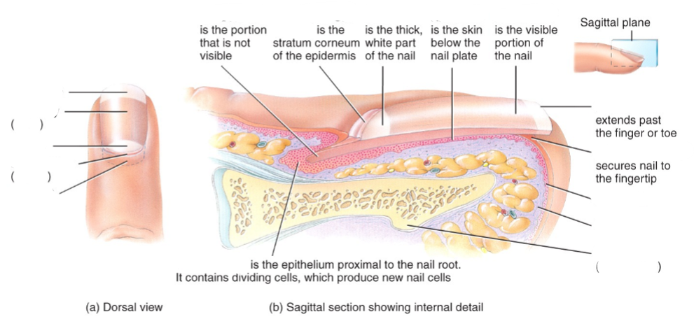
Label the following diagram
What is the follicle of a hair?
The downward continuation of the epidermis which surrounds the hair root
What are arrector pili?
Muscles which pull the hair shaft perpendicular to the skin surface (goose bumps)
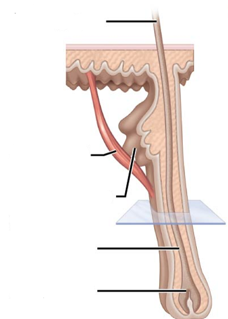
Label the following diagram
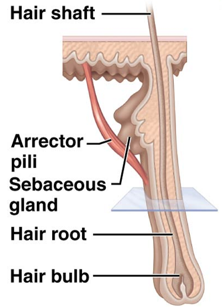
What are sebaceous glands?
Glands which secrete oily substances (sebum) & are connected to hair follicles
What are sweat glands?
Exocrine glands which release sweat into hair follicles or the skin surface via pores
What are ceruminous glands?
Modified sweat glands which produce ear wax in the ear canal
How do somatic sensations arise in the skin?
From the stimulation of sensory receptors in the dermis of the skin
What are the 4 modalities of cutaneous nerves & sensory receptors?
Tactile: touch, pressure, vibration, itch, tickle
Thermal: warm, hot, cold
Pain
Proprioceptive: sensory organs located mainly in muscle & joints
What receptors are located superficially vs deep in the skin?
Superficially: discs which mediate tactile modalities
fine touch, pressure, & vibration
Deep: crude touch & stretching of the skin
Deepest: pressure & fast vibration
Describe the embryological development of dermatomes
Relationship b/w nerves & skin is established during development
Tissue which gives rise to muscle, bone & dermis are organised segmentally into somites
Somites → limb buds → drag segmental innervation w/ them during growth
Describe the difference b/w somatic & visceral pain
Somatic pain: pain from the skin
Sharp & well-localised
Visceral pain: dull & poorly localised
Accompanied by referred pain
Referred to somatic area by the same dermatome
What is referred pain?
When you feel pain in one area of the body, but the actual source of the pain is located elsewhere
Provide an example of referred pain?
Radiation of chest pain down the arms during a heart attack b/c of these somatic areas supplied by the same dermatome which supplies the pericardium of the heart
How are stretch marks formed?
Overstretching of skin → disruption of lateral bonding b/w collagen fibres in the dermis + rupturing of small blood vessels
Rupturing of small blood vessels → leaked blood in the dermal layer → reddish streaks → turn silver when (poorly vascularised) scar tissue appears
Scar tissue replaces where collagen fibres were located @ the epidermal layer
Why are burns life-threatening?
One of the roles of the skin is to maintain hydration, so severe damage to the skin → catastrophic loss of body fluids → dehydration
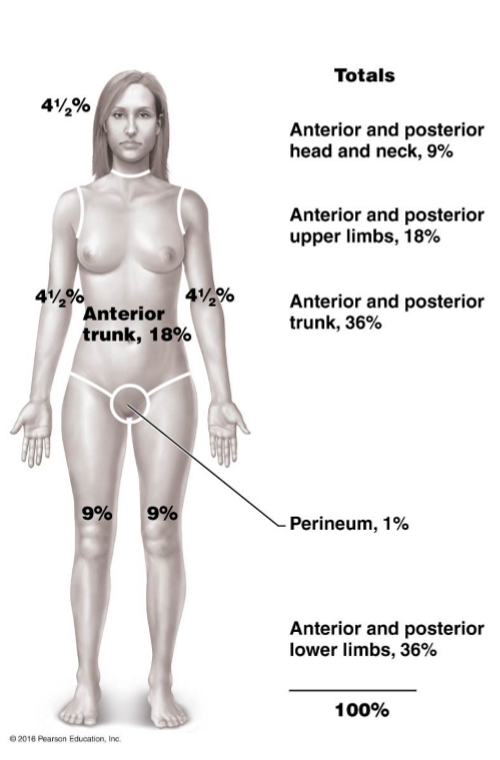
How can the % of fluid loss when a person is burned be determined?
By evaluating the % of the body surface burned
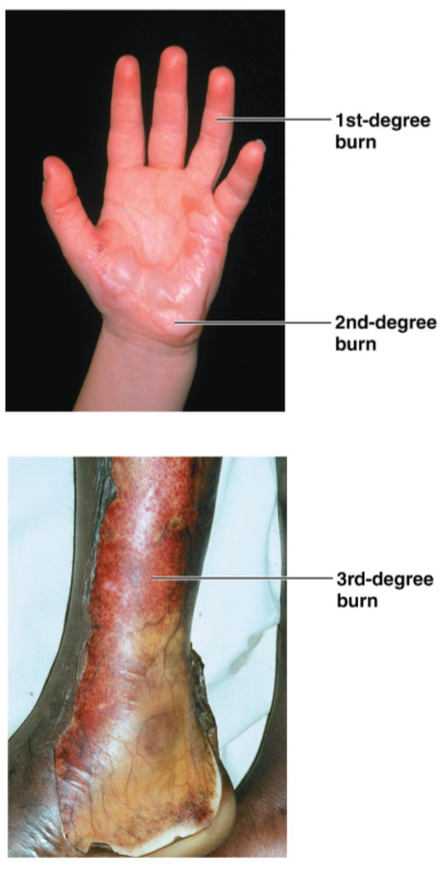
What are first, second & third degree burns?
1st: injured epidermis only
2nd: injured epidermis & 1st layer of dermis
3rd: injury to full thickness of skin