uta biol 2457 practical 2
1/202
There's no tags or description
Looks like no tags are added yet.
Name | Mastery | Learn | Test | Matching | Spaced | Call with Kai |
|---|
No analytics yet
Send a link to your students to track their progress
203 Terms
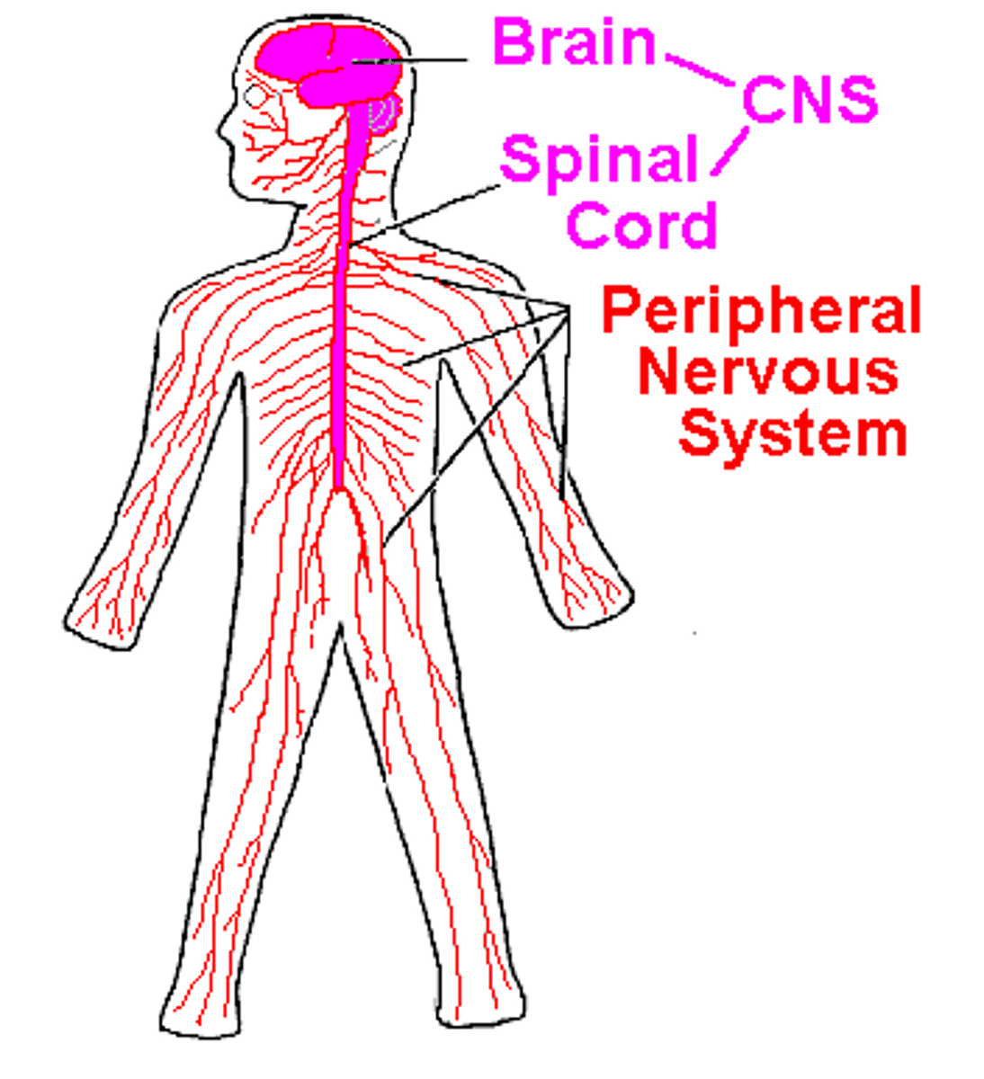
central nervous system
(CNS) brain and spinal cord.
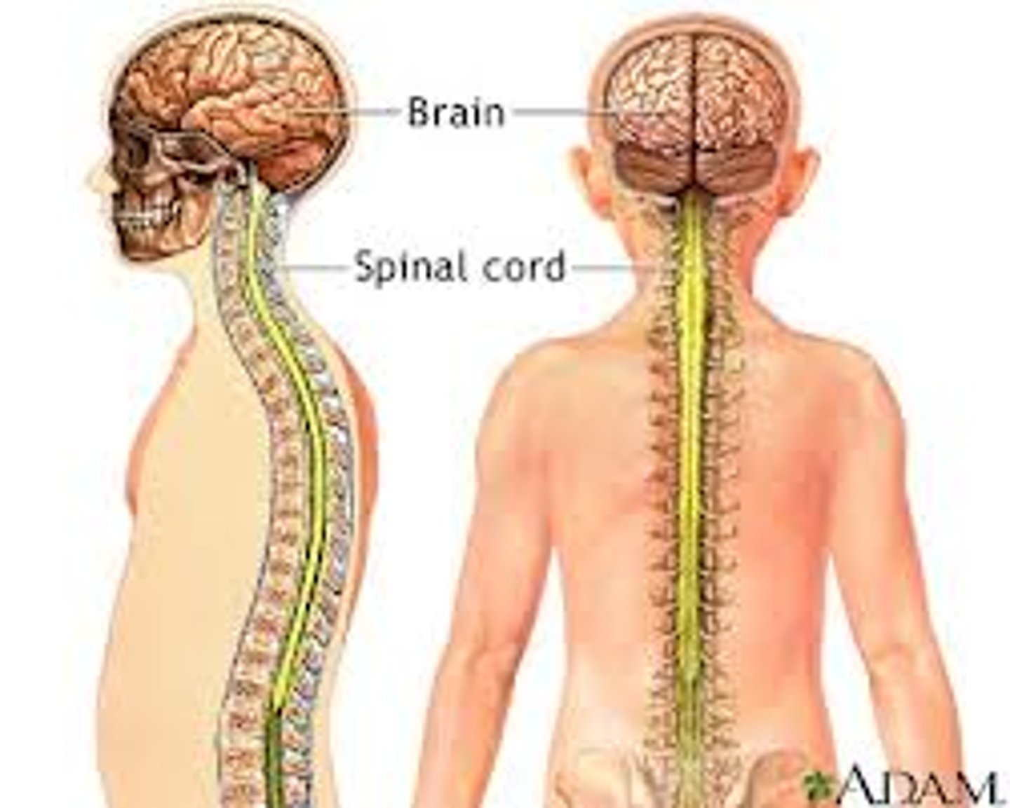
peripheral nervous system
(brain and spinal cord) Connects the central nervous system to the body's organs and limbs.
sensory (afferent) neurons
neurons that carry incoming information from the sensory receptors (sight, smell, sound etc.) to the brain and spinal cord
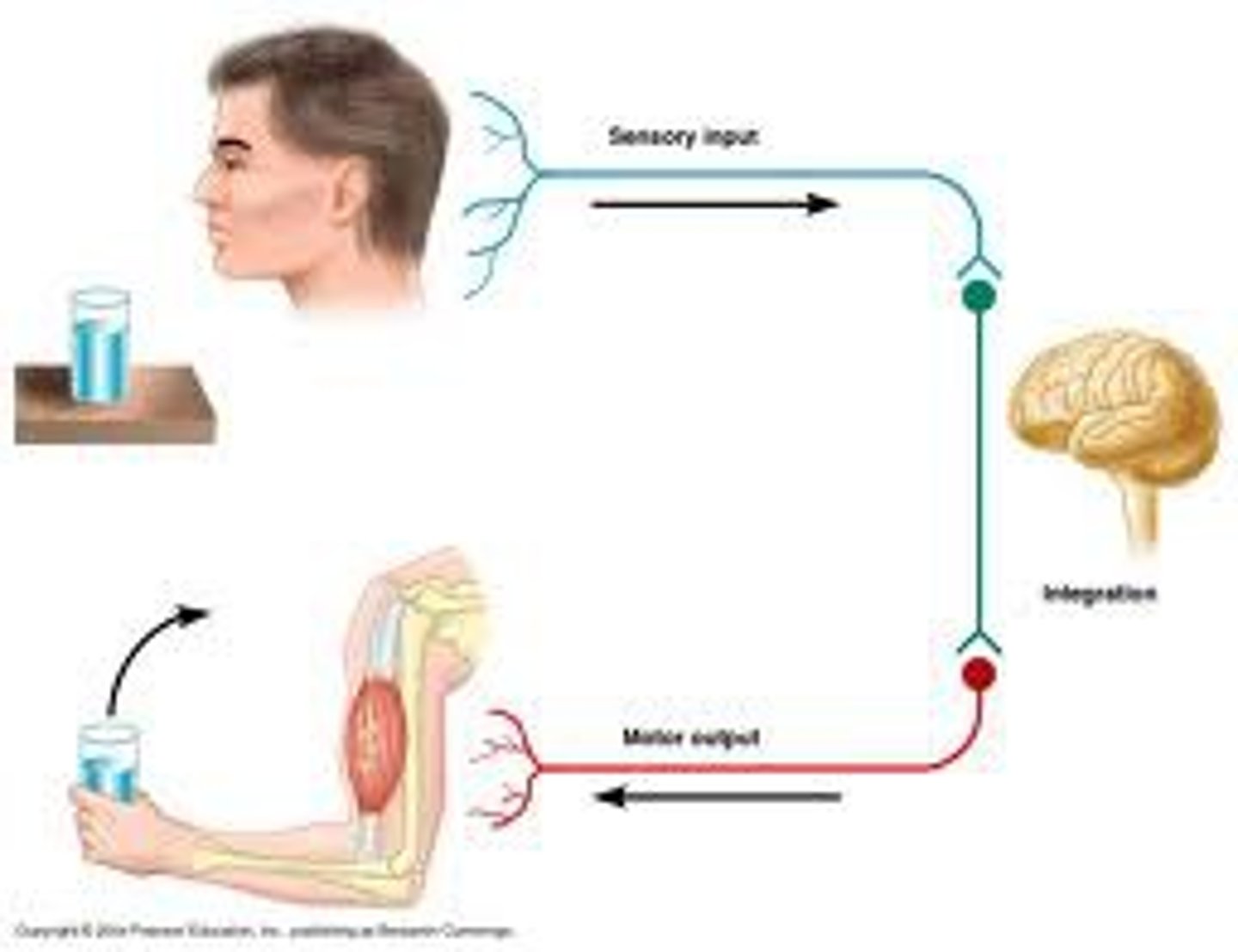
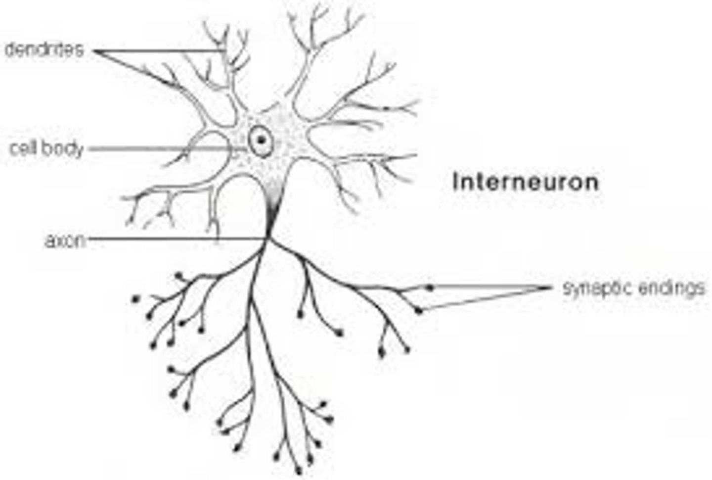
Interneurons
Central nervous system neurons that internally communicate and intervene between the sensory inputs and motor outputs
motor (efferent) neurons
Nerve cells responsible for making an action or movement happen.Multipolar Shaped Neurons
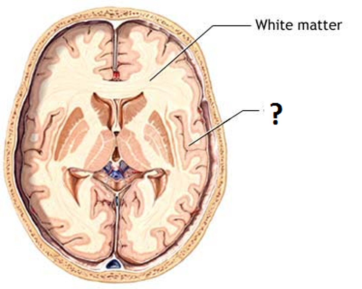
white matter
myelinated axons
grey matter
unmyelinated neuron cell bodies and short, unmyelinated axons
meninges
three protective membranes that surround the brain and spinal cord. dura mater, arachnoid mater, pia mater
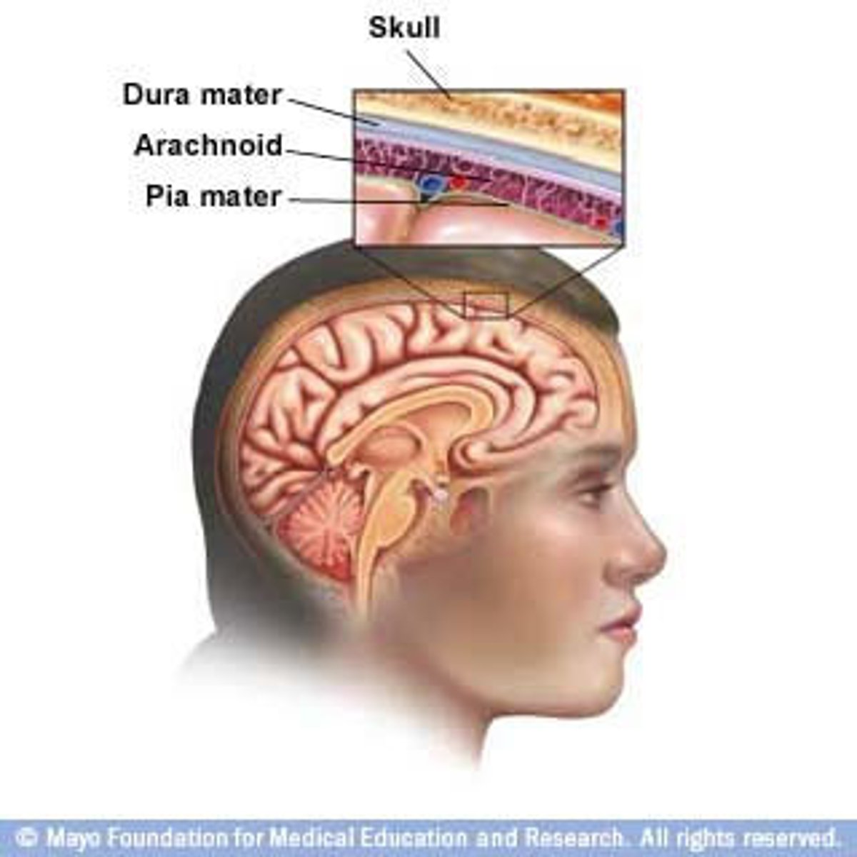
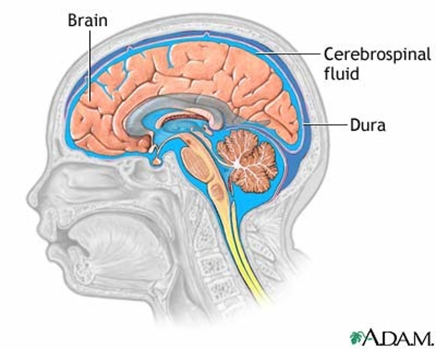
dura matter
thick, outermost layer of the meninges surrounding and protecting the brain and spinal cord
arachnoid mater
weblike middle layer of the three meninges
pia matter
thin, delicate inner membrane of the meninges
cerebrospinal fluid
Fluid in the space between the meninges that acts as a shock absorber that protects the central nervous system.
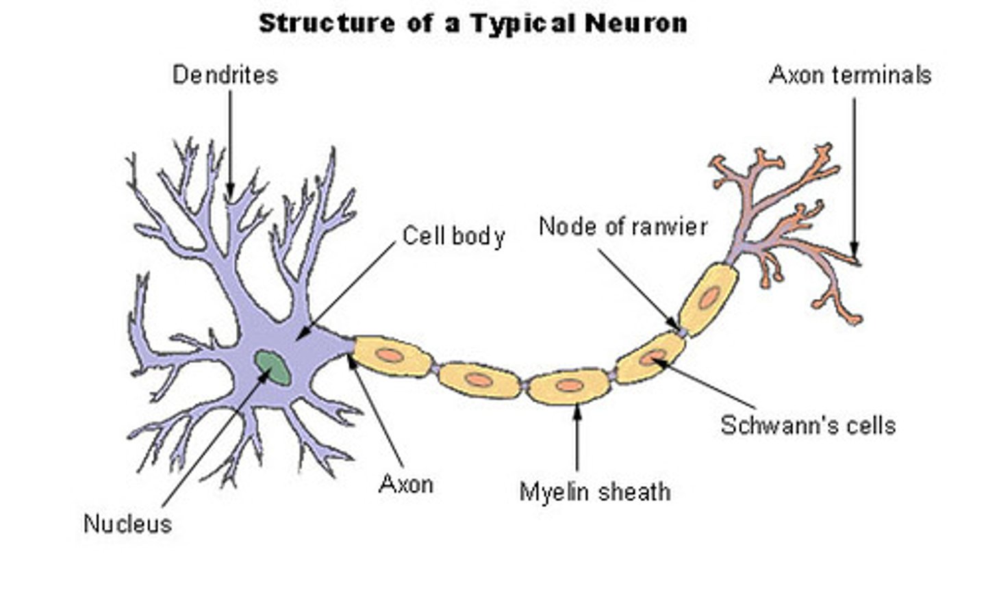
dendrites
Branchlike parts of a neuron that are specialized to receive information.
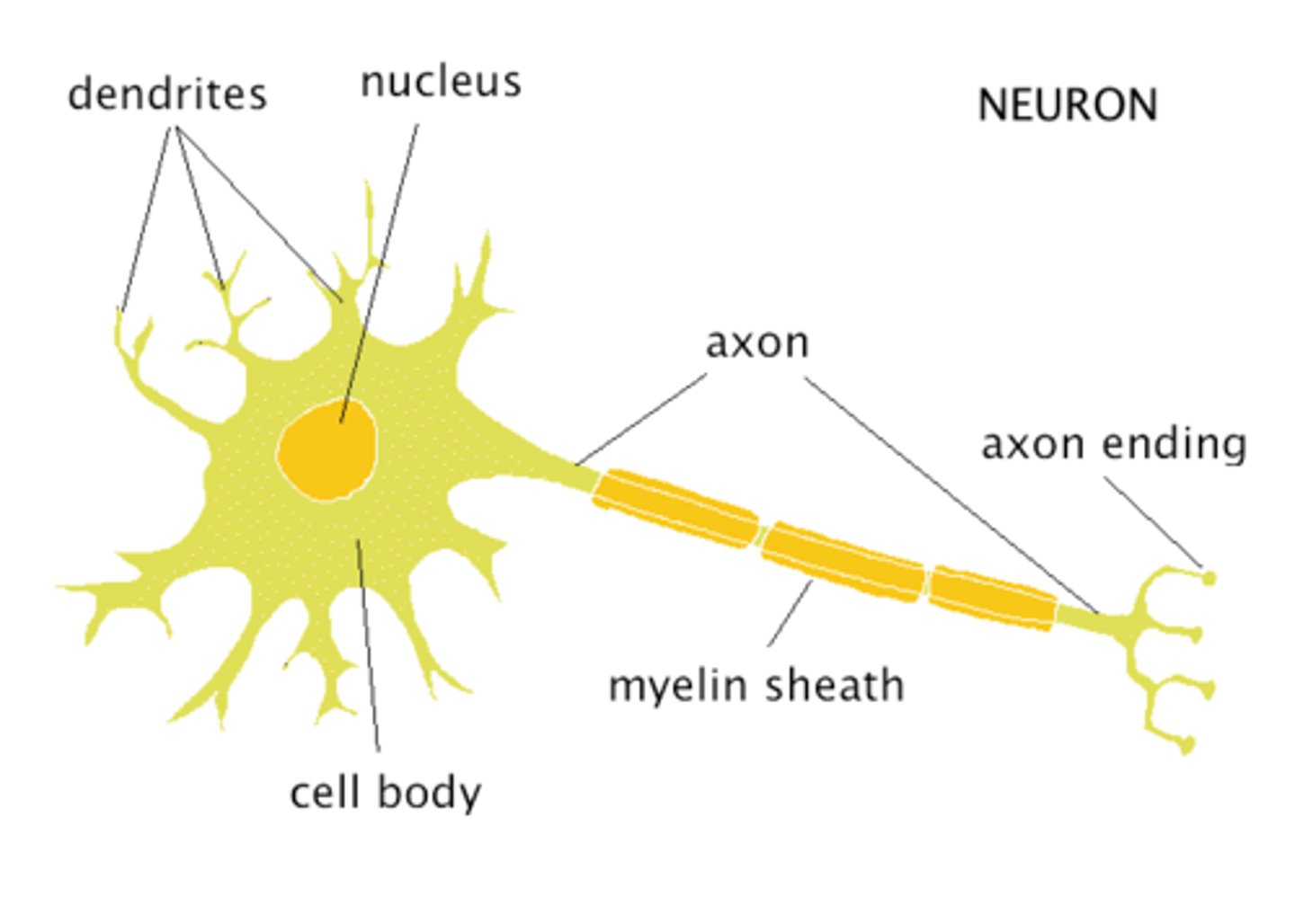
cell body (soma)
the part of a neuron that coordinates information-processing tasks and keeps the cell alive
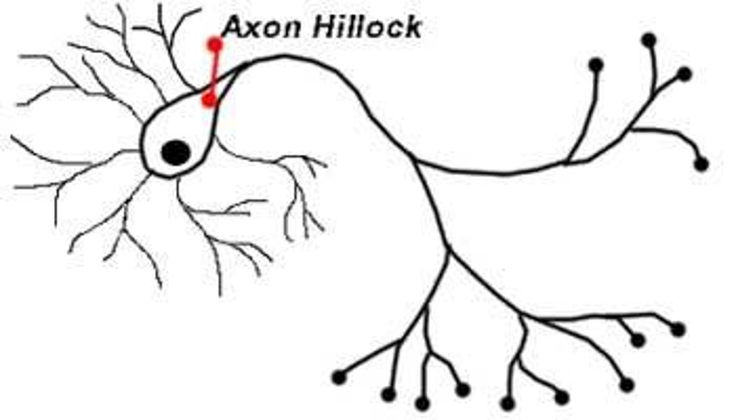
axon hillock
the cone-shaped area on the cell body from which the axon originates
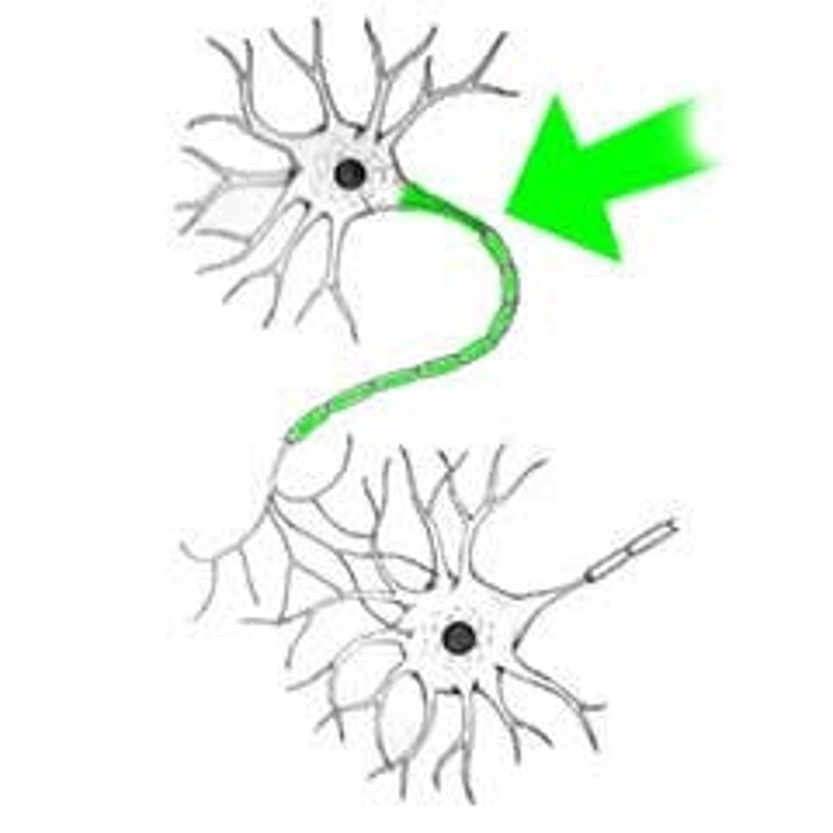
axon
the neuron extension that passes messages through its branches to other neurons or to muscles or glands
Schwann cells
Type of glia in the PNS, Supporting cells of the peripheral nervous system responsible for the formation of myelin.
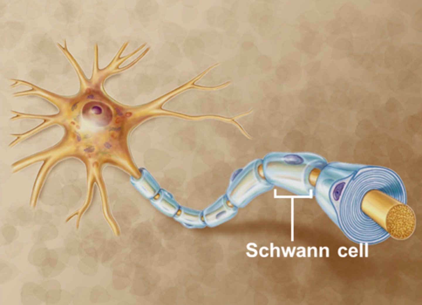
myelin sheath
covers the axon of some neurons and helps speed neural impulses
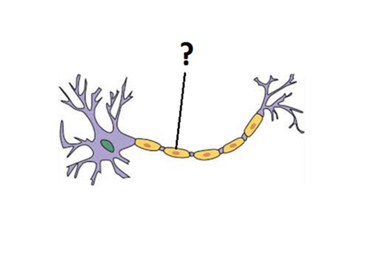
Nodes of Ranvier
Gaps in the myelin sheath to which voltage-gated sodium channels are confined.
axon terminals
Branches at the end of the axon that contain tiny pouches, or sacs, called synaptic vesicles.
Cerebrum
Largest part of the brain; responsible for voluntary muscular activity, vision, speech, taste, hearing, thought, and memory.
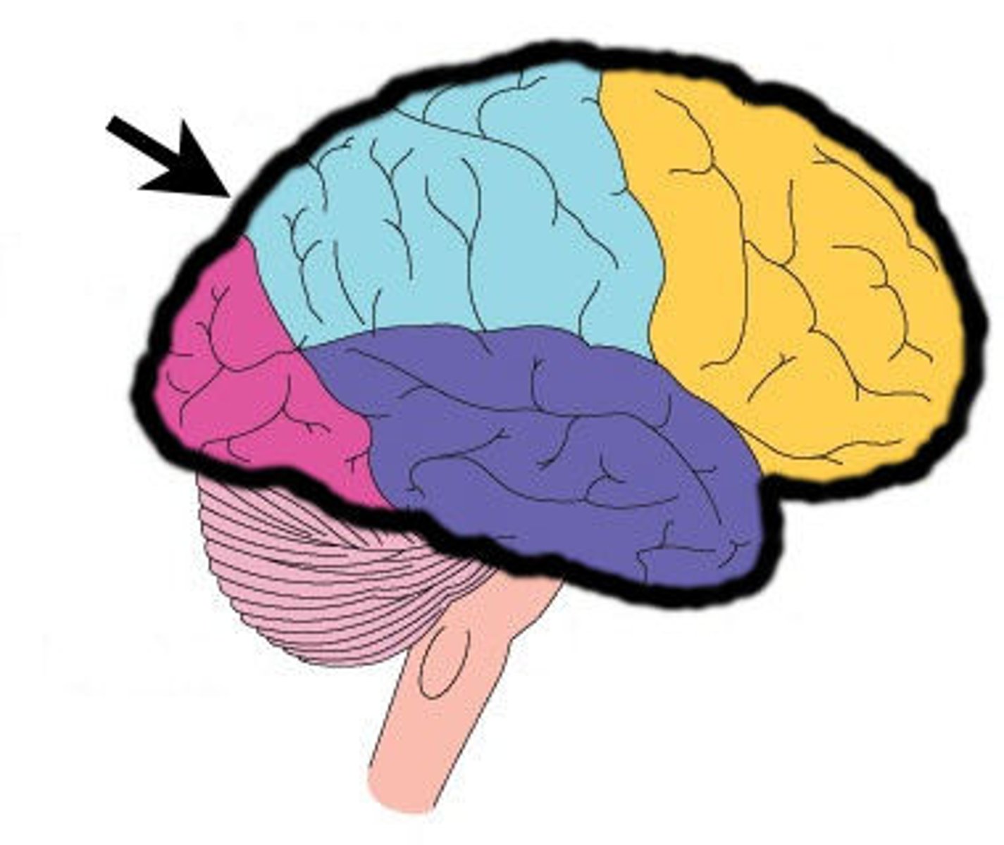
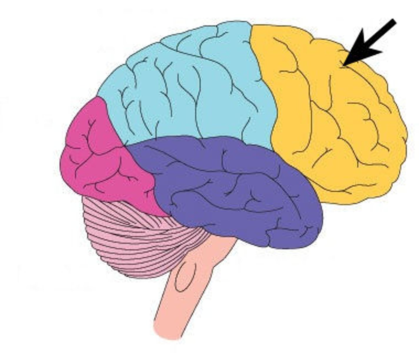
frontal lobe
A region of the cerebral cortex that has specialized areas for movement, abstract thinking, planning, memory, and judgement
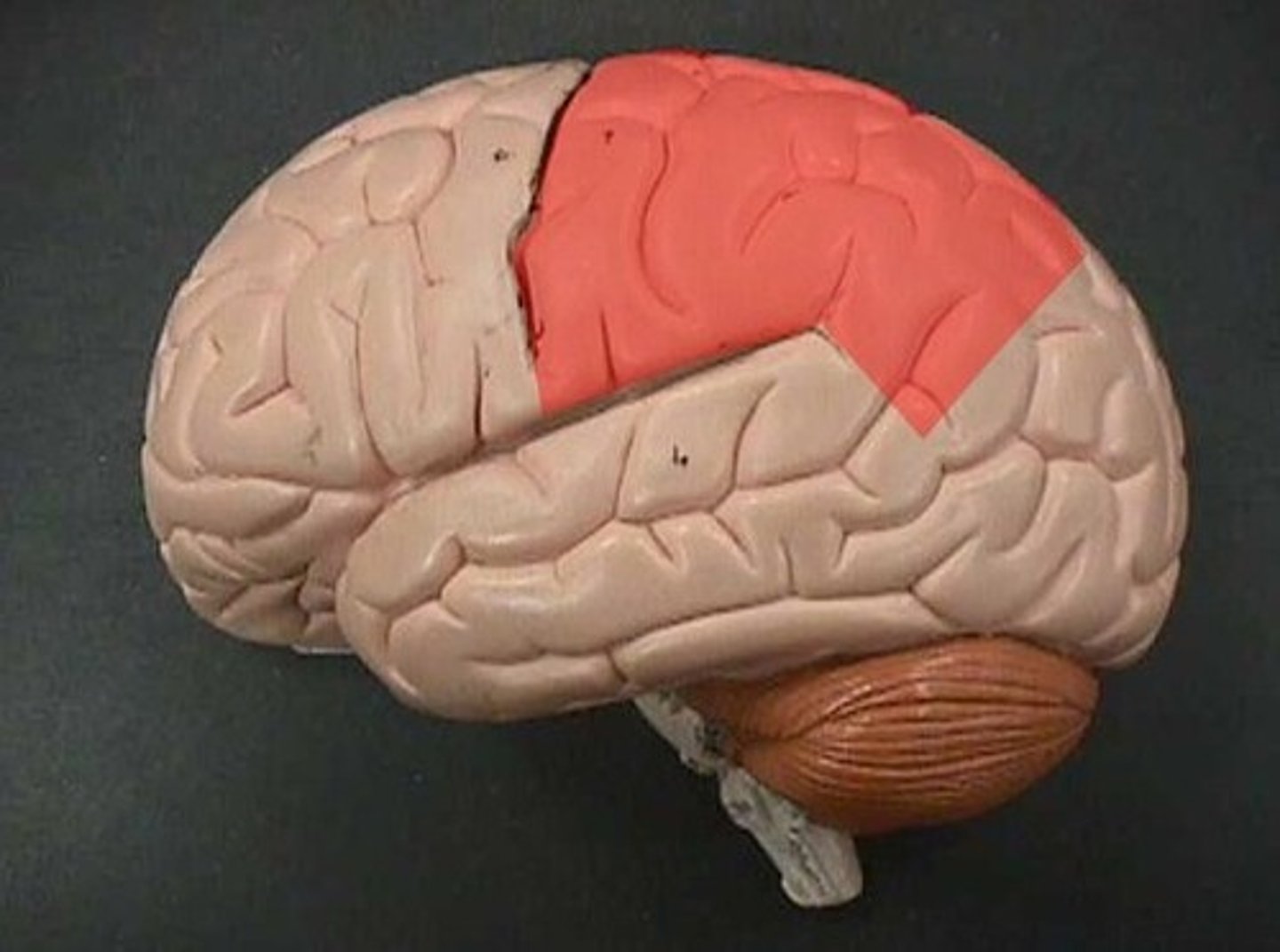
parietal lobe
portion of the cerebral cortex lying at the top of the head and toward the rear; receives sensory input for touch and body position
temporal lobe
A region of the cerebral cortex responsible for hearing and language.

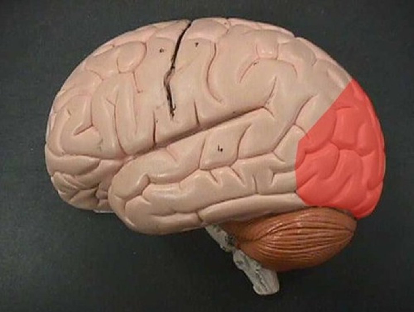
occipital lobe
A region of the cerebral cortex that processes visual information
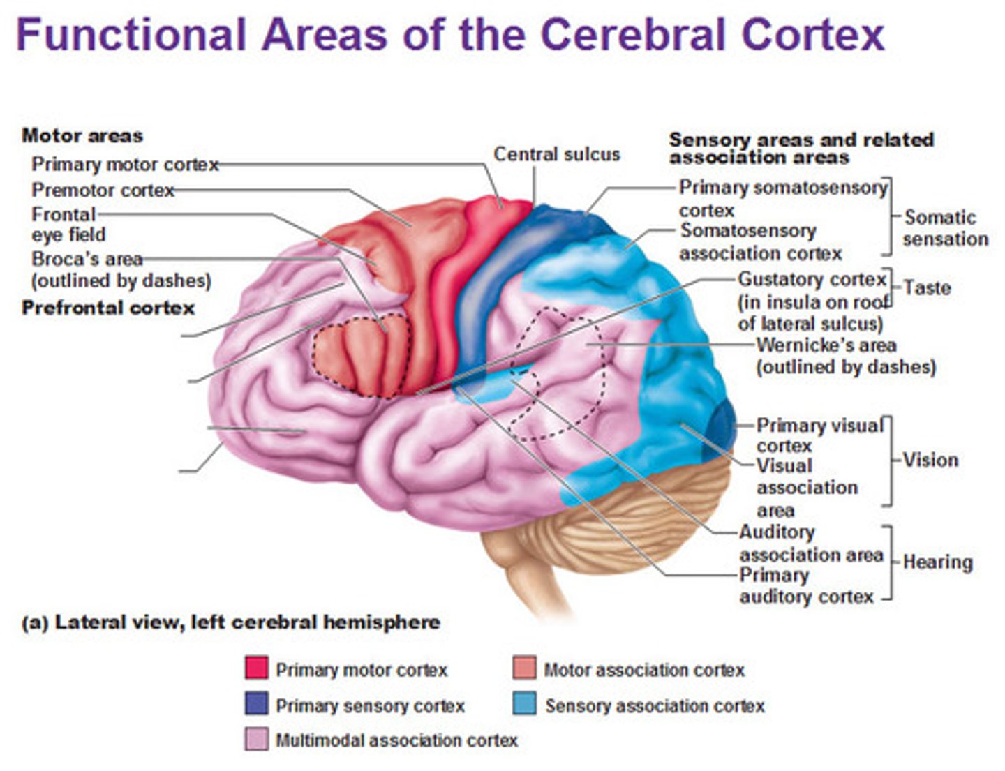
functional areas
areas of the brain that are specialized in the production of certain tasks
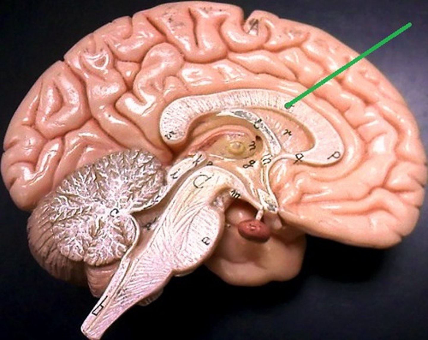
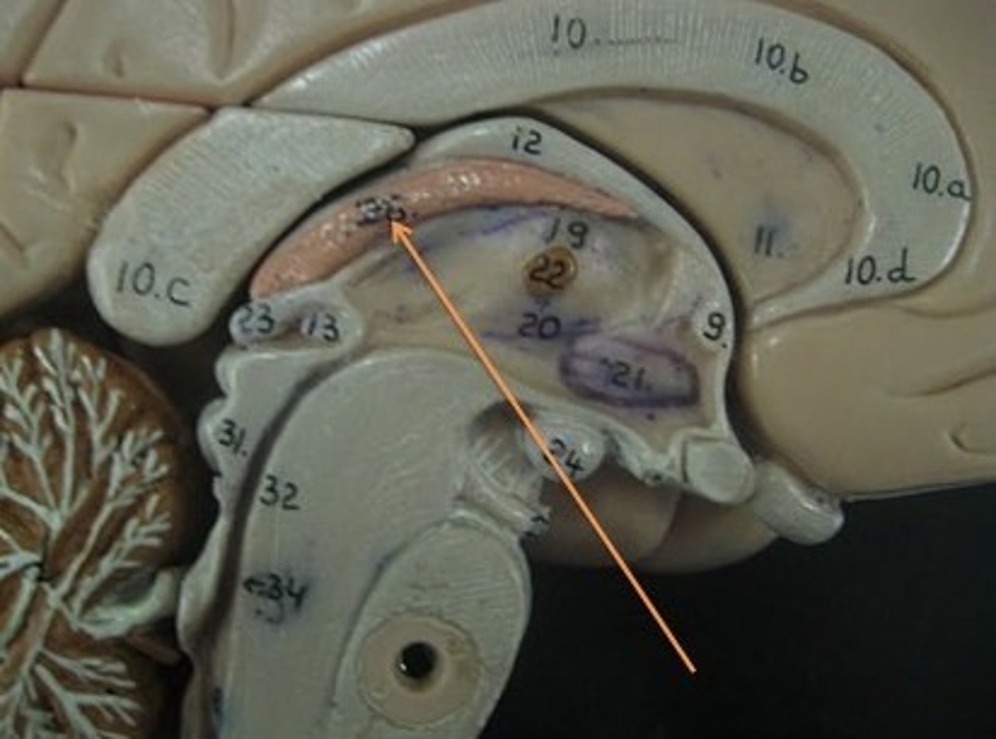
corpus callosum
the large band of neural fibers connecting the two brain hemispheres and carrying messages between them
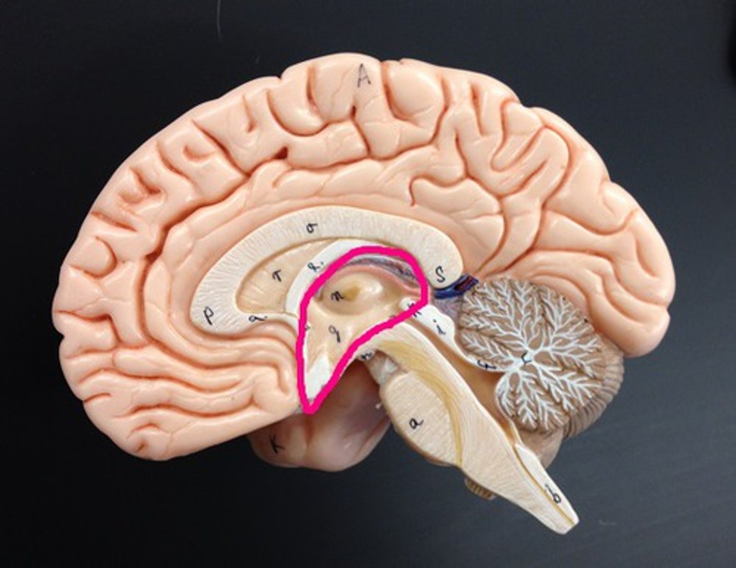
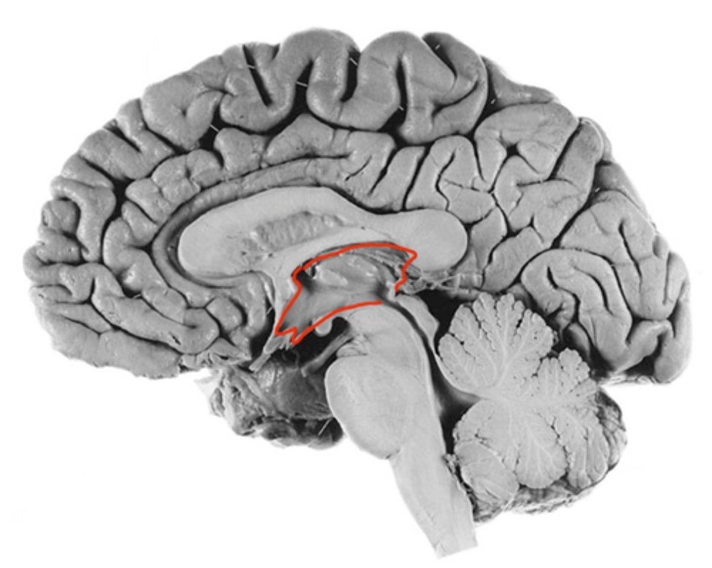
Diencephalon
thalamus, hypothalamus, epithalamus
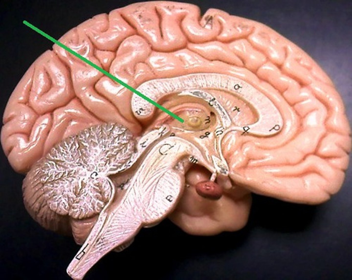
thalamus
the brain's sensory switchboard, located on top of the brainstem; it directs messages to the sensory receiving areas in the cortex and transmits replies to the cerebellum and medulla
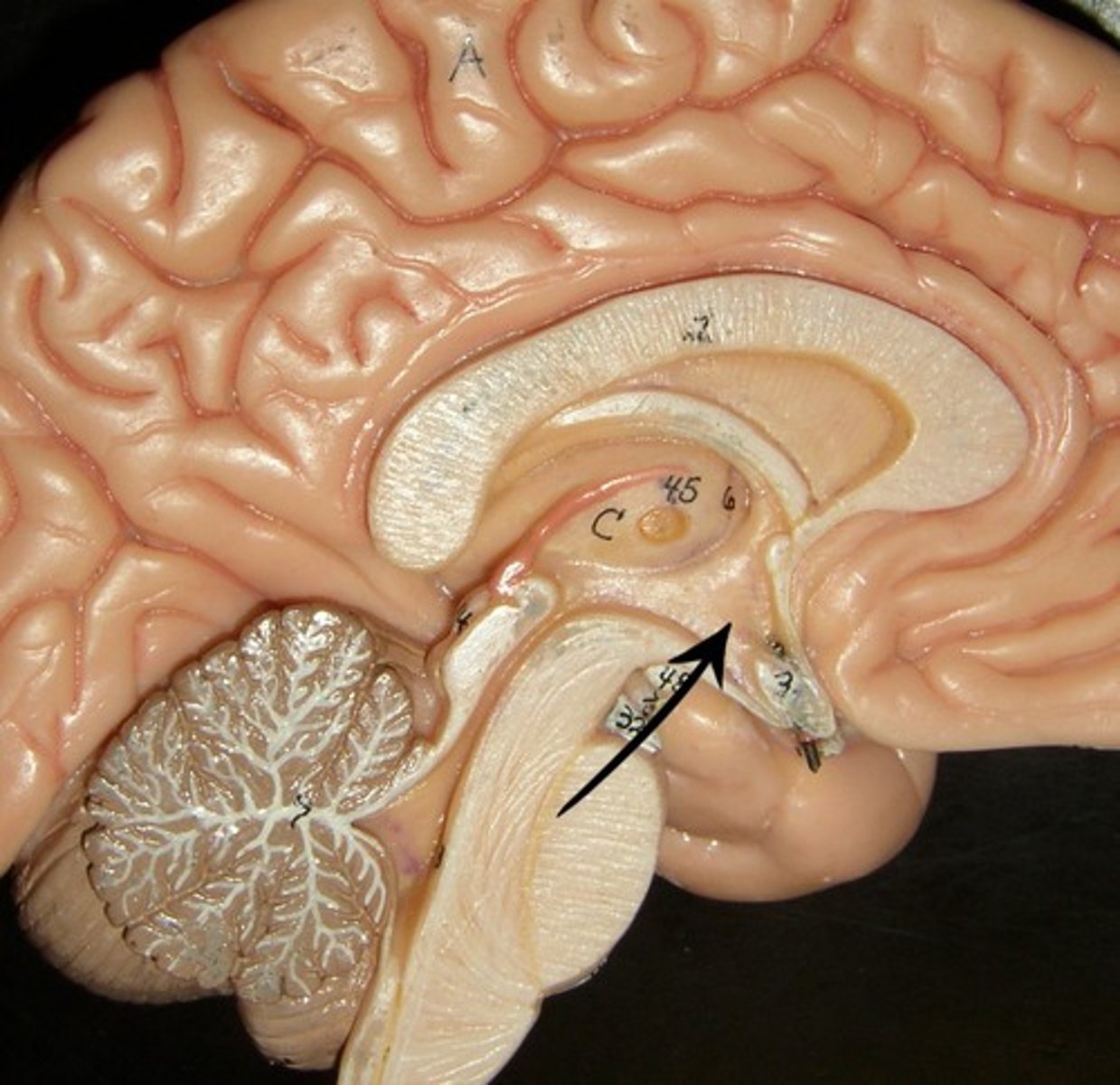
Hypothalamus
a neural structure lying below the thalamus; directs eating, drinking, body temperature; helps govern the endocrine system via the pituitary gland, and is linked to emotion
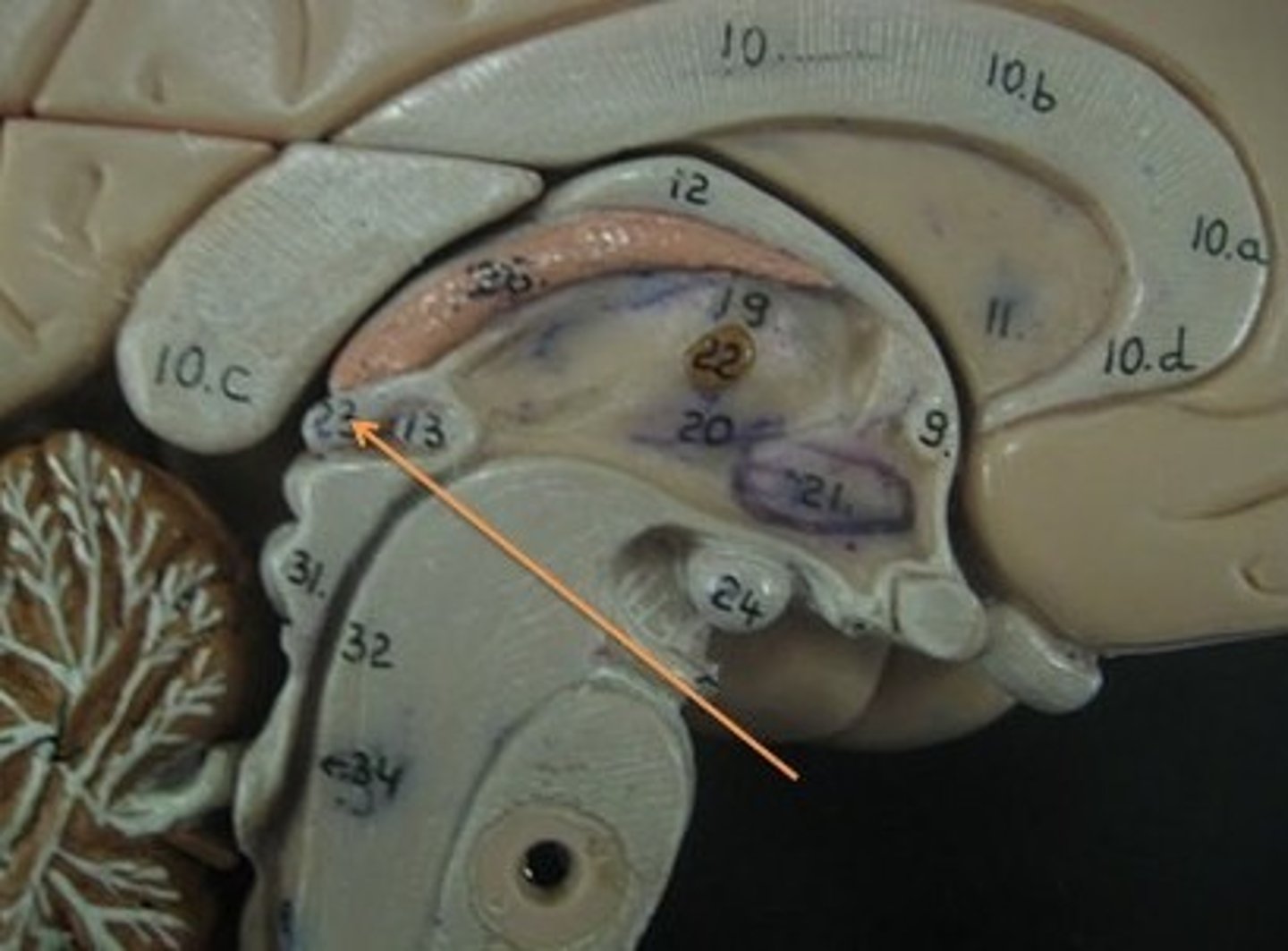
pineal gland
produces melatonin
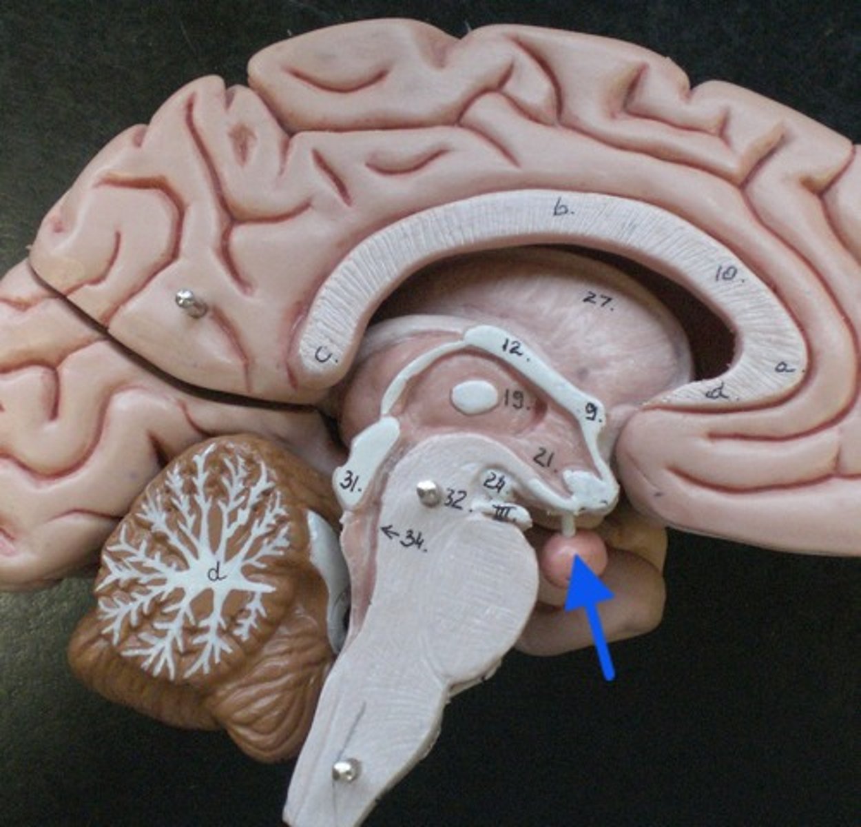
pituitary gland
The endocrine system's most influential gland. Under the influence of the hypothalamus, the pituitary regulates growth and controls other endocrine glands.
pituitary gland anterior
- Growth hormone
--- Drugs for growth hormone deficiency: somatrem (Protropin), somatropin (Humatrope)
--- Drugs for growth hormone excess: bromocriptine (Parlodel), octreotide (Sandostatin)
- Thyroid-stimulating hormone
--- Thyrotropin (Thytropar)
- Adrenocorticotropic hormone
--- Corticotropin (Acthar)
pituitary gland posterior
antidiuretic hormone and oxytocin
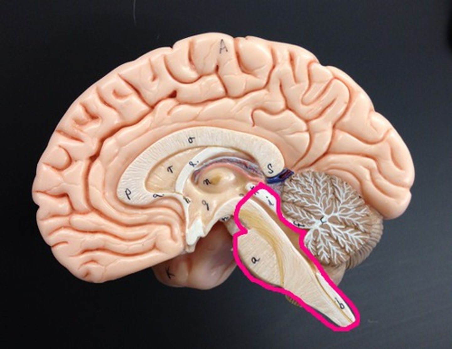
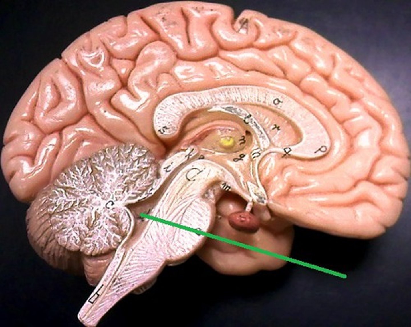
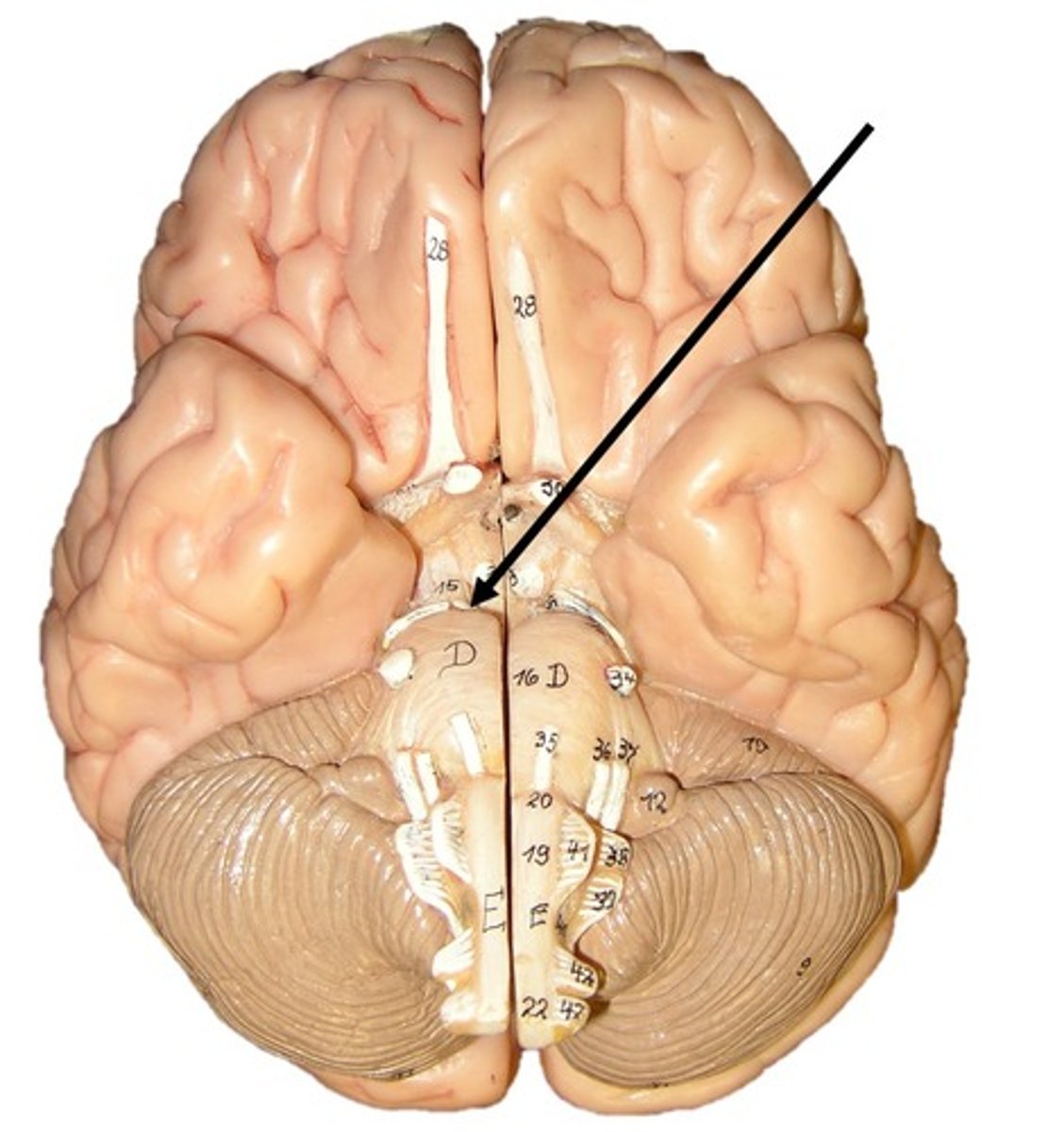
brain stem
midbrain, pons, medulla oblongata
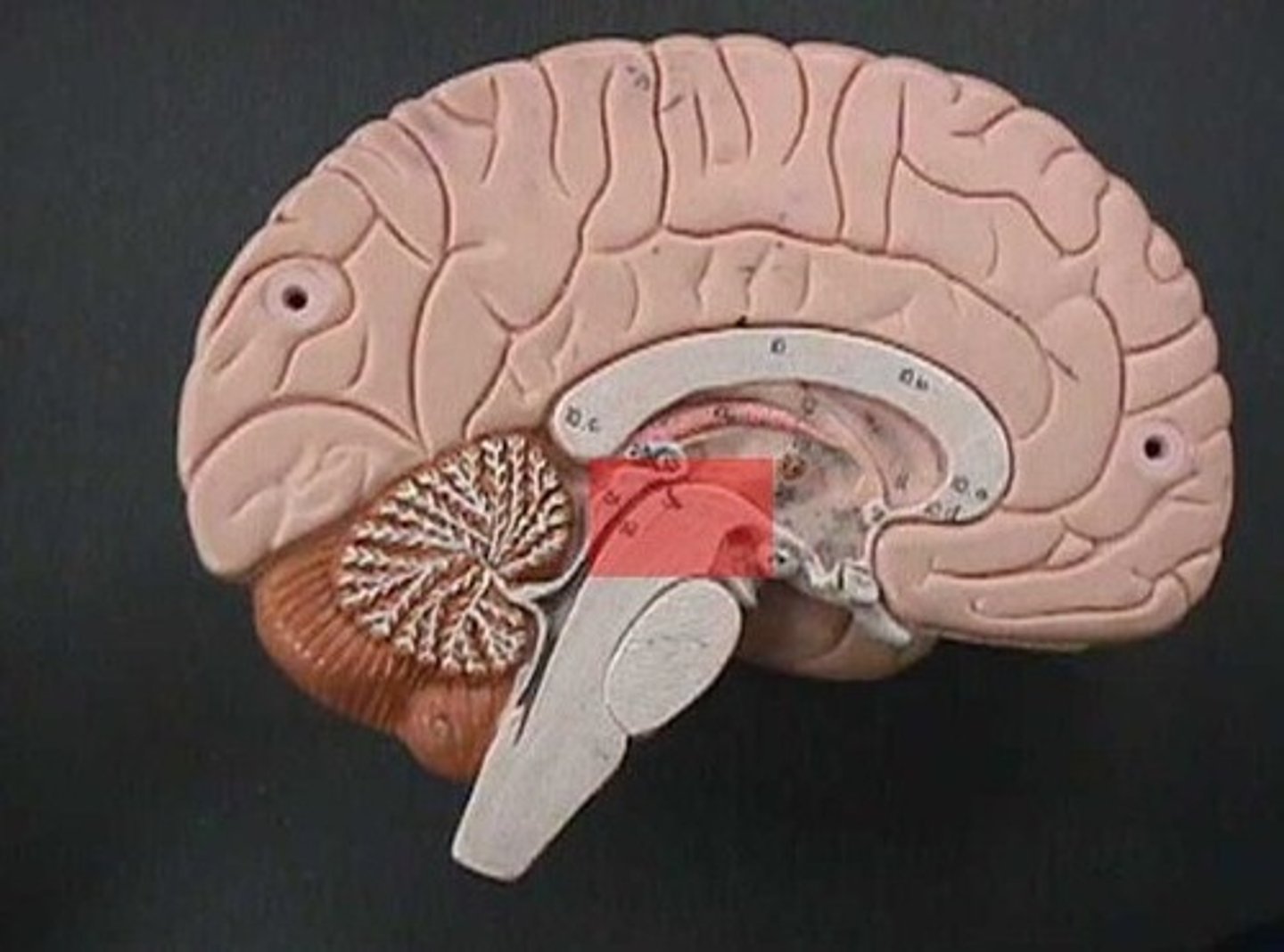
midbrain
Region between the hindbrain and the forebrain; it is important for hearing and sight.
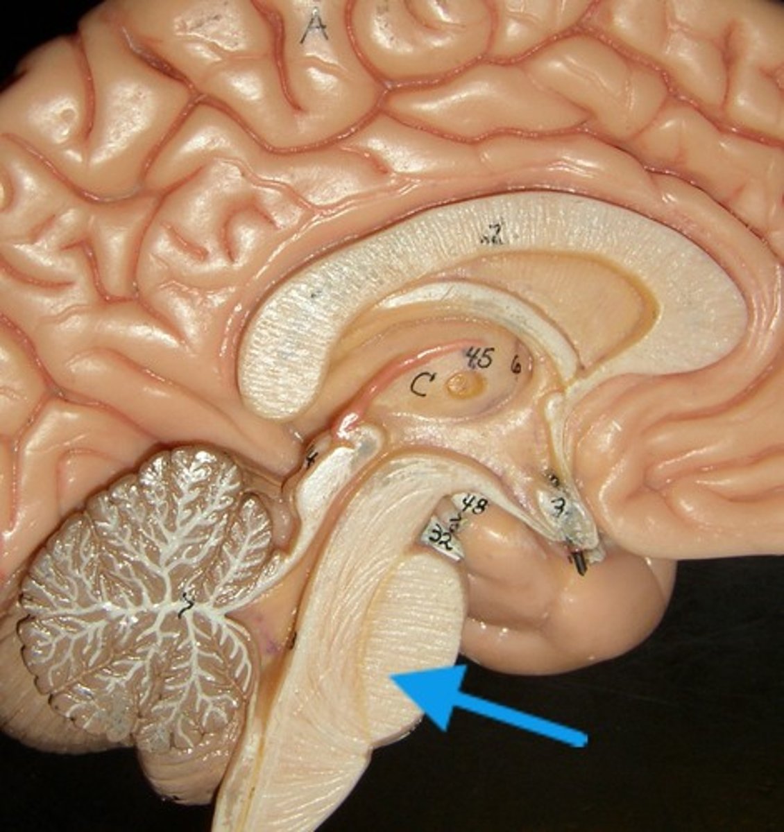
pons
sleep and arousal
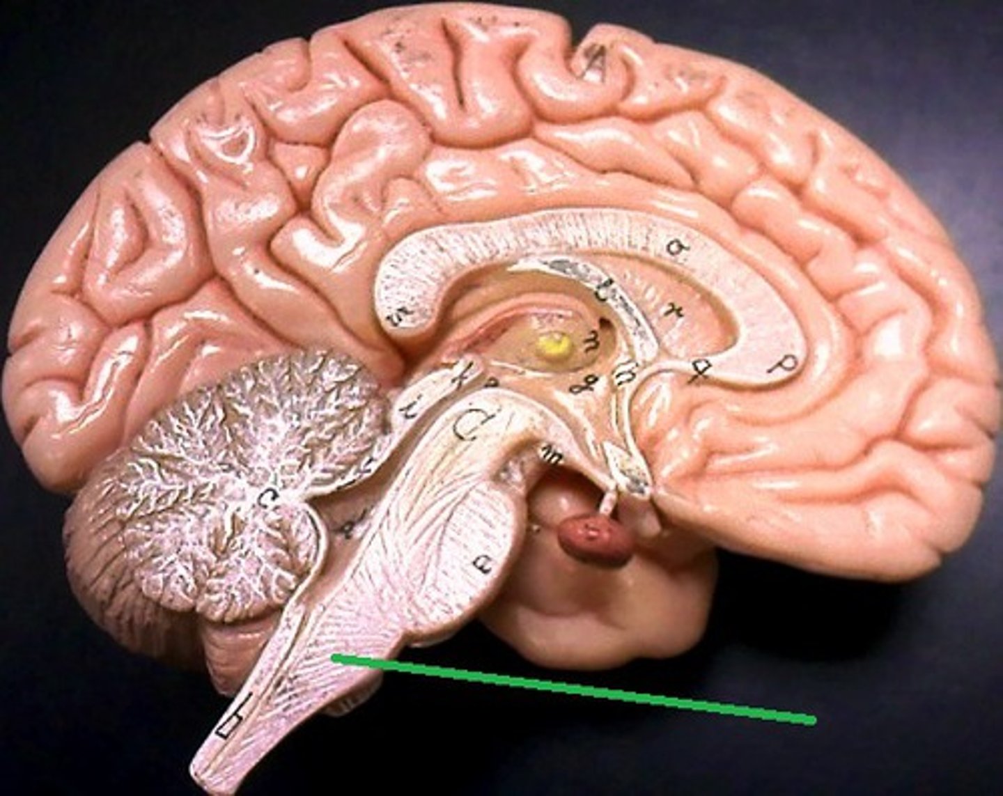
medulla oblongata
Part of the brainstem that controls vital life-sustaining functions such as heartbeat, breathing, blood pressure, and digestion.
reticular formation
a nerve network in the brainstem that plays an important role in controlling arousal
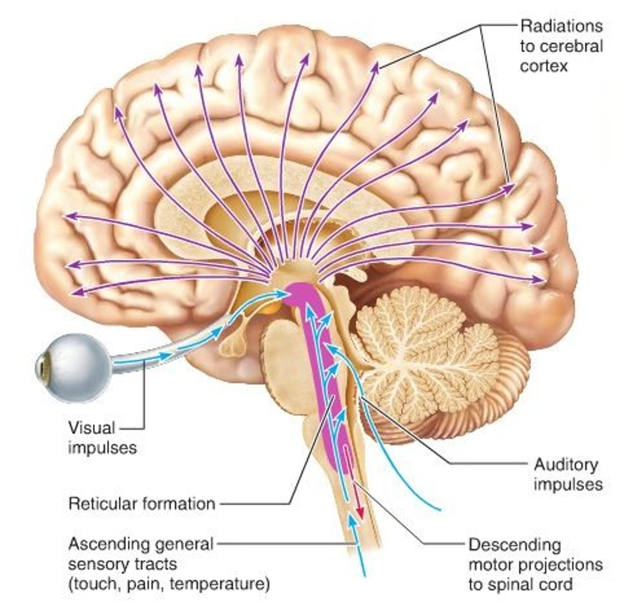
reticular activating system
Located in the upper brain stem; responsible for maintenance of consciousness, specifically one's level of arousal.
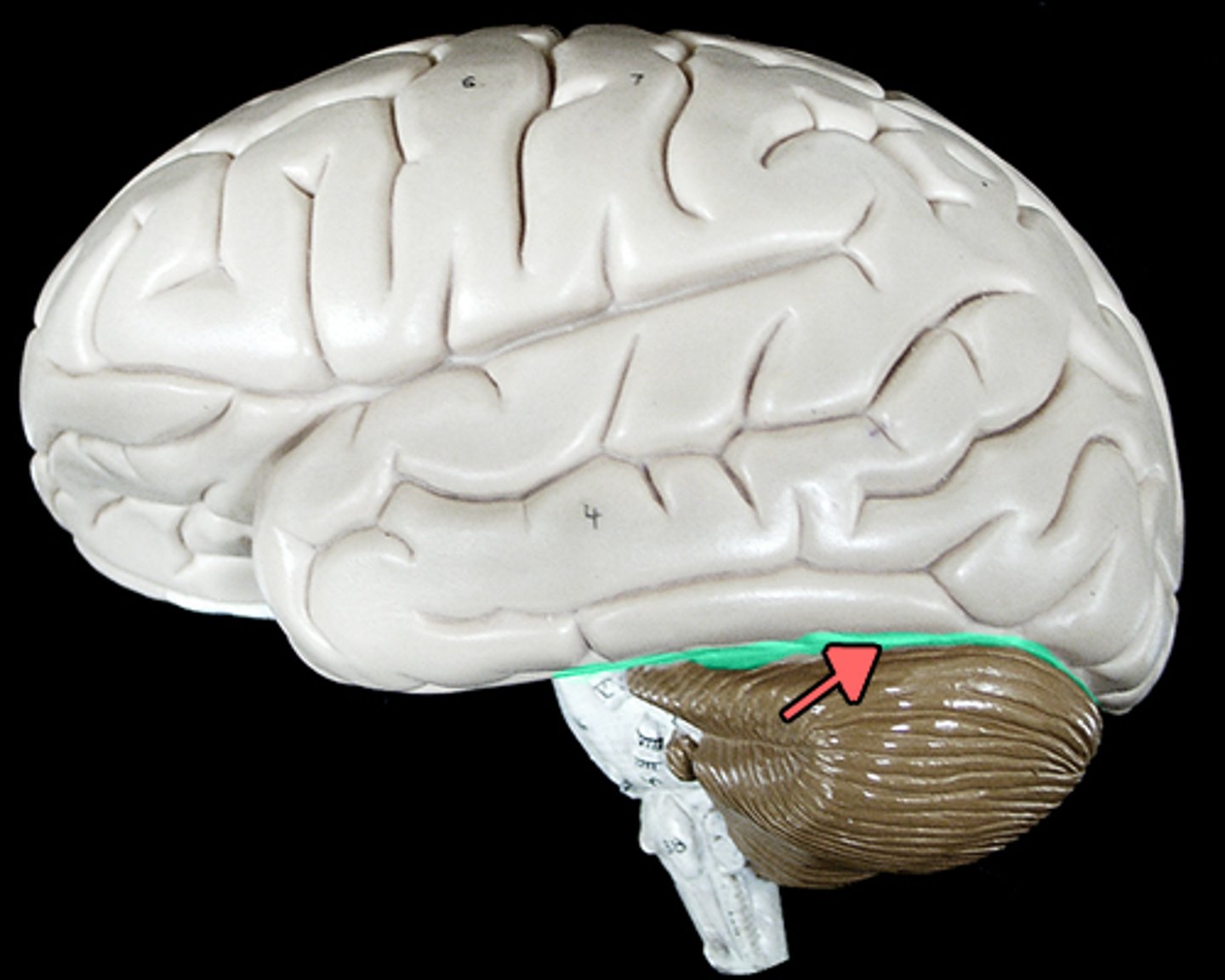
cerebellum
A large structure of the hindbrain that controls fine motor skills.
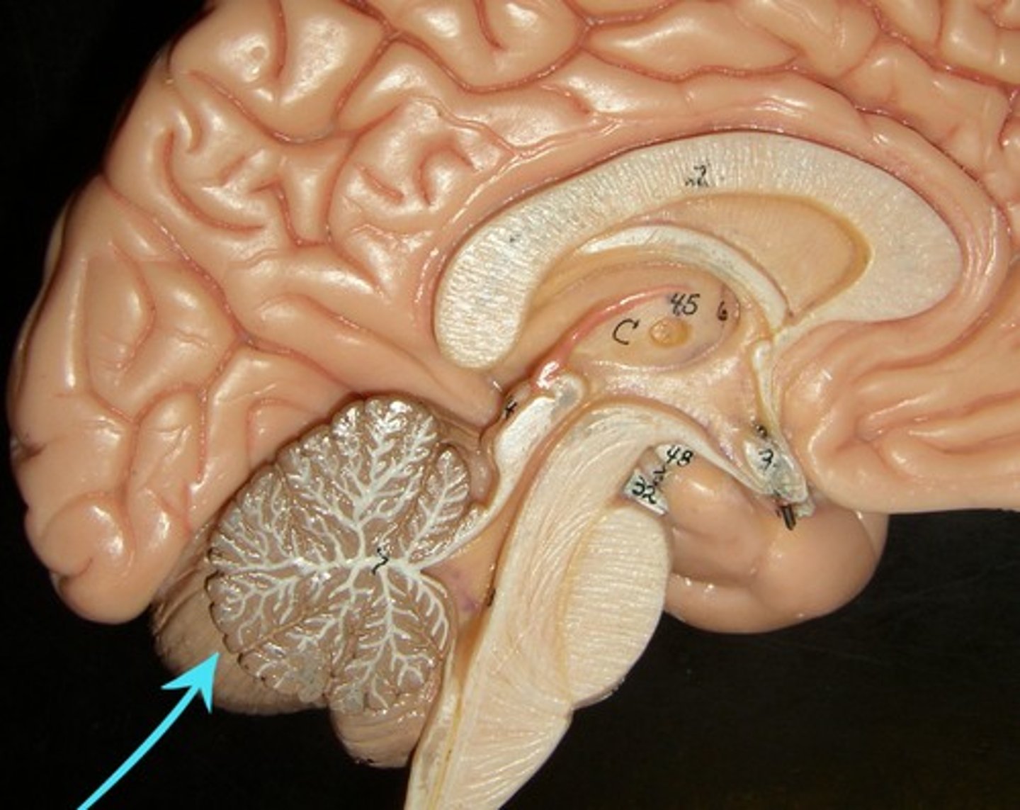
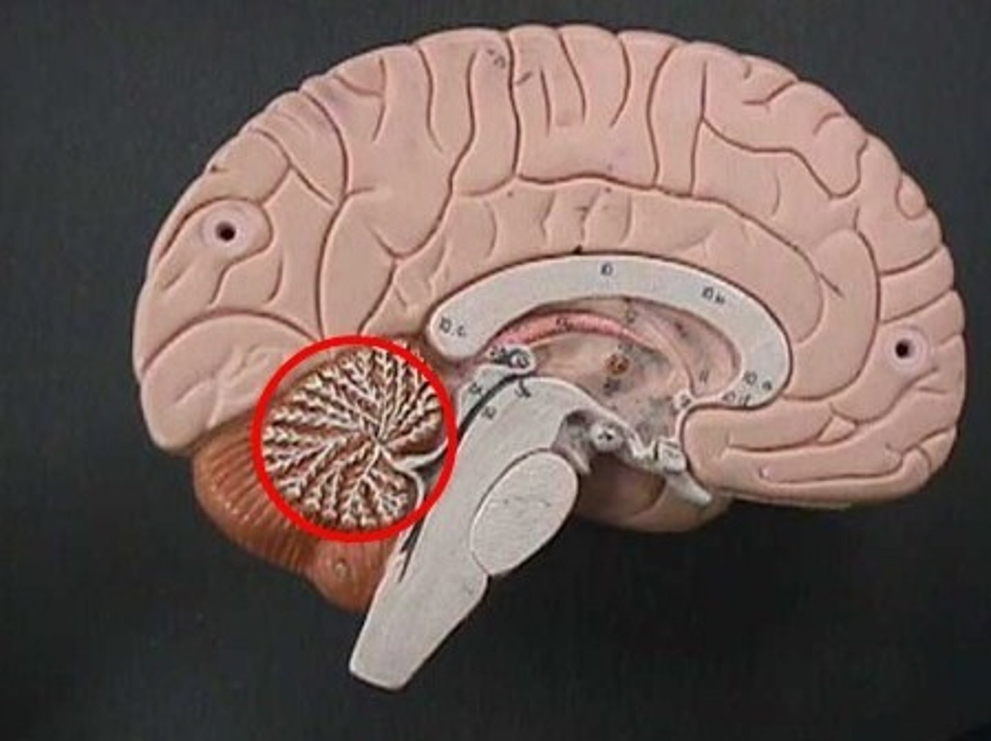
arbor vitae
white matter of the cerebellum
tree of life
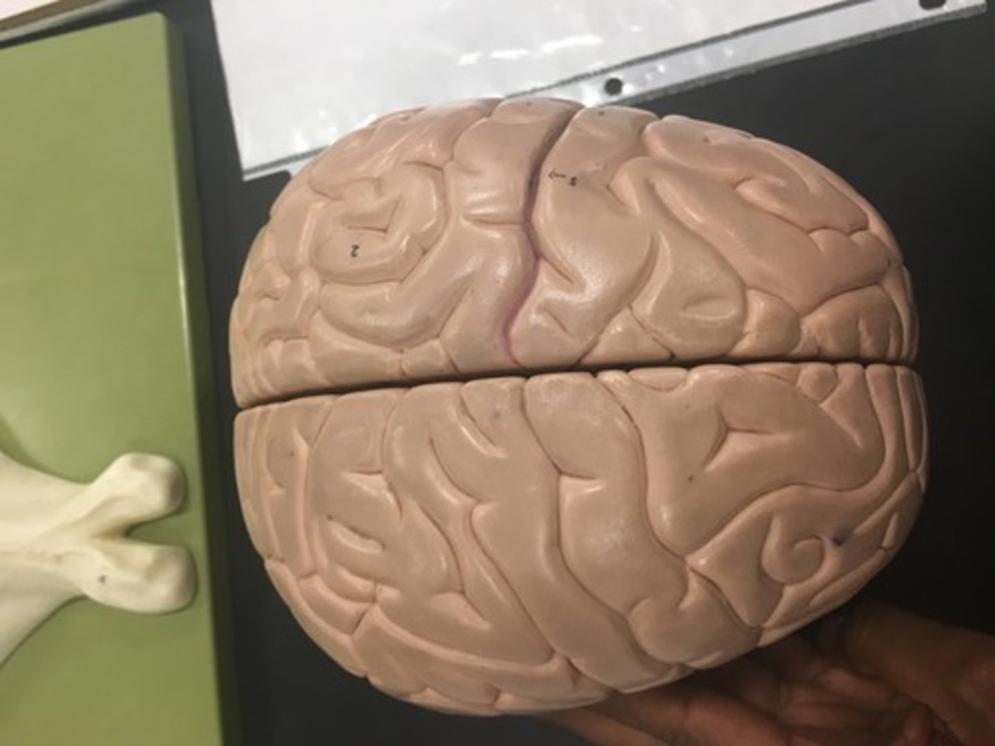
gyri
ridges of the brain
sulci
shallow grooves that separate gyri
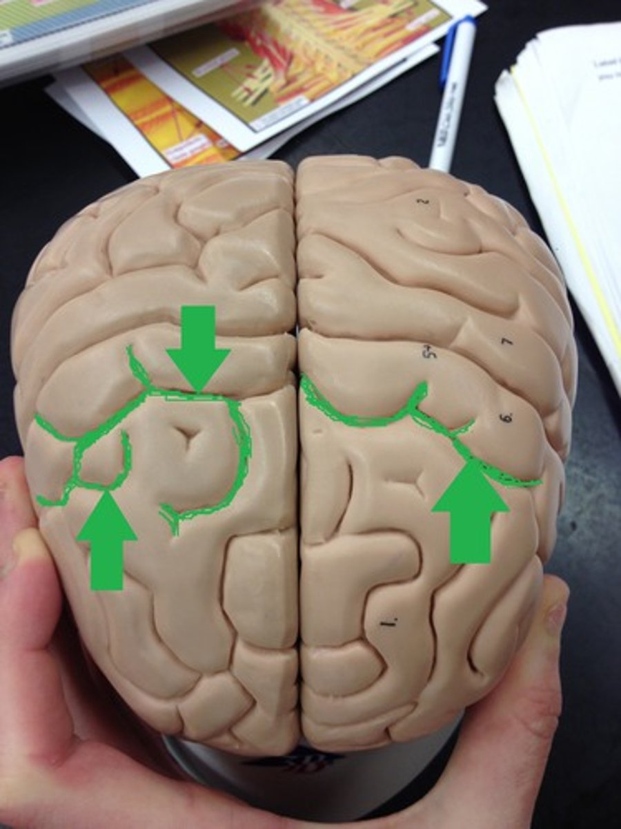
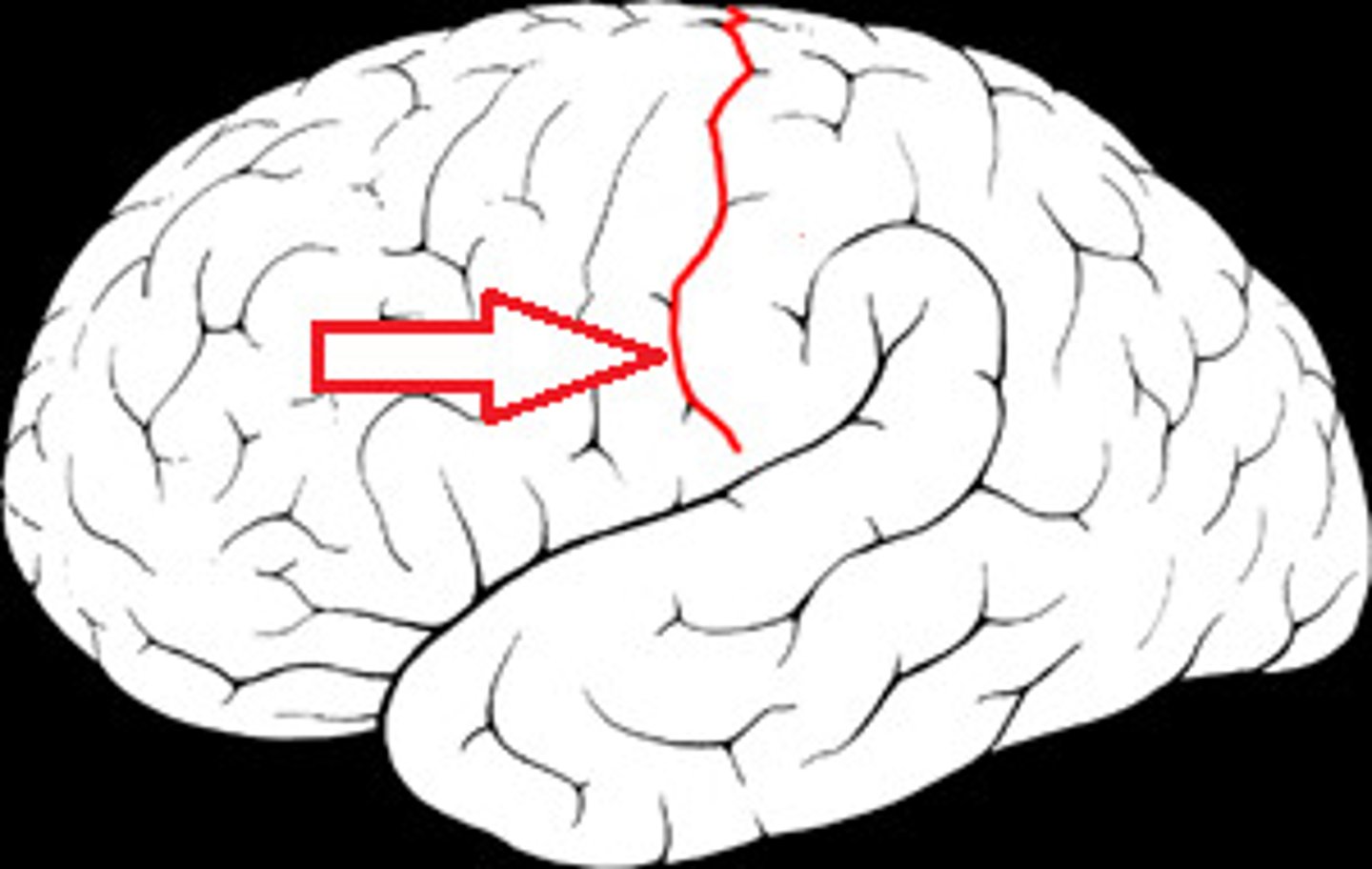
central sulcus
Separates frontal lobe from parietal lobe
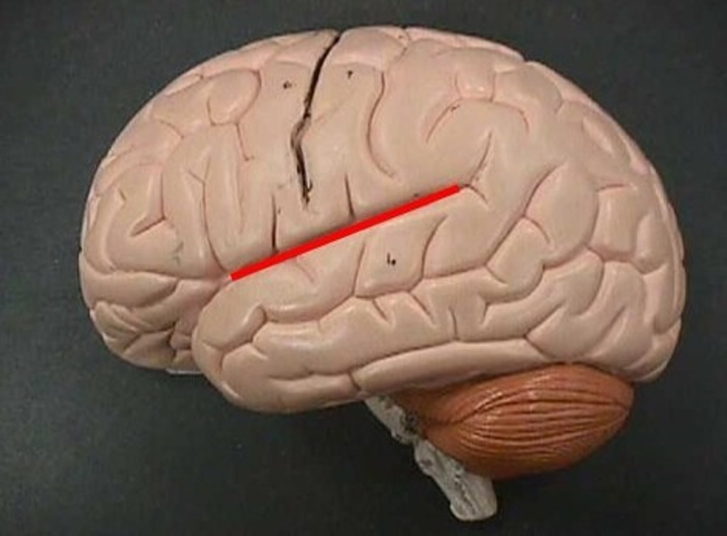
lateral sulcus
separates the parietal and temporal lobes
parieto-occipital sulcus
separates parietal and occipital lobes
Fissures
deep grooves in the brain
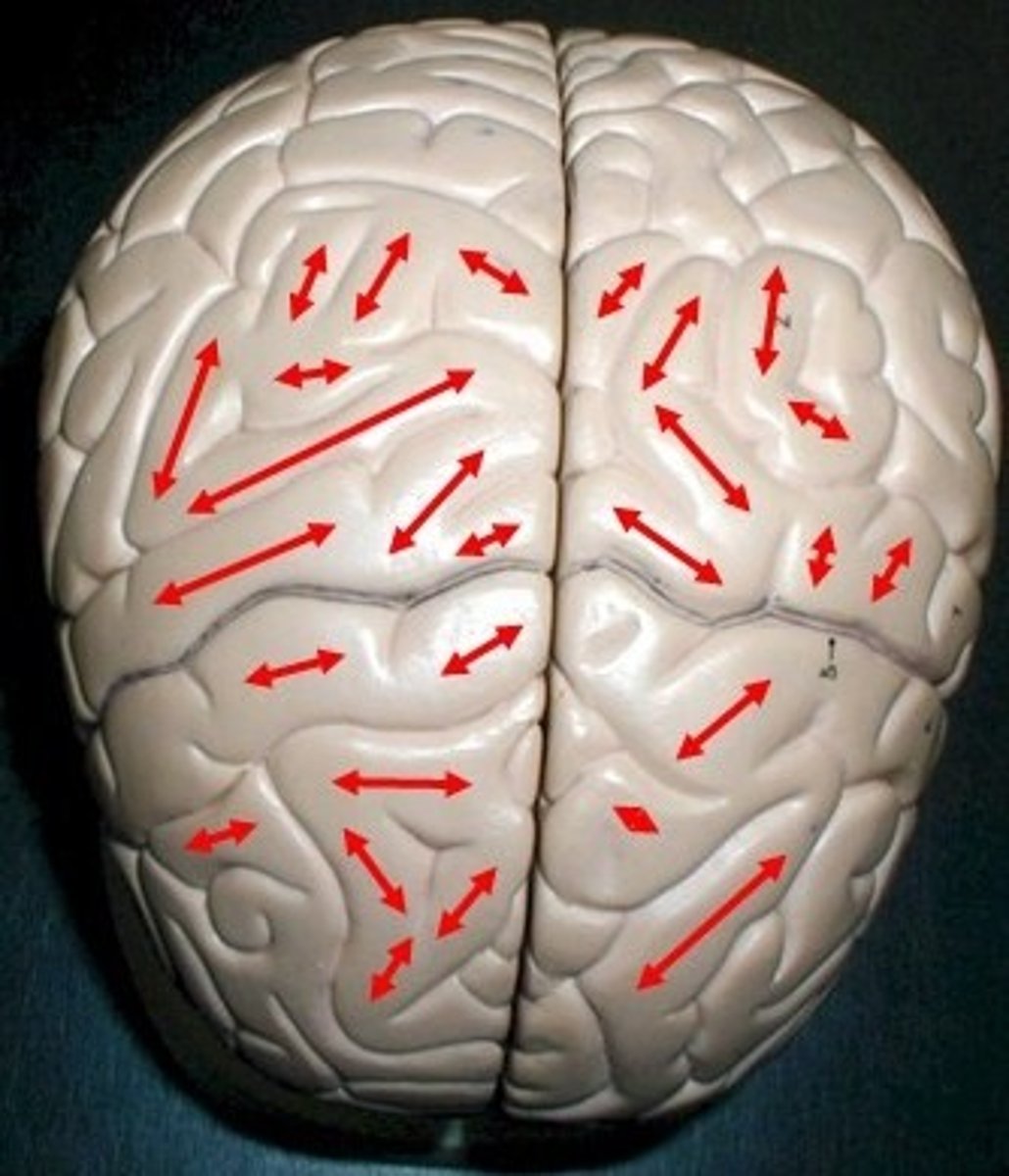
longitude fissure
Crease/line that divides the brain in to left and right side hemisphere.
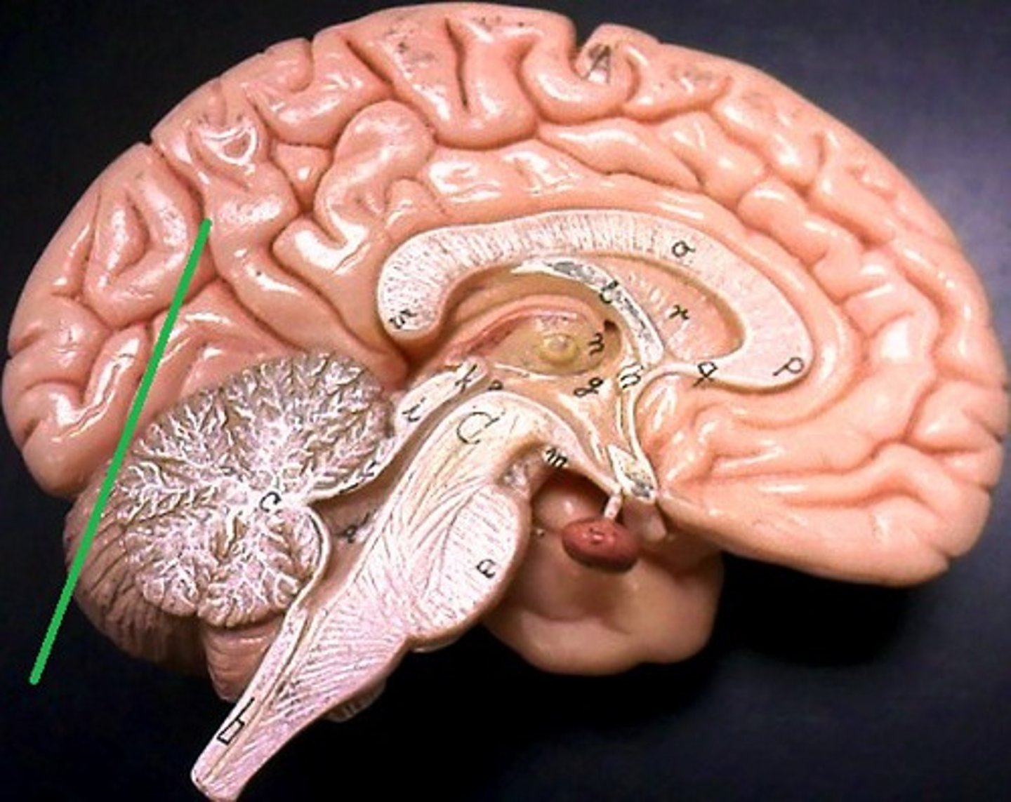
transverse cerebral fissure
separates cerebrum from cerebellum
ventricles
the two lower chambers of the heart, and they pump blood out to the lungs and body.
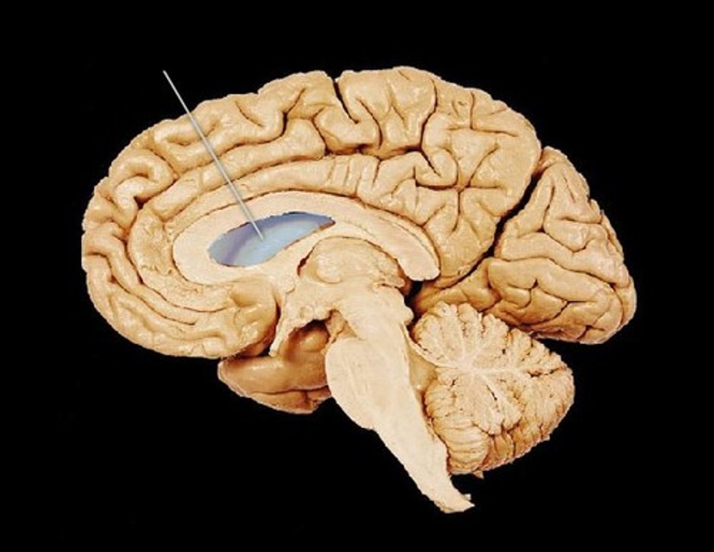
Lateral (1st & 2nd) Ventricles
within cerebral hemispheres; separated by septa pellucida
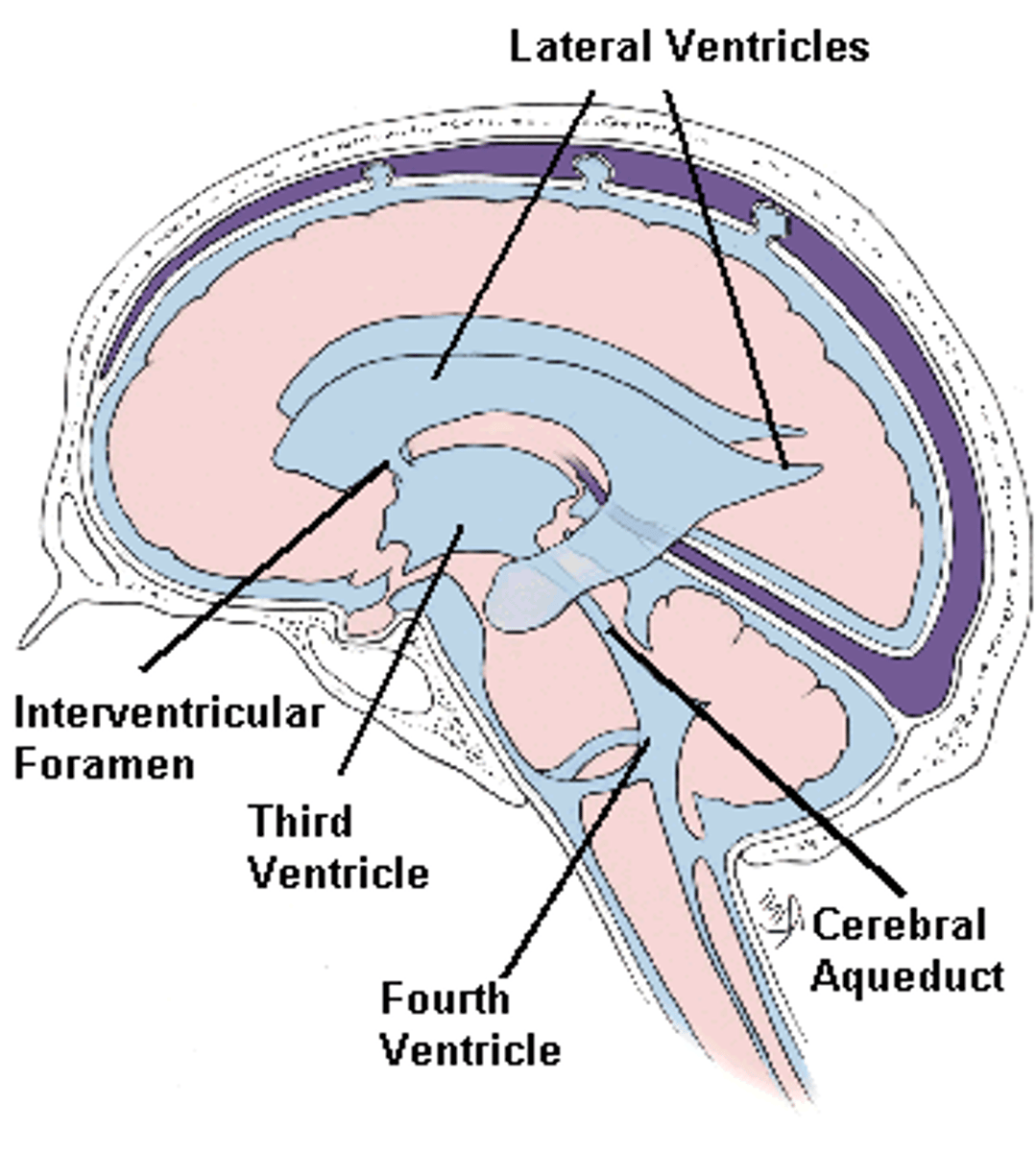
3rd ventricle
found in the diencephalon and communicates with lateral ventricles via intraventricular foramen
4th ventricle
between pons and cerebellum
choroid plexus
A highly vascular portion of the lining of the ventricles that secretes cerebrospinal fluid.
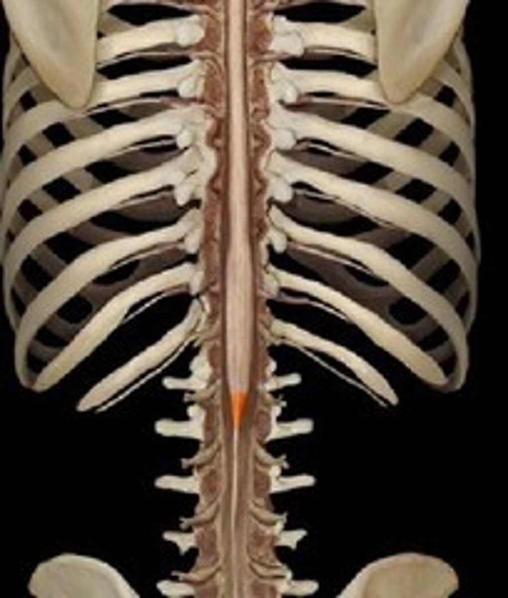
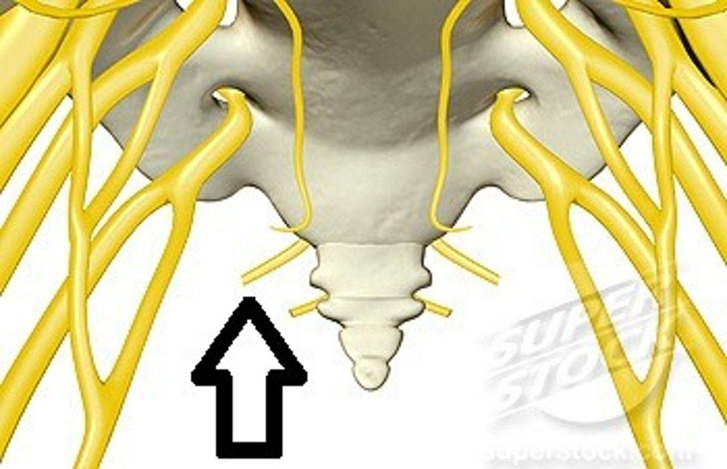
conus medullaris
tapered end of spinal cord
if the conus medullaris is damaged
Conus medullaris syndrome is a type of incomplete spinal cord injury that is less likely to cause paralysis than many other types of spinal cord injuries. Instead, the most common symptoms include: Severe back pain. Strange or jarring sensations in the back, such as buzzing, tingling, or numbness.
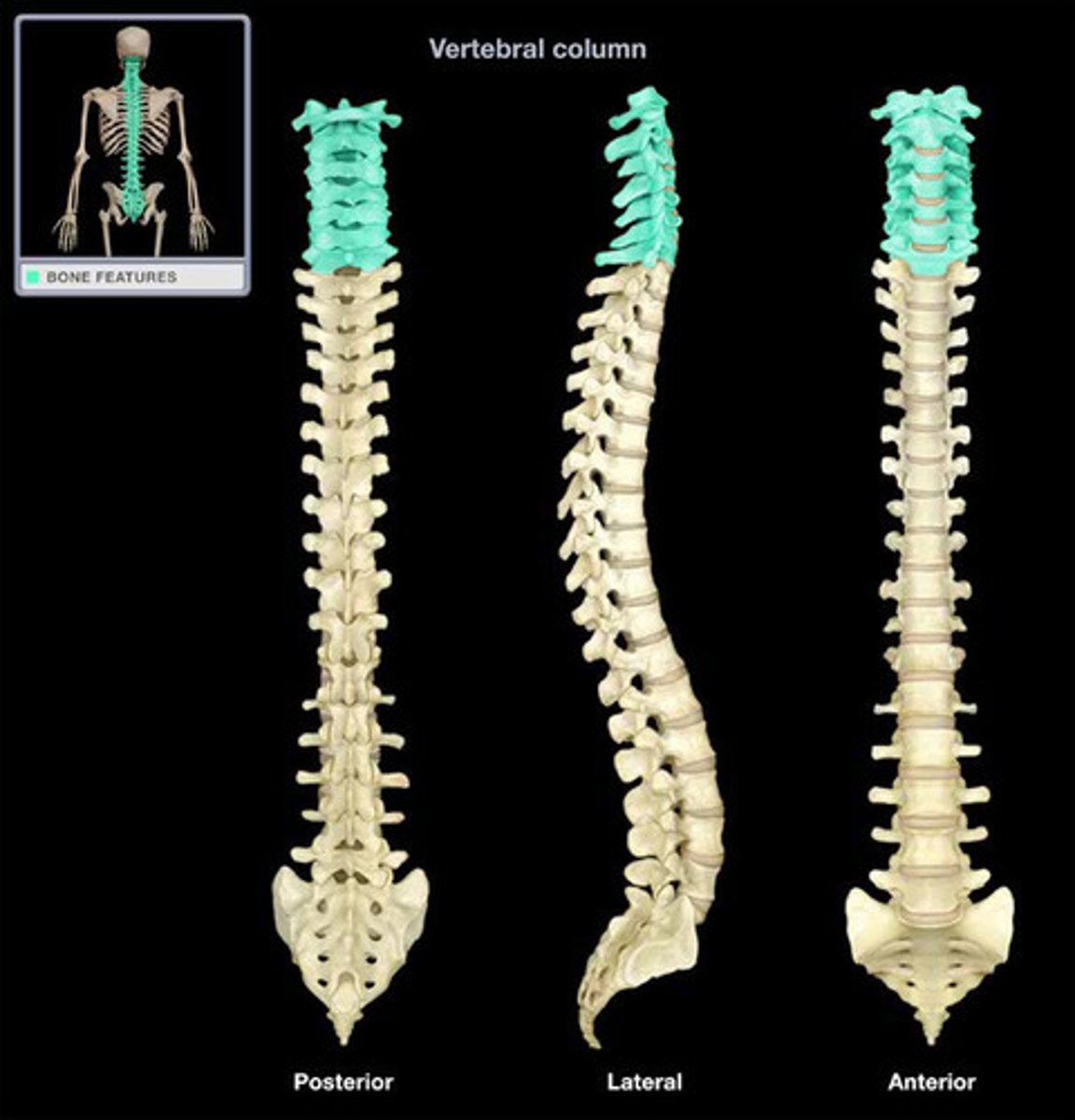
cervical spinal nerves
C1-C8
may cause pain that radiates into the shoulder and/or arm, as well as muscle weakness and numbness if damaged
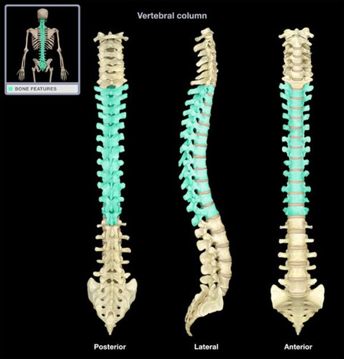
thoracic spinal nerves
T1-T12
back problems
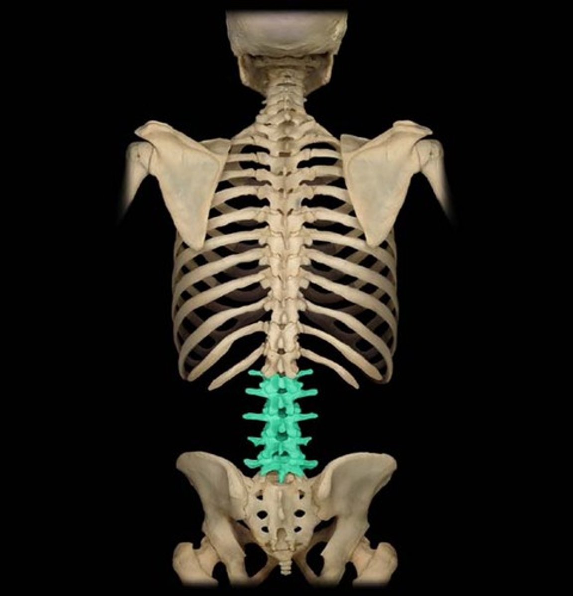
lumbar spinal nerves
L1-L5
injuries generally result in some loss of function in the hips and legs
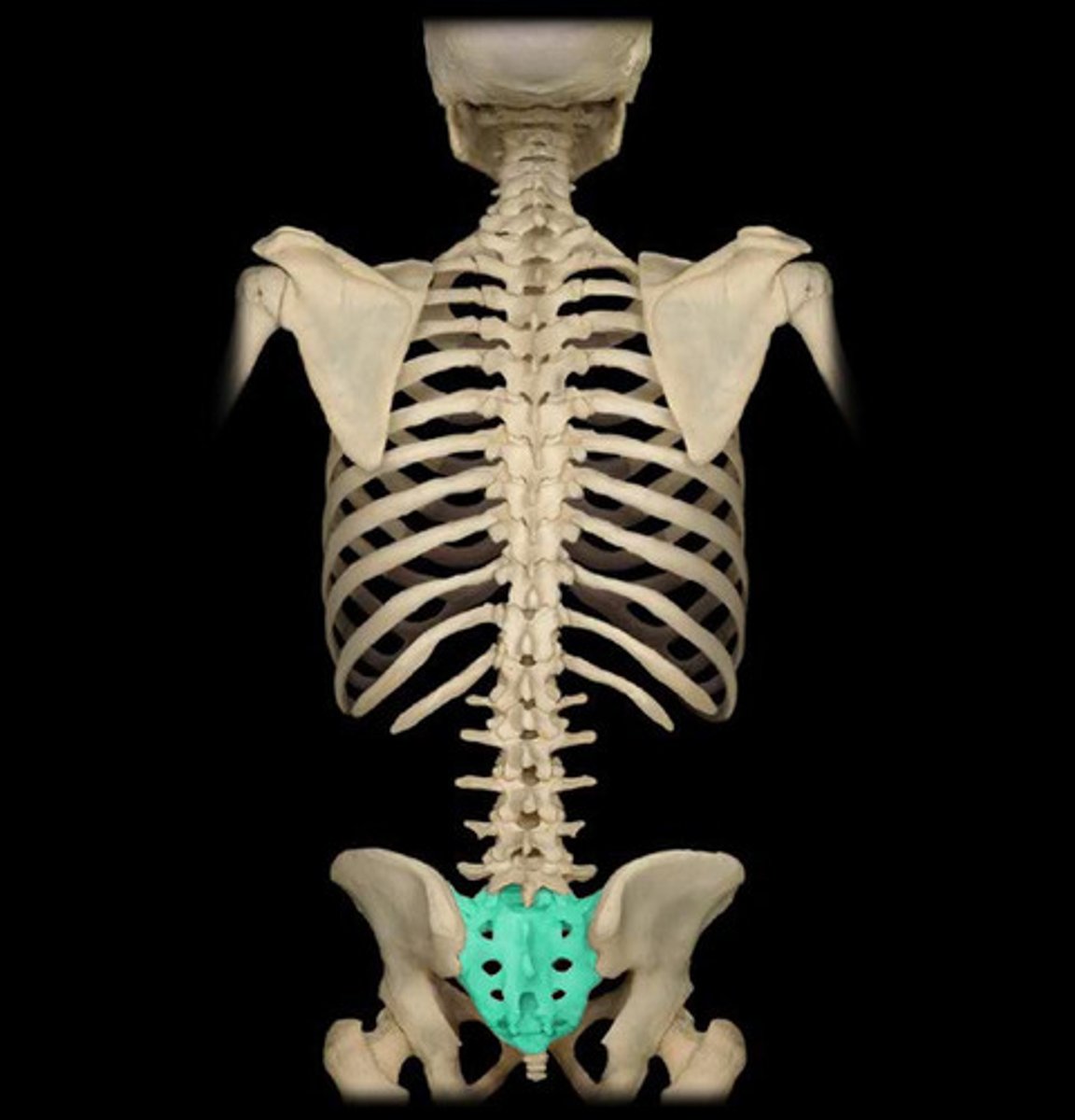
sacral spinal nerves
S1-S5
If you have sacral nerve damage, you may experience symptoms on one or both sides of the body. Meanwhile, damage to the sacral spine may cause you to lose some function in your legs or hips. You could find it difficult to walk or drive a car
coccygeal spinal nerves
1 pair
Achy or piercing pain in the tailbone
Dorsal, Ventral, & Lateral Horns
Inner part of spinal cord that's made of GRAY matter
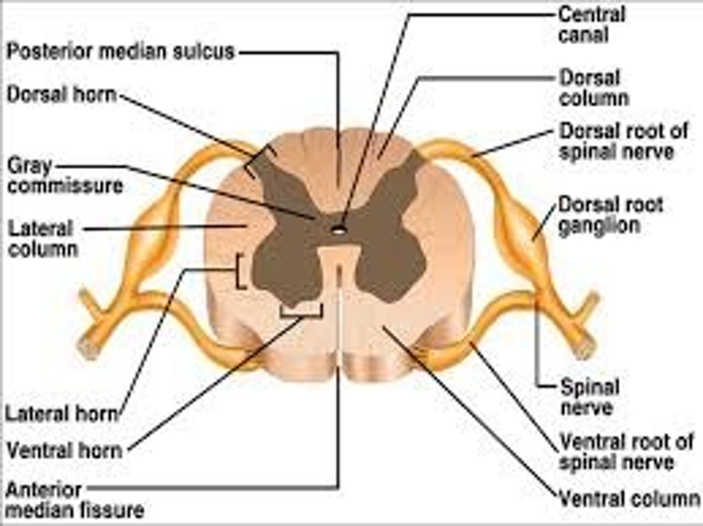
dorsal root
the sensory branch of each spinal nerve
dorsal root ganglion
contains cell bodies of sensory neurons
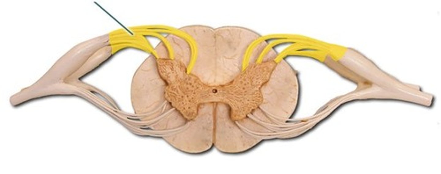
dorsal rootlets
sensory
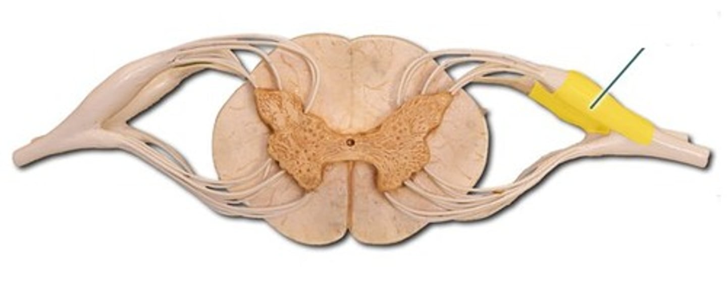
ventral root
contains axons of motor neurons
If the ventral root of a spinal nerve was severely damaged or cut, it would cut off the pathway of motor information from the spinal cord to the spinal nerve. Therefore, whatever effectors that spinal nerve controlled would no longer work; it would be paralyzed.
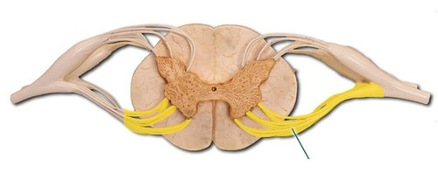
ventral rootlets
motor
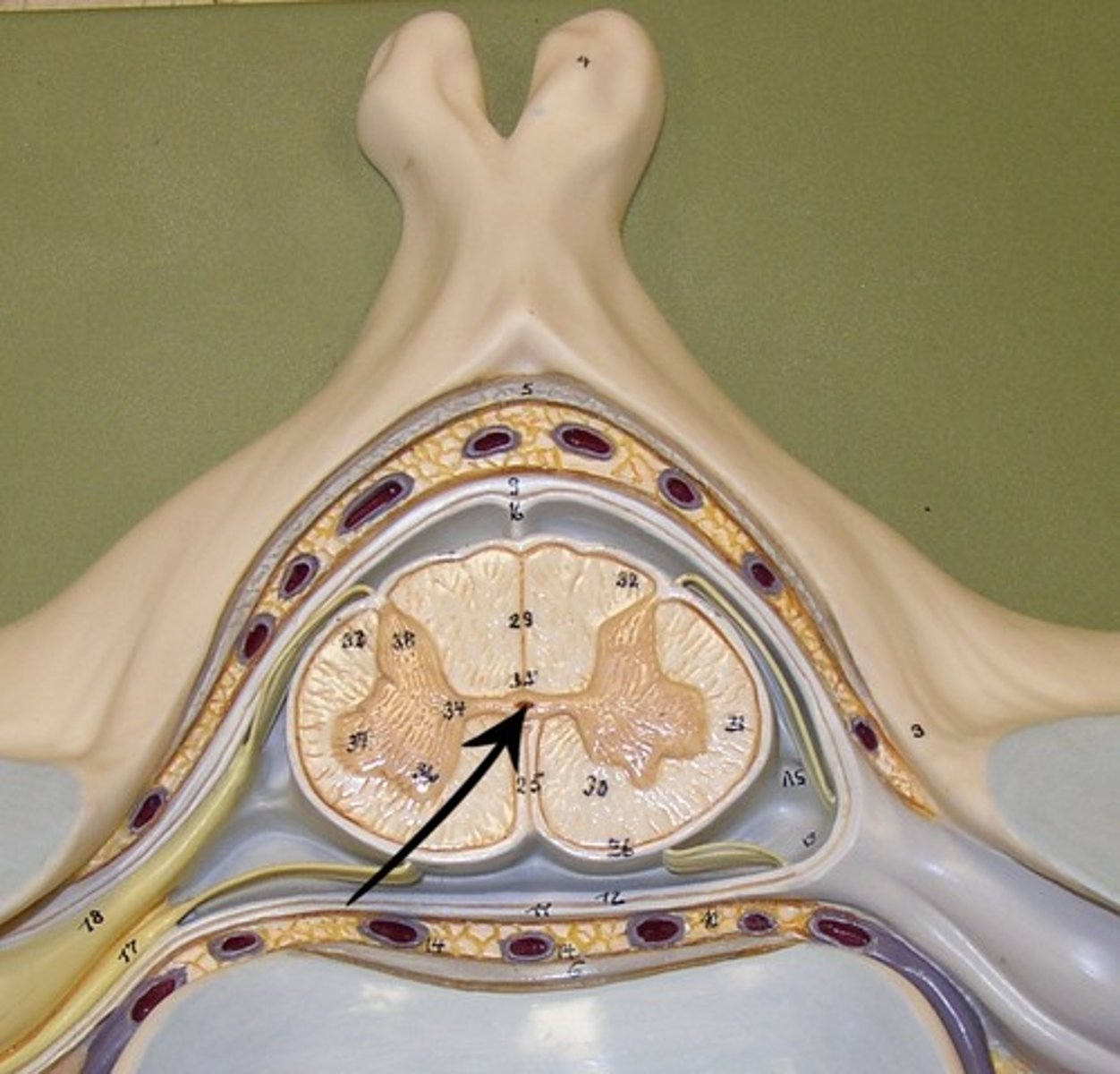
central canal
transports cerebrospinal fluid (CSF)
if the central canal is damaged
Paralysis or loss of fine control of movements in the arms and hands, with relatively less impairment of leg movements. Loss of or change in sensation below the site of the injury. Loss of bladder control
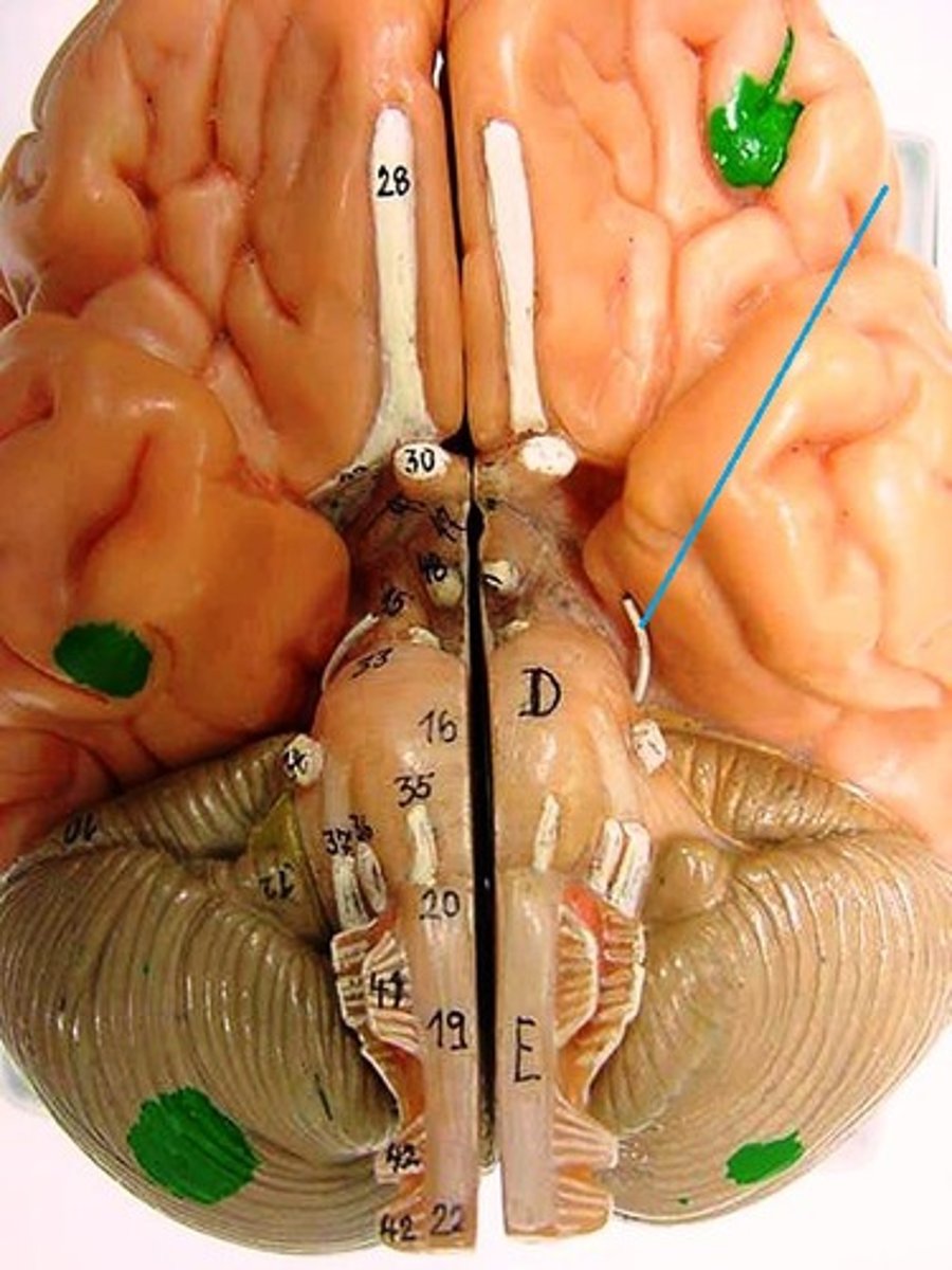
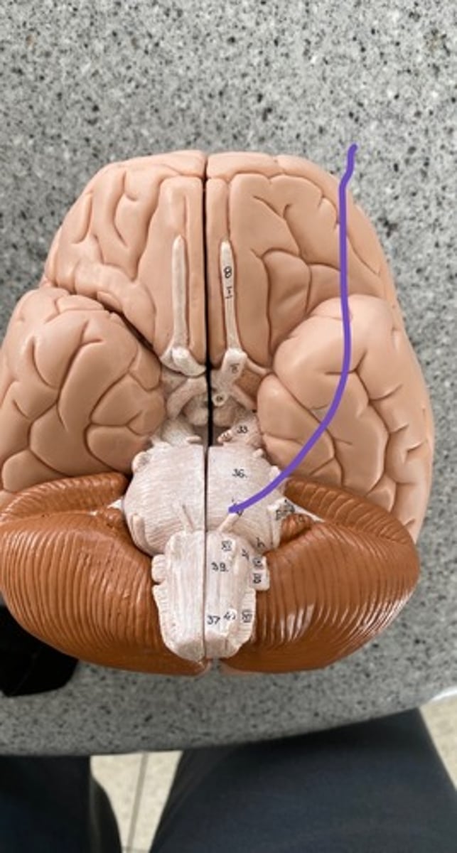
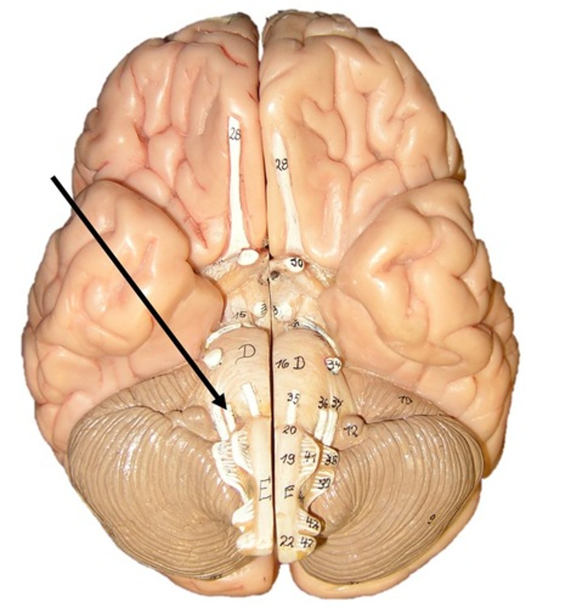
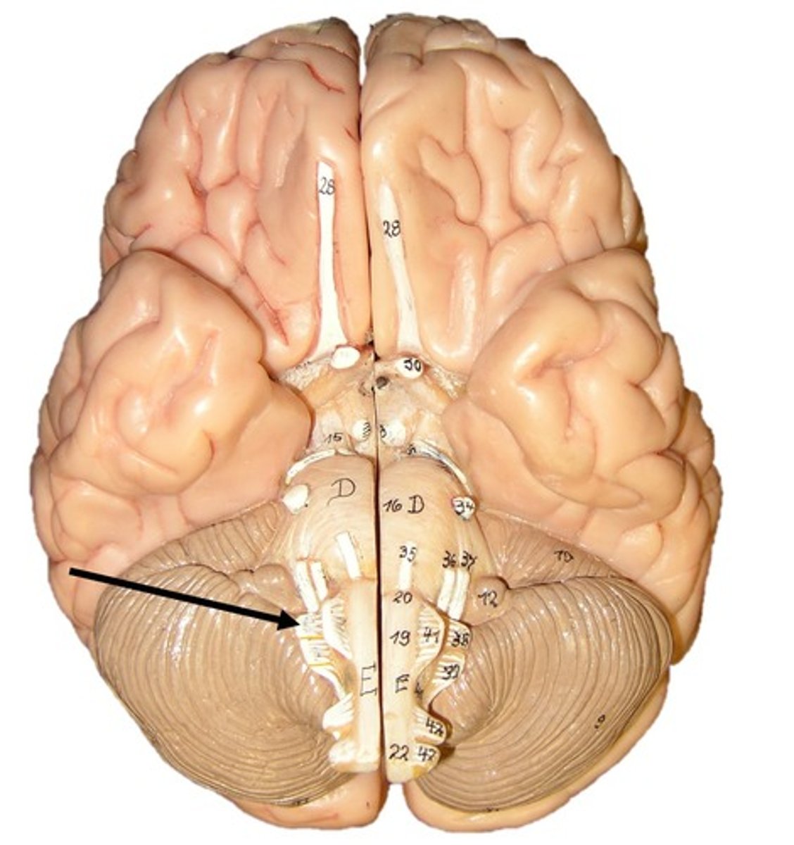
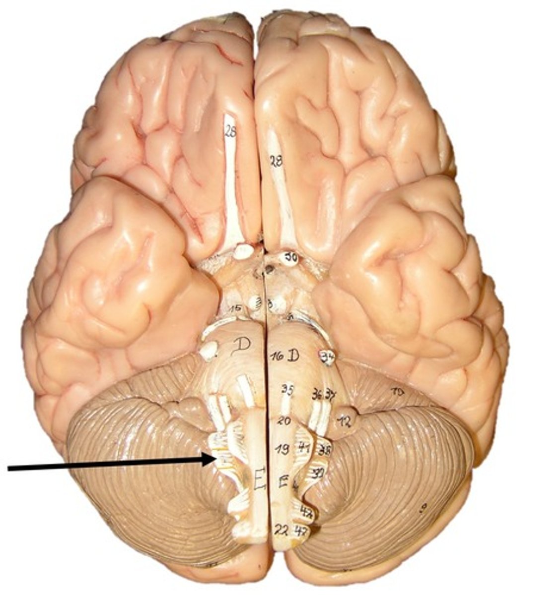
Olfactory Nerve (I)
sensory, smell
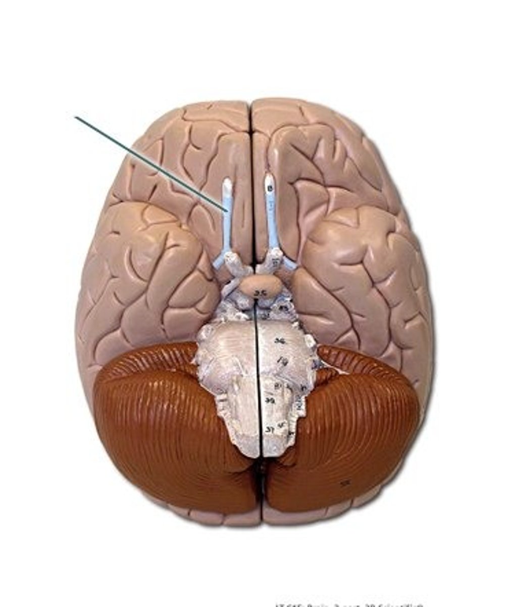
Optic Nerve (II)
vision, sensory nerve

Oculomotor Nerve (III)
- motor
- eye movement narrows pupil and focuses lens
Trochlear Nerve (IV)
- motor
- directs the eyeballs.
Trigeminal Nerve (V)
sensory and motor
- Largest cranial nerves, extending from the pons to the face
- Ophthalmic (V1): nose to eyes to forehead.
- Maxillary (V2): maxillae to temples.
- Mandibular (V3): mandible to frontof ears - Convey sensory impulses fromvarious areas of face (V1 and V2).
- Supply motor fibers (V3) for mastication.

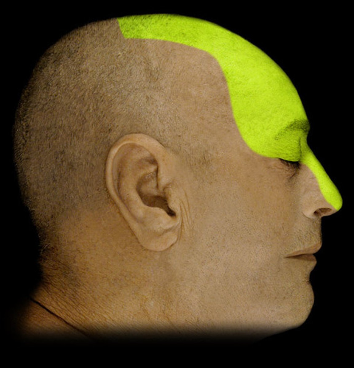
opthalmic
Ophthalmic (V1): nose to eyes to forehead
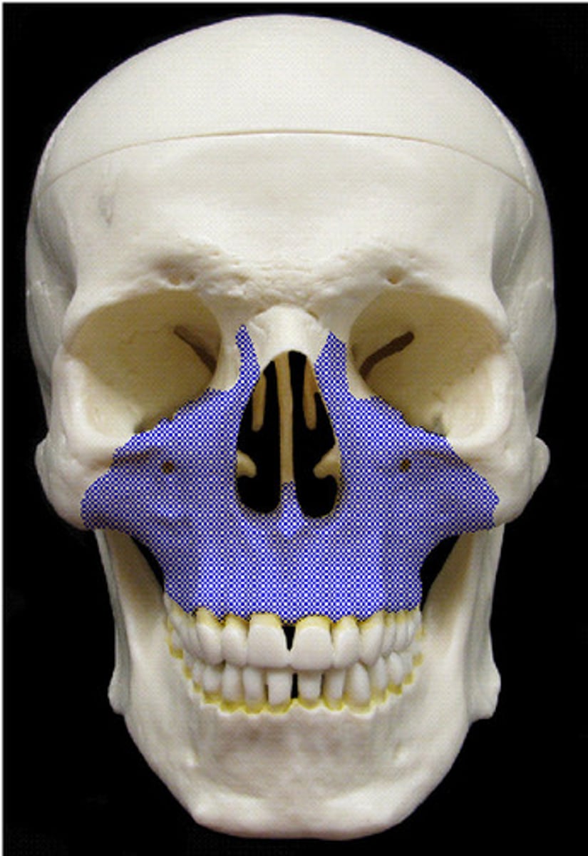
maxillary
Maxillary (V2): maxillae to temples
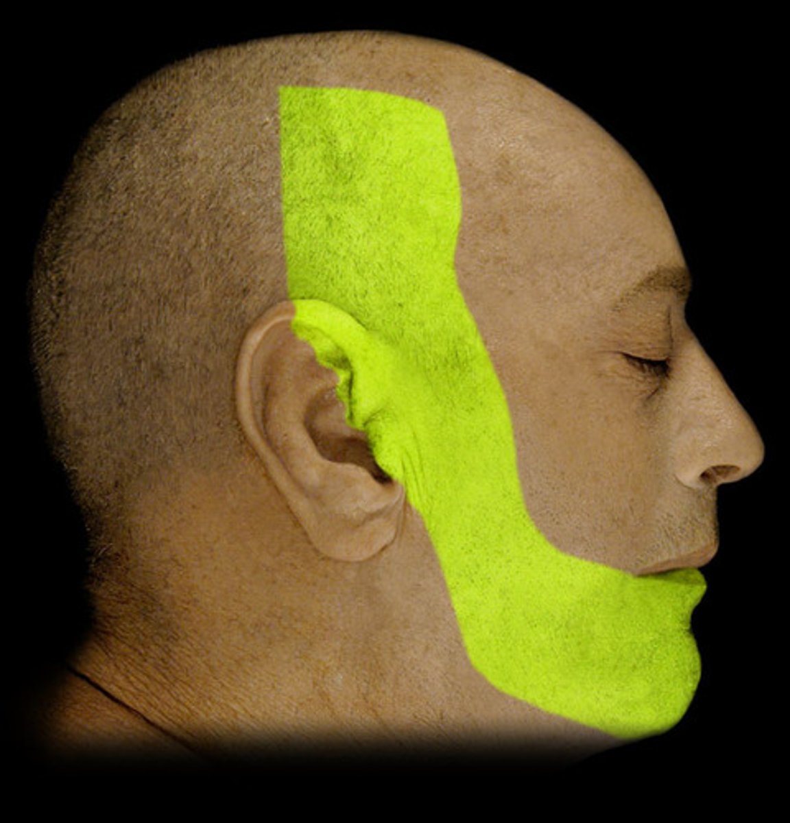
mandibular
Mandibular (V3): mandible to front of ears
Abduscens Nerve (VI)
- motor
- extend from just below pons to lateral rectus muscle of eyeballs.
- primarily a motor nerve that directs the eyeballs
Facial Nerve (VII)
sensory and motor; provides motor innervation of facial muscles that are responsible for facial expression, parasympathetic innervation of the glands of the oral cavity and the lacrimal gland, and sensory innervation of the anterior 2/3 the tongue
Vestibulocochlear Nerve (VIII)
sensory
hearing and equilibrium

Glossopharyngeal Nerve (IX)
sensory and motor
taste and swallowing
Vagus Nerve (X)
sensory and motor
Reception of blood pressure and blood gas chemistry, controls smooth movements in digestive system, decreases cardiac muscle contractions, increases secretion of digestive juices
Accessory Nerve (XI)
- motor
- swallowing, head, neck, and shoulder movements
- trapezius and sternocleidomastoid muscles
Hypoglossal Nerve (XII)
motor: muscles of the tongue contribute to swallowing and speech

parasympathetic nervous system
- Rest-and-Digest
- Supports functions that conserve and restore energy during rest and recovery.
- When body is at relaxed.... parasympathetic input dominates over sympathetic.
- 3 decreases... heart rate, bronchial diameter, and pupil diameter
sympathetic nervous system
- "Fight-or-Flight"
- Supports functions that use energy and reduces functions that store energy.
- When body is in vigorous activity.... sympathetic input dominates parasympathetic.
- 3 increases... heart rate/stroke volume, bronchial diameter, and pupil diameter.• Also involved with fear, embarrassment, nervousness, and rage.
- "E situations"... exercise, emergency, excitement, embarrassment
dual innervation
organs that receive instructions from both sympathetic and parasympathetic divisions
dynamic antagonism
between two divisions maintains homeostasis
autonomic ganglia
Collections of nerve cell bodies, belonging to the autonomic division of the peripheral nervous system, that are found in various locations and innervate the major organs.
autonomic reflexes
smooth muscle regulation, heart and blood pressure regulation, regulation of glands, digestive system regulation
pupillary reflex
The automatic process by which the iris contracts and relaxes to control the size of the pupil, in response to the relative brightness of light entering the eye
salivary reflex
food odor detection causes salivation
sweat reflex
Regulates body temperature, in repsonse to stimuli
Gastric Reflex
initiated by presence of food in stomach
micturition reflex
spinal reflex that partly controls urination
defecation reflex
activated by stretch receptors stimulated by filling of the rectum
somatic reflexes
activation of skeletal muscles
gag reflex
A normal reflex mechanism that causes retching; activated by touching the soft palate or the back of the throat.
withdrawal reflex
a spinal reflex that pulls a body part away from a source of pain
stretch reflex
muscle contraction in response to stretching within the muscle
patellar reflex
a reflex extension of the leg resulting from a sharp tap on the patellar tendon; knee jerk