Sensory Organs - unit 3
1/196
There's no tags or description
Looks like no tags are added yet.
Name | Mastery | Learn | Test | Matching | Spaced | Call with Kai |
|---|
No analytics yet
Send a link to your students to track their progress
197 Terms
Ear
The ear is a transducer. It converts electric acoustic energy (sound waves) into electrochemical energy that is transmitted to the auditory nerve in the brain.
Outer Ear
What you can see, the passageway into the inner ear. Energy is acoustic.
Middle Ear
Contains the ossicles, the energy is mechanical vibrations.
Inner Ear
Vestibular System and cochlea, energy is hydrodynamic wave motion.
Pinna/Auricle
In the outer ear, cartilaginous sound collector, collects sound from the environment. Whole outer ear, the part you see.
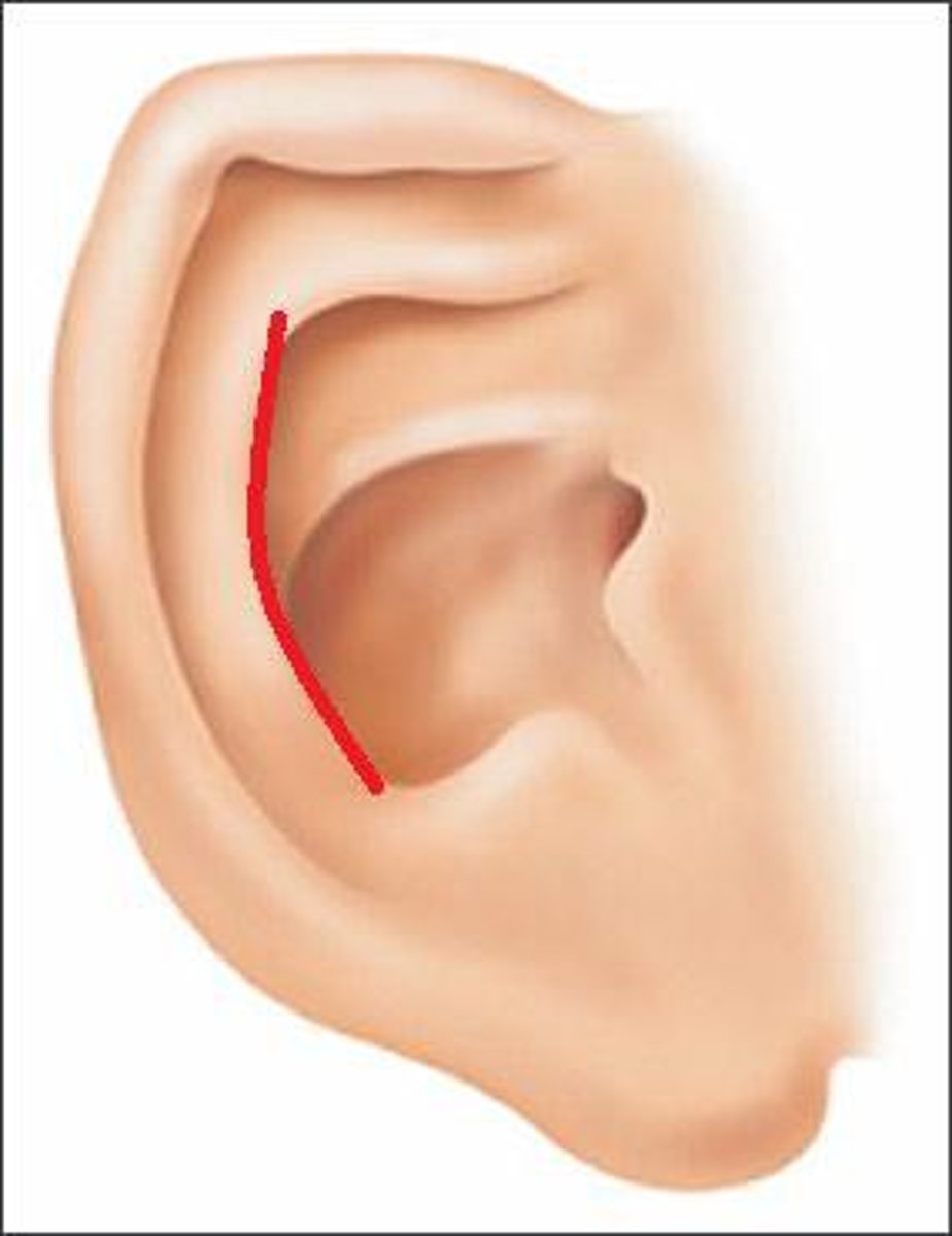
Helix
In the pinna, curved part around the outside.
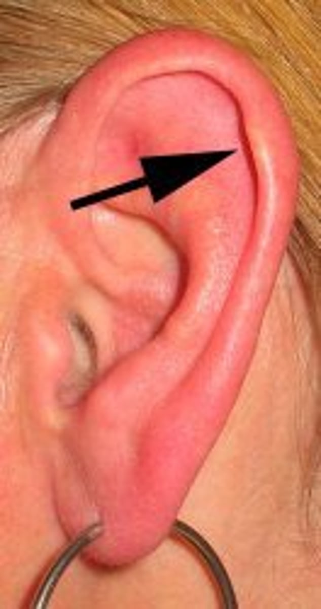
Antihelix
Fold anterior to the helix, curved part on the inside next to helix, bifurcates by splitting into 2.
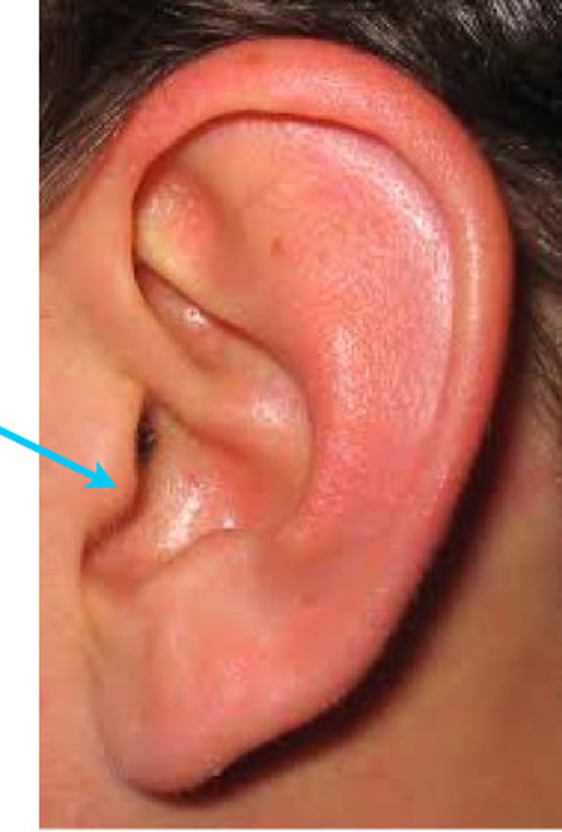
Tragus
Flap covering the entrance to the ear canal, cartilage is covered by epithelium.
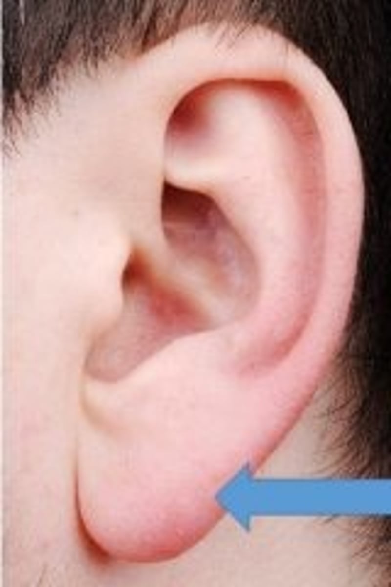
Ear Lobe
Non-cartilaginous skin flap.
what is the External Auditory Meatus (EAM)
Ear canal, diameter: ~7mm, length: ~25mm, concha>eardrum.
Lateral 1/3 is cartilage, medial 2/3 is bone. Angle is ~55 degrees at the EAM and tympanic membrane (eardrum).
Ciliated epithelium gets the gunk out, and cue-tips push the gunk back in. Could cause the eardrum to stop moving.
Tympanic Membrane
Boundary between the outer and middle ear, functions like a drum, vibrates. If the membrane can't move, it can't transmit sounds, slightly concave when viewed from EAM.
Ossicles
In the middle ear, transmit energy from TM to inner ear, very tiny bones and all 3 fit on a dime. Malleus, Incus, Stapes.
Malleus
Hammer, contains manubrium (long part on the bottom), head (roundish part on top), and neck (separates manubrium and head is connected here). Malleus joins the TM along length of manubrium. Head articulates with the incus, largest of the ossicles.
Incus
Contains long process (parallel with the manubrium of malleus), body (articulates with malleus at the articular facet) and lenticular process (end of long process, articulates with stapes).
Stapes
Contains head (at the top, articulates with incus), neck (below head), footplate (articulates/joins oval window at bottom). Smallest of ossicles.
Oval Window
Part of the middle ear, fenestra vestibuli, connects with the stapes.
Round Window
Part of middle ear, fenestra cochlea, leads to scale tympani in inner ear.
Eustachian Tube
Runs from middle ear to the pharynx, drains middle ear, vestibular function is to keep air pressure equalized. This is happening when your ears pop.
Middle Ear Muscles
Reduce transmissions of high intensity sounds, part of auditory reflex.
Stapedius Muscle
Middle ear muscle that keeps high intensity sounds from entering the cochlea. More effective against high intensity, low frequency sounds.
Tensor Tympani
Middle ear muscle, when it contracts, it pulls the malleus which tightens the TM. Connected to the ossicles by a tendon and also protects cochlea from sounds. More effective against high intensity, low frequency sounds.
Bony Labyrinth
Bone and membrane.
Membraneous Labyrinth
Bone and membrane that is generally in the osseous labyrinth.
Semicircular Canals
Inner ear, sense balance, movement of the body in space.
Cochlea
Inner ear, helps with hearing.
Vestibule
Inner ear, in between the semicircular canals and cochlea. The entry to the inner ear space.
Anterior/Superior Semicircular Canals
On top, sense when the head moves to the shoulder.
Posterior/Vertical Semicircular Canals
Sense when your head moves up and down, nodding yes.
Horizontal/Lateral Semicircular Canals
In the middle, sense shaking no, side to side.
Ampulla
Wider part of the canal that contains fluid, fluid senses the movement.
Modiolus
Core, finely perforated bone at the core. Where fibers of the 8th vestibulocochlear nerve pass through.
Osseous Spiral Lamina
Bony shelves that extend from modulus, divide the cochlear labyrinth into 2 chambers: scala tympani and scala vestibuli.
Perilymph
Fluid that fills in the scala vestibuli and tympani.
Endolymph
Fluid that fills the scala media
Basilar Membrane
Floor of the scala media, organ of hearing is located along the basilar membrane, structures that arise from BM are inner ear hair cells.
Organ of Corti (spiral organ)
Includes auditory receptors
Outer Hair Cells (OHC)
Contains 3 rows.
Inner Hair Cells (IHC)
Contains 1 row near modulus, don't come in contact with tectorial membrane.
Tectorial Membrane
Has some functionality to the way we hear sound, related to proximity of outer hair cells.
Stereocilia
Sensory hairs on the surface of cells.
Cilia
Cilia are linked if stereo cilia move on one cell, bump into others making them move as well.
aqueous and vitreous humors
the watery fluid that fills the anterior and posterior chambers of the eye
choroid
the middle layer of the wall of the eye
ciliary body
the structure between the choroid and the iris that anchors the lens in place
cones
sensory cells in the retina that are sensitive to bright light and provide color vision
conjunctiva
a delicate external membrane that covers the exposed eyeball and lines the eyelid
cornea
a transparent tissue over the anterior center of the eye
extrinsic muscles
muscles attached to the outer surface of the eye that are responsible for changing the direction of viewing
iris
the anterior portion of the choroid, which gives the eye its color
lacrimal glands
tear secretors
lens
a transparent, flexible structure that is curves outward on both sides
optic chiasma
the point at which the optic nerves cross
pupil
the opening through which light rays enter the eye
retina
the innermost layer of the eye, containing light sensitive nerve endings that send impulses through the optic nerves to the brain
rods
sensory cells in the retina that are activated in dim light
sclera
the tough, fibrous outer layer of the eye
diplopia
double vision
astigmatism
blurred vision due to an irregular curvature of the cornea or lens
strabismus
crossed eyed
myopia
nearsightedness
hyperopia
farsightedness
vitreous humor
A
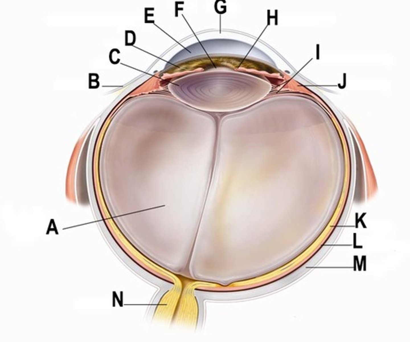
conjunctiva
B

lens
C

iris
D

aqueous humor
E

pupil
F

cornea
G

retina
K

choroid layer
L

sclera
M

The olfactory apparatus
a group of organs used for smell
Nasal cavity
is a large air filled space inside the nose, above and behind the nostrils.
Olfactory membrane, aka olfactory epithelium
is a specialized tissue inside the nasal cavity involved in smell. About the size of a postage stamp.
Mucus
a slimy substance secreted by mucous membranes and glands for lubrication, protection, etc.; odorant molecules dissolve into this and then they bind to the olfactory receptors
olfactory receptors
in the olfactory epithelium; these bind to odorant molecules
chemoreceptors
a sensory cell or organ responsive to chemical stimuli, found in the olfactory epithelium.
Olfactory bulb
a bulbous structure, an extension of the olfactory nerve, that connects the olfactory nerve to the olfactory membrane.
Olfactory nerve
It relays olfactory data to the brain.
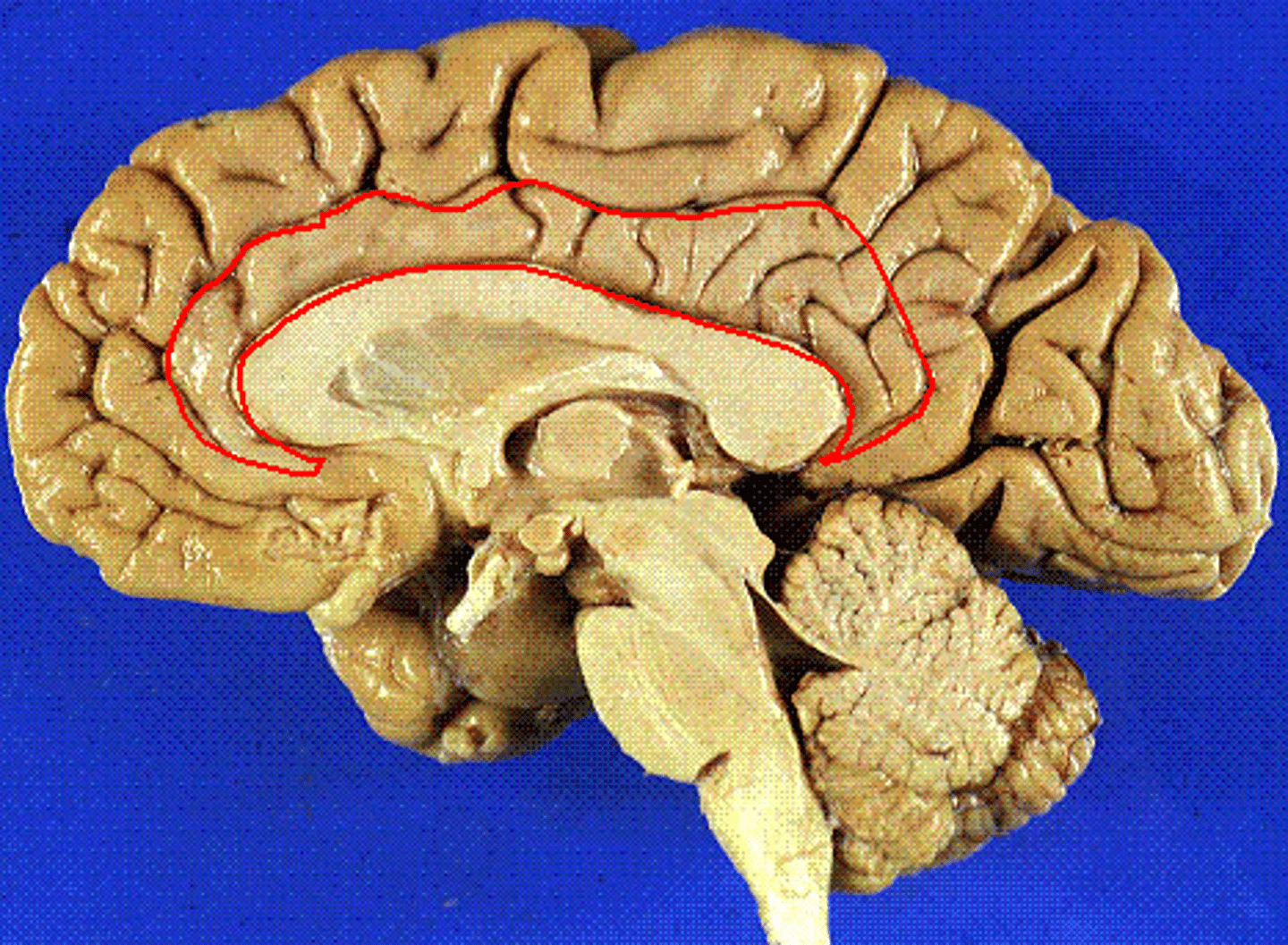
Limbic system
a region of the brain concerned with instinct, memory and emotion. Some olfactory information passes through this, thus linking smells and emotional memory.
Frontal lobe
The frontal lobe, located at the front of the brain, is one of the four major lobes of the cerebral cortex in the mammalian brain. Where smell stimulus information is processed and interpreted.
Odorant molecules
Airborne particles that enter the nasal cavity and stimulate the olfactory receptors in the epithelium.
Gustation
the action of tasting, the ability to taste
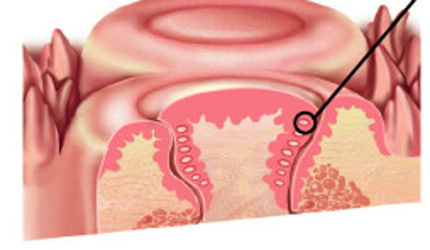
Papillae
Small, bumpy structures on the upper surface of the tongue that are covered with taste buds. These give the tongue its characteristic rough texture.
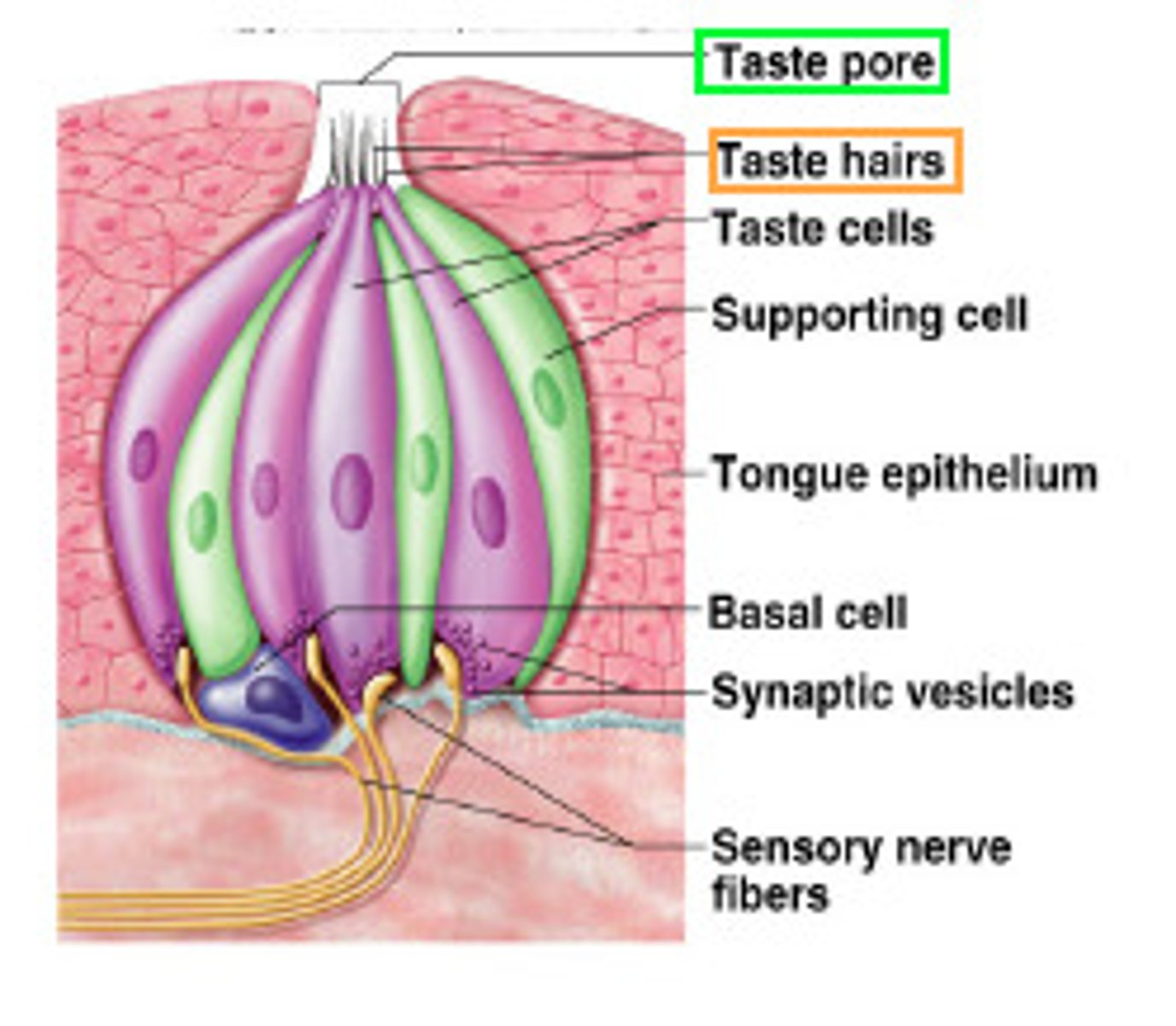
Taste buds
Contain the taste receptor cells. Located on papillae on the upper surface of the tongue, soft palate, and the cheek.
Taste receptor cells
The gustatory system consists of these cells in the taste buds. Taste buds, in turn, are contained on structures called papillae. These cells contain sensory nerve fibers that bind with food molecules.
Sensory fibers
Microscopic hairs that bind to food molecules and begin the process of transduction of taste sensation to the brain.
The five taste sensations
salt, bitter, sour, sweet, umami
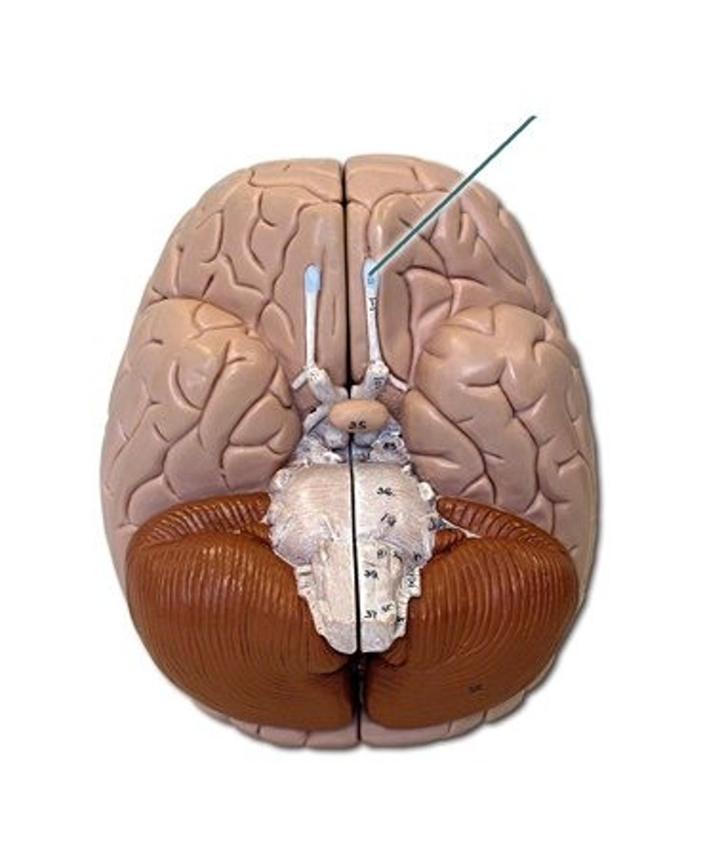
What is this?
olfactory bulb
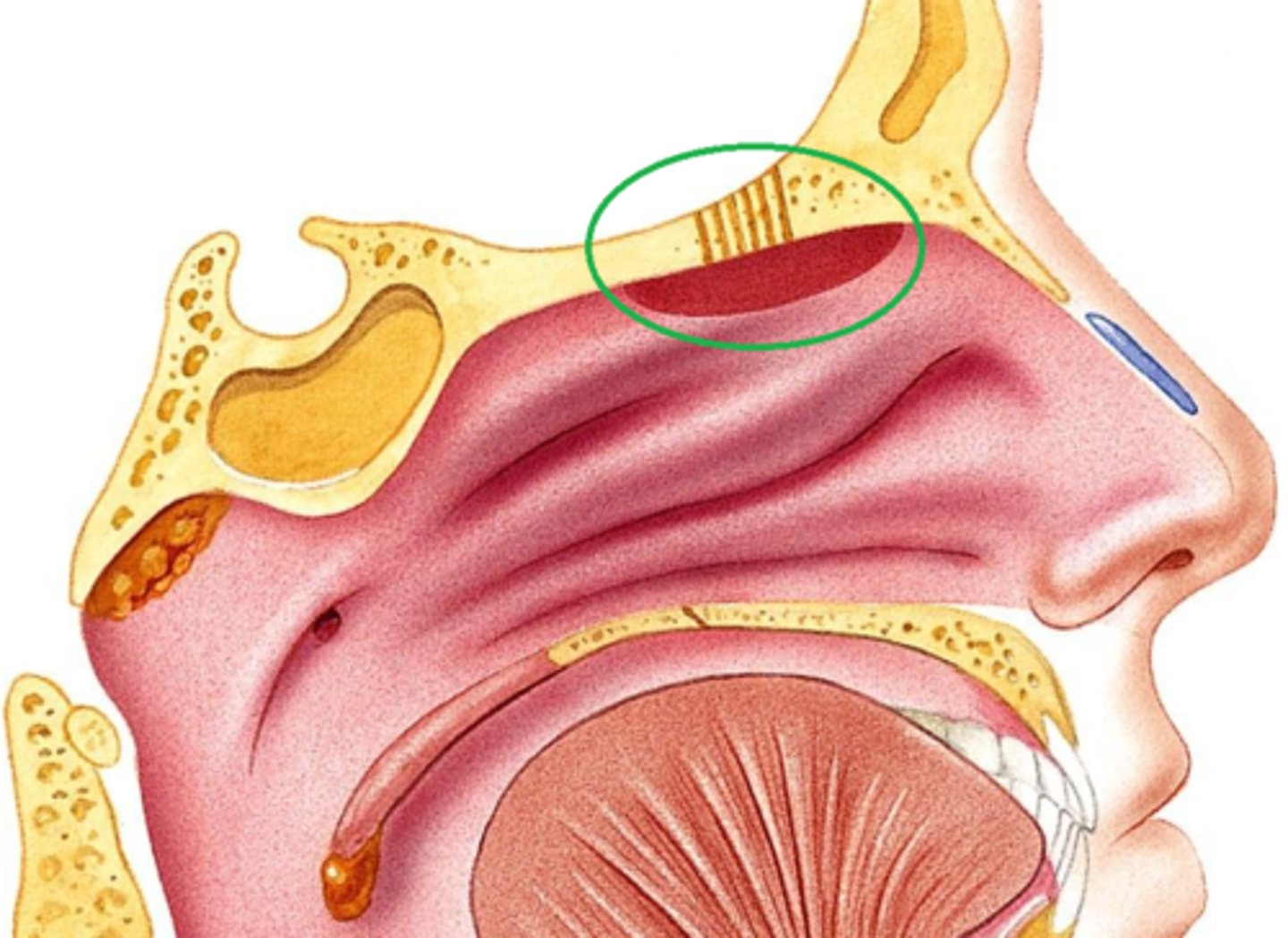
What is this?
olfactory epithelium
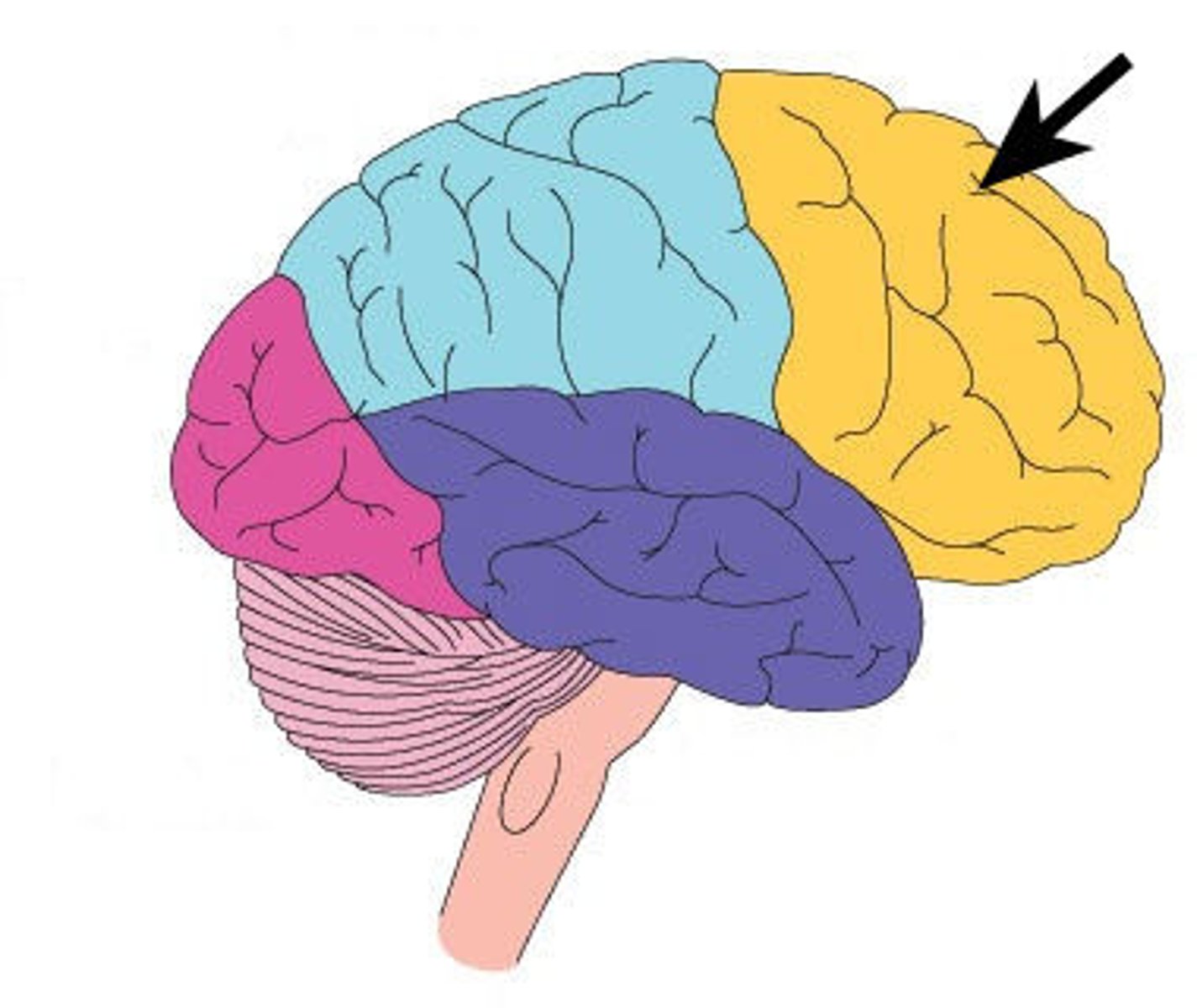
What is this?
frontal lobe
What is this?
lymbic system
What is this?
taste bud
What is this?
chemoreceptors
the sense of taste
depends on the sense of smell; taste is thought to be 80% smell.
Olfactory adaptation
this occurs when a smell becomes less noticeable over time. Also known as olfactory fatigue, olfactory habituation, and odor fatigue.
Receptors
Stimulated by stimuli and they in turn stimulate sensory neurons which send messages to the brain for interpretation. Grouped according to the kind of stimulus they receive
Photoreceptor
Respond to visible wavelength of light
Mechanoreceptor
Sensitive to mechanical energy
Thermoreceptor
Sensitive to heat and cold