MSK Problem Areas
1/37
There's no tags or description
Looks like no tags are added yet.
Name | Mastery | Learn | Test | Matching | Spaced | Call with Kai |
|---|
No analytics yet
Send a link to your students to track their progress
38 Terms
Equine: A horse with suspected subsolar hoof abscess is brought immediate relief after drainage @ white line that released moderate amount of black exudate. What additional treatment
Tetanus Toxoid
Risk of tetanus associated w/ deep penetrating wounds
Systemic ABX are not necessary for uncomplicated subsolar abscess
Equine: A horse that is lame with short choppy gait in front limbs + swelling in both fetlock joints. Pain on palpation + flexion of the fetlock joints. What is the most likely DX
Osselets
Inflammation of the periosteum on the dorsal distal epiphyseal surface of MC 3 and fetlock joint
Can progress to osteoarthritis so early intervention is critical
TX: Rest, anti-inflam, intra-articular injections
Equine:The horse has pain and swelling along the coronary band and is barely toe touching in the left hind leg. You note that there is a marked change to the shape of the hoof. Which of the following conditions, caused by excessive strain on the common digital extensor tendon where it attaches to the front of the coffin bone, is the most likely diagnosis?
Buttress foot (Pyramidal Disease/Extensor process disease)
Excessive strain on P3—> periostitis in pyramidal process
± fracture of the process
As the disease progresses, lameness worsens and skin of the coronet becomes thickened and indurated. In addition, the wall of the hoof protrudes at the toe
Equine: What is a hygroma
A fluid filled swelling at the carpus usually due to repeated trauma leading to bursitis
Horses are not lame but have reduced range of motion of the koint
Equine: Treatment of choice for a carpal hygroma
Surgical exploration and drain placement
surgical excision of the bursal lining may be indicated when recurrence is a problem
Equine: What treatments is considered useful for chronic laminitis condition when refractory to conventional medical management?
Deep digital Flexor tenotomy
Especially when the distal phalanx is rotated or pedal bone penetration is present
may also be refractory
Equine: A horse w/ stiff gait, intermittent hind limb lamness, bony swelling on the hock of both hind limbs, one leg the toe drags the other leg lands toe first. Positive flexion test, What is this and how to treat
Bone Spavin
osteoarthritis of the distal intertarsal joint and/ or the tarsometatarsal joint
Ultimately you will have to treat by doing arthrodesis of the joint as it will progress
Equine: How to treat a horse with stringhalt
Lateral digital extensor tenectomy
Equine: What is the prefered treatment for sacroiliac luxation/subluxation
Sacroiliac luxations in horses are generally considered untreatable
the condition often stabilize w/ time and supportive care w/ rest and NSAIDs
Total Hip arthroplasty is reserved for severely injured horses that do not improve with supportive care
Equine: What is the most appropriate treatment for clubfoot
Clubfoot (Deformity of distal interphalangeal joint)
Treatment of choice is desmotomy of the accessory ligament of the deep digital flexor tendon (inferior check ligament)
Equine: What surgical treatment for Bowed tendon
Superior check ligament desmotomy
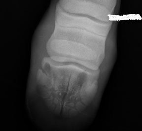
After performing a nerve block of the abaxial sesamoid region resolves lameness what is the the diagnosis
here is an incomplete fracture of the left front third phalanx through the sagittal plane. The fracture plane extends to the subchondral surface but does not appear to go through the articular cartilage, as there is no displacement at the articular surface
Equine: What will be seen on a navicular bone radiograph in a horse w/ navicular syndrome
Osteophyte formation
Bone remodeling
Enlarged vascular channels
Equine: Club foot is the result of
Deep digital flexor tendon contracture
Equine: what are Splints (metacarpal exotosis)
Periostitis of the interosseous ligament between the third and second metacarpal (or metatarsal) bone
Equine: What is thoroughpin
Effusion of the tarsal sheath
sheath of the deep digital flexor
Equine: On physical exam, you note that the foal has a flexural limb deformity affecting both forelimbs, with the right forelimb more severely affected than the left forelimb. The affected limbs both have excessively upright hooves with elongated heels, which is preventing the foal from walking normally.
Administer IV oxytetracycline
The most common flexural limb deformity in foals is caused by a relatively short/tight deep digital flexor tendon. This tendon pulls on the coffin bone, causing it to rotate downward in the hoof.
Equine: What is the most common slab fracture of the horse
3rd carpal bone
Equine: An 8-year-old Warmblood gelding is presented to you for repeated but intermittent clinical signs of exercise intolerance, weakness, muscle fasiculations and a stiff abnormal hind-limb gait. The owners do not ride their horse regularly, but notice these clinical signs most often at the start of a trail ride. Based on the history, signalment and clinical signs, which of the following diseases to you suspect?
A.Polysaccharide Storage Myopathy (PSSM)
B.Glycogen Branching Enzyme Deficiency (GBED)
C.Immune-Mediated Myositis
D.Hyperkalemic Periodic Paralysis (HYPP)
E.Malignant Hyperthermia
Based on the breed and clinical signs, PSSM should be a top consideration. A subset of horses have a storage disorder in which there is an accumulation of glycogen and abnormal polysaccharide within the skeletal muscle. PSSM has been linked to an autosomal dominant mutation of the glycogen synthase gene in Quarter Horses. However, other breeds, such as Paint, Appaloosas, Warmbloods and draft horses can also be affected. Horses with PSSM often have elevations in creatine kinase and aspartate aminotransferase; rhabdomyolysis in PSSM likely results from an energy deficiency within the contracting muscles.
Equine: What is the most common cause of sesamoiditis
Tearing of the ligamentous attachments
Radiographic findings in sesamoiditis can include new bone formation or osteolytic lesions and radiolucent lines which are prominent vascular channels. Treatment involves long term rest and NSAIDs. Prognosis is guarded to poor.
Equine: What would you expect an endurance horse after a long race where it was sweating profusely be on blood work
Metabolic Alkalosis
Horse sweat is high in chloride, potasium, calcium, and magnesium
They develop hypochloremic, hypokalemic metabolic alkalosis with low Ca and Mg. Renal retention of bicarbonate leads to the metabolic alkalosis.
Equine: Which nerve block would be most specific for alleviating pain associated with laminitis?
Abaxial sesamoid block
Equine: What is the best way to treat severe ringbone in a horse
Surgical arthrodesis of the pastern joint
it is curative and can restore a young horse back to performance status
Equine: A horse shows no improvement in lameness after doing an abaxial nerve block. what is the location of the horse’s lameness
The injury involves the metacarpophalangeal joint or a more proximal structure
Equine: Which condition is associated with apical fracture of the proximal sesamoid bones, avulsion fractures of the palmar aspect of the third metacarpal bone, or fractures of the distal third of the small metacarpal bones in horses?
Suspensory ligament desmitis
present with these fractures often
Equine: What is stringhalt
Stringhalt is a myoclonic disease affecting one or both pelvic limbs. It causes spasmodic hyperflexion of the leg.
The etiology is unknown but sweet pea poisoning is thought to be associated with the condition.
Diagnosis is based on clinical signs, but electromyography can be used to confirm the diagnosis. Treatment involves tenectomy of the lateral digital extensor tendon; however, not all cases respond to the treatment.
Bovine:What type of soil is associated with low selenenium/low vitamin E
Volcanic soil
Cattle grazing are at high risk of white muscle disease
Bovine: With a cow that is diagnosed with lumpy jaw, what treatment is not recommended in pregnany animals as it can cause abortion
Intravenous sodium iodide
Lumpy Jaw
The prognosis is poor so most should be culled however sodium iodide(Better) and penicillin are successful at arresting the disease in very early causes
Bovine:1-year old unvaccinated Angus steer presents as a result of acute lameness and depression. He has historically been healthy and one of the best animals in the group. On physical exam, there are no signs of trauma and the steer is febrile. A crepitant, edematous swelling is seen on the muscles of the left shoulder. It is hot and painful to the touch. Assuming there are no signs of trauma, what is the most likely diagnosis?
Clostridium chauvoei (blackleg)
Bovine: At what age should cattle be vaccinated for Clostridium chauvoei (blackleg)
At 3 to 4 months against this and other clostridium diseases
Bovine:What is the most common direction of a coxofemoral luxation in cattle?
Cranial and dorsal displacement
Usually these cows will be down after parturition and will fail to respond to hypocalcemia therapy.
Dystocia and sciatic or obturator paralysis will increase the risk of luxation.
Bovine: How would a calf with spastic paresis (Elso heel)
Produces a continuous stiffness of the hocks
Bilateral or Unilateral
No signs of trauma, unable to walk or hocks wont flex
Continuous gastrocnemius
Bovine: How to treat spastic paresis (AKA Elso heel)
Tibial neurectomy
gastrocnemius tenectomy
Bovine: The term spondylosis refers to what condition of older bulls?
Degenerative intervertebral joint space disease
Bovine:Which muscle group is not likely to be damaged when a cow performs a spread eagle as a result of a slippery surface?
A.Pectineus
B.Semimembranosus
C.Adductor magnus et brevis
D.Gracilis
Semimebranosus : its an abductor muscle
The others listed are adductors
Bovine:A 2-year old Holstein cow presents for right hind limb lameness. On physical exam, you are able to extend the hock and concurrently flex the stifle. What is your diagnosis?
Ruptured peroneus tertius
Bovine: Ruptured ligaments/tendons result in
Gastrocnemius
Ruptured gastrocnemius will result in flexion of the hock with concurrent extension of the stifle.
Serratus ventralis
A ruptured serratus ventralis muscle is on the front end of the cow.
This muscle originates at the lateral thoracic wall and inserts on the medial surface of the scapula.
If the serratus ventralis ruptures you will see a diagnostic "flying scapula".
Cruciate ligament
A ruptured cruciate ligament would just produce a drawer movement at the level of the stifle.
These are difficult to diagnose because it is difficult to perform a drawer test on cattle.