Human Immune System OAT
1/70
There's no tags or description
Looks like no tags are added yet.
Name | Mastery | Learn | Test | Matching | Spaced | Call with Kai |
|---|
No analytics yet
Send a link to your students to track their progress
71 Terms
Immune system
The immune system is the body’s defense system
Pathogens
Harmful microorganisms that cause disease
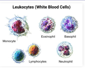
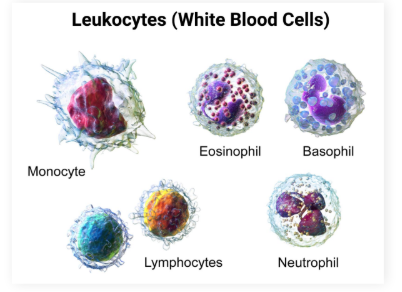
Leukocytes
White blood cells
Lymphocytes
White blood cells that originate from bone marrow, primarily found in lymphatic organs
T cells
B cells
Natural Killer Cells
Where do T cells mature
The thymus
Where do B cells mature
Bone marrow
What is the Innate Immune System
First line of defense, generates a nonspecific (generalized) immune response
External Immunity: Innate immune system
Examples
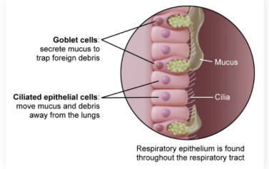
External immunity: Physical/physiological barriers that prevent pathogen entry
Includes skin, hair, cilia, mucous membranes, chemical secretions, and symbiotic bacteria
Internal Immunity: Innate immune system
Internal immunity: Internal defenses activated by the innate immune system to neutralize pathogens that have entered
Composed of the inflammatory response, complement proteins, and phagocytic and natural killer cells
Inflammatory Response: Innate immune system
What triggers it
main activations
Mast Cells
Swelling
Loss of function
increased heat
pain
Mast Cells in immune response
Leukocytes responsible for the first part of the inflammatory response
Known as the rally signal
Sit in the tissue in preparation for injury
In the presence of injury, mast cells release histamine, which dilates blood vessels and increases their permeability, allowing immune cells to enter the tissue
Mast cells also release heparin, an anticoagulant that prevents blood clotting
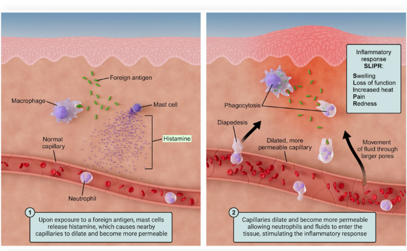
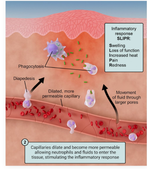
All steps of inflammatory response
What do they do
Pnemonic
Swelling: Permeable capillaries result in fluids leaking into tissues
Loss of function: Body part with inflammation becomes less usable
Increased heat: Increased blood flow increases cutaneous temperature
Pain: Throbbing pain caused by swelling, which puts continuous pressure on nerve endings
Redness: Increased blood flow causes redness of skin
SLIPR
Diapedesis
Definition
What is directed by
The process by which cells move from capillaries to the tissues in order to fight pathogens
Directed by chemokine signaling
Diapedesis Steps
Tissue injury
Damaged cells release cytokines (inflation) and histamine (increases permeability)
Cytokines trigger inflammation
Blood vessels dilate, increasing blood flow, and become more permeable
Endothelial cells express adhesion molecules
White blood cells stick to vessel walls
Chemokines (in charge of chemotaxis) form a gradient
White blood cells follow the chemical trail toward the site of injury (chemotaxis)
Diapedesis occurs
White blood cells squeeze through vessel walls into the interstitial space
At the injury site, inflammation continues to amplify cytokine and chemokine release, attracting more immune cells until the threat or damage is cleared
Chemotaxis
organisms direct their movement toward or away from specific chemicals in their environment
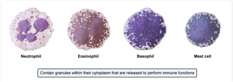
Granulocytes
Cells in the innate immune system with specific granules in their cytoplasm
Include neutrophils, eosinophils, basophils, and mast cells
everything minus Monocytes and Lymphocytes
Abundance of Leukocytes + mneomonic
Neutrophils
Leukocytes
Monocytes and macrophates
Eosinophils
Basophils
Neva Let Monkeys Eat Bananas
Neutrophils
Neutrophils: Innate immunity phagocytes
Most common leukocyte found in the blood, accounting for over half of all leukocytes
One of the first cells recruited to a site of inflammation
Lymphocytes
Three subsections
What is their mechanism of attack
B cells, T-cells, and NK cells (natural killer)
B and T cells are part of adaptive immunity and must be activated. They are the most common type of leukocyte found in lymph
NK cells are part of innate immunity, attacking virally infected and cancerous cells
NK cells lyse target cells using perforin (create holes) and granzymes (stimulate apoptosis)
Monocytes and Macrophages
Phagocytes in innate immunity
Monocytes: Immature form found in the blood vessels
Macrophages: Mature form following diapedesis with secondary ability to act as antigen-presenting cells, activating adaptive immunity
Eosinophils
Eosinophils: Innate immune cells with granules that can be released to kill pathogens (particularly parasites)
Basophils
Contain granules with histamine (dilates vessels) and heparin (anticoagulant)
Least numerous leukocyte
Very similar to mast cells, except basophils circulate as mature cells while mast cells circulate as immature cells
Dendritic Cells
Innate immune cells that scan tissues using pinocytosis (cell “drinking” of fluids and solutes) and phagocytosis (cell “eating” of solid particles)
Antigen-presenting cells that migrate to the lymph nodes to activate adaptive immunity
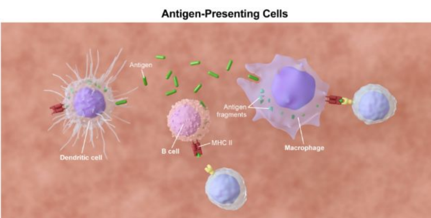
Antigen presenting immune Cells
Macrophages and dendritic cells use toll-like receptors (TLRs) to recognize conserved parts of the microbes
Binding to these receptors triggers phagocytosis and activates the innate immune system
Specifically, they make sure to bind to conserved parts of microbes (parts that microbes must have to survive)
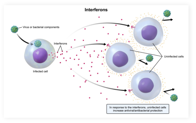
Interferons
Secreted by virally or bacterially infected cells, binding to non-infected cells to prepare them for attack
RED FLAG, WE NEED TO DEFEND, INTERFERE PLEASE
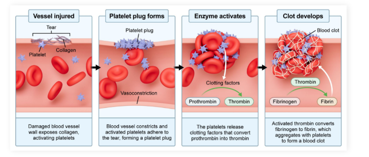
Platelets
Platelets: Anucleate cell fragments that are involved in blood clotting and in activating the innate immune system
Help regulate macrophages and dendritic cells
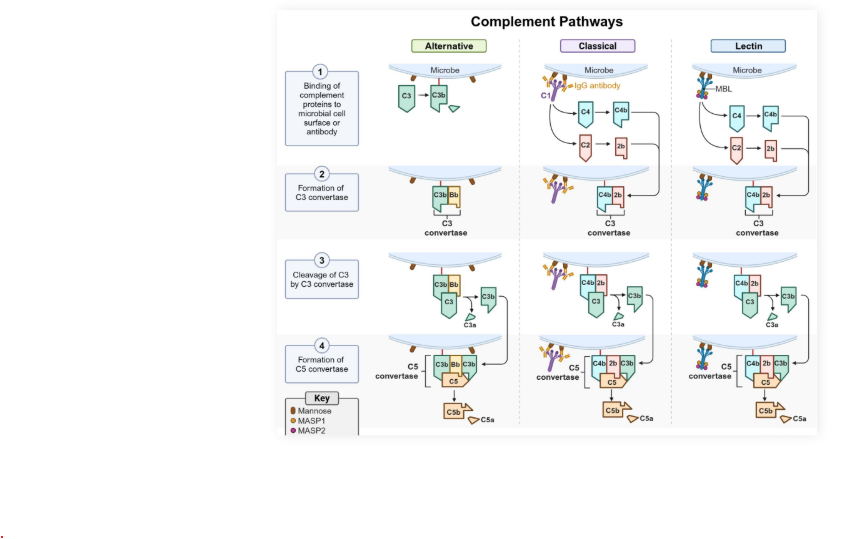
Complement System
Complement system: ~30 proteins that aid immune cells in fighting pathogens
Proteins turn each other on through the activation of a complement cascade, which produces a large effect
Upon recognizing a pathogen, a chain reaction of protease activity is triggered for the proteins to activate each other
Complement Protein Actions
Complement protein actions:
Tag antigens for phagocytosis in a process called opsonization
Increase histamine release and inflammatory response via mast cells
Membrane attack complex (MAC) pokes holes in and lyses pathogens
Adaptive Immune system
Adaptive immunity is a specific immune response
Specific antigens are targeted
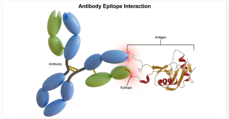
Antigen:
Antigen: An immunogenic foreign molecule that is the target of the immune response
Epitope
Epitope: A portion of the antigen that is recognized by the immune cells
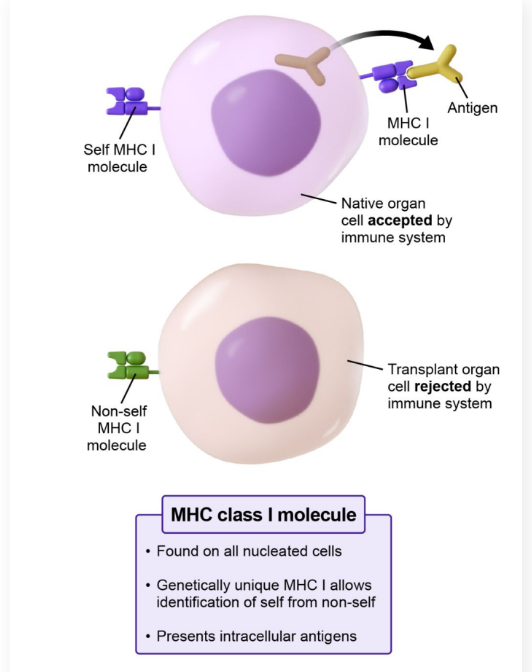
MHC Molecules
Major histocompatibility complex (MHC): Molecules that allow the immune system to recognize foreign cells and antigens
Located on immune cells
Once recognized, the immune system destroys the foreign or infected cell
MHC can be divided into MHC class I and MHC class II
MHC Class 1
Major histocompatibility complex (MHC) class I: Surface molecule on all nucleated cells that present intracellular antigens
Each genetically different individual will have a unique MHC I molecule
The immune system can utilize this inherent uniqueness to distinguish between self and non-self cells
MHC Class 2
Major histocompatibility complex (MHC) class II: Surface molecule on antigen-presenting cells (dendritic cells, macrophages) that present extracellular antigens
Organ Transplants and difficulties with MHC1
Individuals receiving transplants where MHC I molecules do not match may lead to failure or rejection
Immunosuppressants are given to transplant patients to try to prevent this
Autoimmune diseases occur when the immune system attacks self MHC I
Identical twins have identical MHC molecules, allowing these individuals to transplant to each other without the need for immunosuppressants
The donated organ cells will not be marked as foreign and attacked
b Cells
Control antibody-mediated immunity (humoral immunity)
Manage the production and release of antibodies
Can also act as antigen-presenting cells
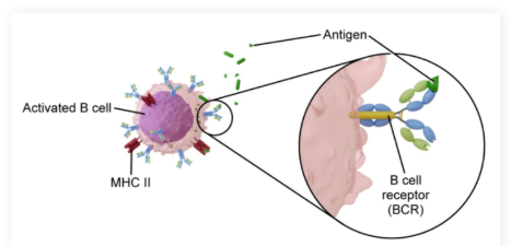
B cell receptors (BCRs): Located on B cells, binding to antigen epitopes either free floating or on the surface of pathogens
Each B cell has a unique BCR
Clonal Selection + Clonal expansion
Types of B cells
Clonal Selection Model: Describes the development of one type of BCR for every B cell
Clonal expansion: B cells divide into either plasma cells or memory B cells
Plasma cells: Produce and secrete antibodies
Memory B cells: Can be activated later in case of another attack
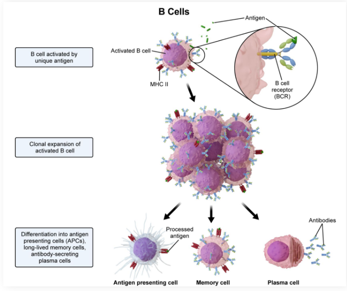
Memory B Cells
Survive for long periods and lie dormant until reactivated by the same antigen that triggered the original clonal expansion
The key to vaccinations
Vaccines stimulate the production of memory B cells, which can later be reactivated
Massive antibody production
Antibodies (immunoglobins)
What are they like structurally
Where do they go around
What do they consist of and what is that bonded by
what is the recognizing region.
Antibodies (immunoglobulins): Structurally identical to BCRs but freely circulate in the blood and lymph
Can tag antigens for phagocytosis, neutralize antigens by coating them, or activate the complement system
Antibodies contain light chains and heavy chains that are linked together by disulfide bonds
The variable region recognizes different antigens while the constant region is the same for antibodies within the same class
5 main antibody classes
● IgM
● IgA
● IgE
● IgD
● IgG
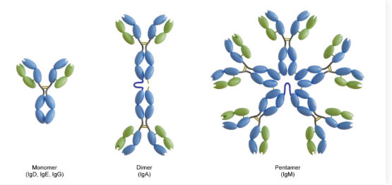
lgM Antibodies
The largest antibody, present in a pentameric form
The first antibody to be produced
Activates the complement system
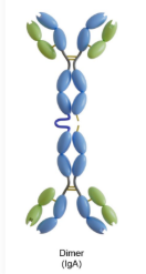
LgA Antibodies
Present in a dimeric form
Most abundant in bodily secretions
Newborns receive passive immunity through breast milk containing IgA
lgE Antibodies
Monomers present on basophils and mast cells as antigen receptors
Triggers histamine release and an allergic reaction when bound to an allergen
Think “Ig-sneEze”
lgD Antibodies
Monomer with very little information known (produced in small amounts)
Dih is mysterious
IgG antibodies:
Monomer that is the most abundant antibody in circulation
The only antibody to cross the placenta, providing a fetus with passive immunity
Aids the complement system in causing opsonization (marking for death) by tagging antigens for phagocytosis
Helps IgM acivate the complement systemt
T Cells
T cells: Control cell-mediated immunity
Directly act on cells rather than releasing antibodies
T cell receptors (TCRs): Unique (similar to BCRs), binding to only one type of antigen per T cell
T cells also undergo clonal selection just like B cells
How are T-Cells Activated
T cells must bind to antigens on antigen-presenting cells (APCs) to be activated
This can occur via MHC I or MHC II
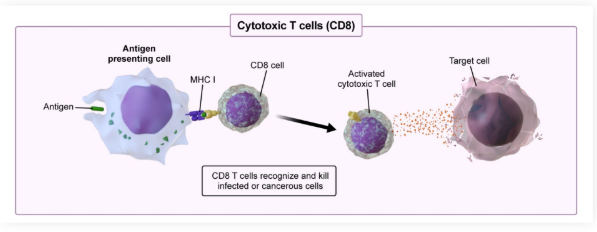
T-Cells with MHC 1
MHC I presentation: T cells differentiate into cytotoxic T cells (CD8+)
Directly kill infected cells through perforin (pokes holes) and granzymes (cause apoptosis)
These are different than NKCs because they are more specific and require antigen presentation
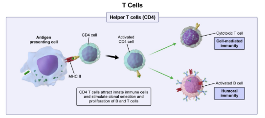
T-Cells with MHC II
MHC II presentation: T cells differentiate into T helper cells (CD4+)
Release interleukins to boost both innate and adaptive immunity
Interleukins help attract innate immune cells and increase proliferation of other T and B cells
Passive Immunity
The immunity one organism gains from receiving antibodies from another organism that already has immunity
Ex: A fetus gain immunity via IgG crossing the placenta
Ex: Newborns gain immunity via IgA in breast milk
The fetus and newborn in these examples are referred to as immuno-naive, as they do not yet have their own active immunity
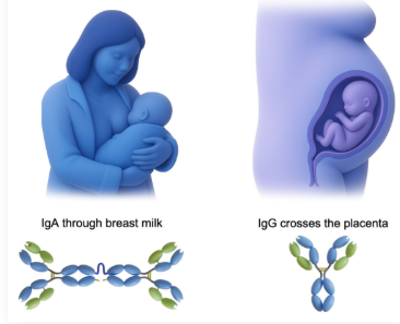
Immunonaive
immuno-naive: An organism without their own active immunity
Active Immunity
Active immunity: The immunity one gains from being infected once already by a pathogen
Vaccination: Introduces antigens or inactivated pathogens to stimulate active immunity
Referred to as artificial immunity
Induces the formation of memory B and T cells
Interleukins
Attract innate immune cells and increase proliferation of other T and B cells
Occurs when T cells differentiate into T helper cells (cd4+)
Bacterial Diseases
Pnemounic
Tuberculosis
Gonorrhea
Leprosy
Syphilis
E. coli
Streptococcus, Bacillus, Staphylococcus, Mycoplasma, Spirochete infections
Tony Go Long, Said Eli
Viral Diseases
Pneuomonic
Influenza
Hepatitis
Herpes
Chicken pox
Human papillomavirus (HPV)
Human immunodeficiency virus (HIV)
Measles
Polio
Icy Hens Hatch Chickens Having Huge Messy Pox
Genetic Diseases
Down syndrome
Cystic fibrosis
Huntington’s disease
Sickle cell
Tay-Sachs
“Down Cyclops!”, Hercules Said Tiredly
Parasitic Diseases
Malaria
Tapeworms
Fungal Diseases
Yeast infections
Athlete’s foot
Fungal and Parasitic Diseases Mneominic
● Please Make Time For Your Aunt
● Parasitic
● Malaria
● Tapeworm
● Fungal
● Yeast infections
● Athletes foot
Humoral Immunity
the aspect of adaptive immunity mediated by antibodies produced by B cells
Cell Mediated Immunity
In the adaptive immune response, T cells are responsible for cell-mediated immunity. Unlike B cells, which release antibodies to attack pathogens (humoral response), T cells directly kill pathogens themselves (cell-mediated response).
Pallor
Abnormal Paleness of the Skin
What are fevers controlled by?
NOT A LOCAL RESPONSE
It is controlled by the brain, and is used to kill temperature sensitive pathogens or to slow down their growth.
Toll Like Receptors
What uses them
What do they trigger
(tlr)
Macrophages and dendritic cells
They are used to trigger phagocytosis and activates immune response.
They recognize the conserved parts of microbes
MAC (membrane attack complex)
Part of the complement system with innate immunity
Allows salts and fluids to enter a pathogen membrane causing to to swell and lyse.
What cells have MHC1 complexes
EVERY NUCLEATE CELL
so no erythrocyte or platelets
What are immunoglobins
Antibodies
Immunogens
antigens
Relationship of BCR’s and Antibodies
THEY ARE THE SAME,
save for the fact that BCR’s are attatched to B cells while antibodies are free floating.
TCR’s
How many antigens can they bind do
Can they clone
Can they recognize free floating antigens?
What cells would cause adaptive immunity to cease to exist?
Helper T cells
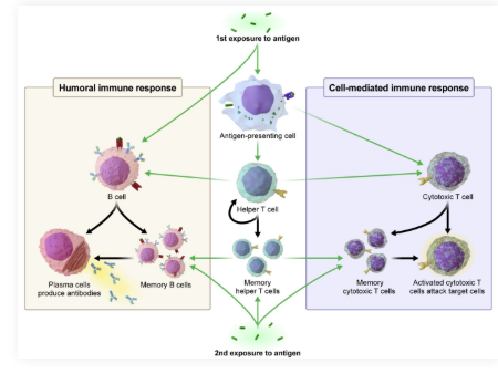
Adaptive immunity has two divisions:
Antibody-mediated immunity (aka humoral immunity) involves B cells that produce antibodies.
B cells encounter and process antigens. Helper T cells (CD4+) subsequently activate B cells by recognizing the antigen-MHC II and releasing interleukins, prompting B cell division into plasma cells.
Cell-mediated immunity relies on cytotoxic T cells (CD8+) to directly attack and eliminate infected or abnormal cells.
Helper T cells recognize antigen-MHC I on antigen-presenting cells and release cytokines, which activate and help proliferate cytotoxic T cells for targeted cell killing.
Helper T cells play critical roles in activating both of these pathways. Without helper T cells, antibody-mediated and cell-mediated immunity would cease to exist.