GW BGZ 2025 Case 1 - At the fitness center
 Call Kai
 Learn
Learn Practice Test
 Spaced Repetition
 Match
Match Flashcards
 Knowt Play
1/39

There's no tags or description
Looks like no tags are added yet.
Name | Mastery | Learn | Test | Matching | Spaced | Call with Kai |
|---|
No analytics yet
Send a link to your students to track their progress
40 Terms
What are the main types of bones based on shape?
here are six major bone types:
Long bones (Ossa longa)
Longer than they are wide.
Function as levers for movement.
Examples: femur, humerus, tibia.
Short bones (Ossa brevia)
Roughly cube-shaped.
Provide stability with limited movement.
Examples: carpals (wrist), tarsals (ankle).
Flat bones (Ossa plana)
Thin, flattened, often curved.
Protect organs and provide muscle attachment.
Examples: ribs, sternum, scapula, skull bones.
Irregular bones (Ossa irregularia)
Complex shapes that do not fit other categories.
Examples: vertebrae, mandible.
Sesamoid bones (Ossa sesamoidea)
Embedded within tendons.
Reduce friction and improve leverage.
Examples: patella, pisiform.
Pneumatic bones (Ossa pneumatica)
Contain air-filled spaces (sinuses).
Examples: frontal bone, ethmoid bone, sphenoid bone, maxilla, temporal bone.
What is laminar bone?
Laminar bone refers to mature bone tissue organized into layers called lamellae.
Characteristics:
Found in compact bone.
Provides strength and resistance to stress.
Represents the normal mature organization of bone tissue.
What are the names of the bones in the body and where are they? (the “bigger” bones)?
Bone / Bone Group | Location in the Body |
|---|---|
Cranium | Upper part of the skull; protects the brain |
Facial bones | Front of the skull; form the face |
Clavicle | Collarbone; between the breastbone and shoulder |
Scapula | Shoulder blade; upper back |
Sternum | Breastbone; center of the chest |
Ribs | Surround the chest and protect the heart and lungs |
Vertebrae | Individual bones of the spine |
Vertebral column | Entire backbone/spine running from neck to pelvis |
Sacrum | Triangular bone at the base of the spine, between the hips |
Humerus | Upper arm bone (shoulder to elbow) |
Radius | Forearm bone on the thumb side |
Ulna | Forearm bone on the little-finger side |
Carpals | Wrist bones |
Metacarpals | Bones of the palm of the hand |
Phalanges (hand) | Finger bones |
Femur | Thigh bone; hip to knee |
Patella | Kneecap |
Tibia | Shinbone; larger lower-leg bone |
Fibula | Smaller lower-leg bone, beside the tibia |
Tarsals | Ankle bones |
Metatarsals | Middle bones of the foot |
Phalanges (foot) | Toe bones |
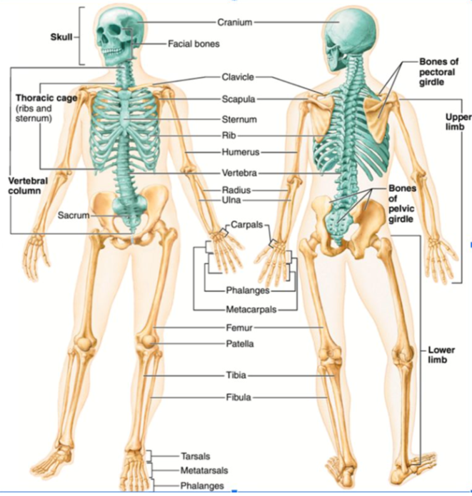
What is bone and what are its main functions?
Bone is a specialized connective tissue that forms the skeleton. Although bone appears hard and lifeless, it is actually a living tissue containing cells, blood vessels, nerves, and an extracellular matrix.
Bone performs several important functions:
1. Support
Bones provide the structural framework of the body and support all soft tissues and organs.
2. Protection
Bones protect vital organs:
Skull protects the brain.
Vertebral column protects the spinal cord.
Rib cage protects the heart and lungs.
3. Movement
Bones act as levers for muscles. When muscles contract, they pull on bones, producing movement at joints.
4. Mineral Storage
Bones store important minerals:
Calcium
Phosphate
These minerals can be released into the bloodstream when needed.
5. Blood Cell Production
Red bone marrow produces:
Red blood cells
White blood cells
Platelets
This process is called hematopoiesis.
6. Fat Storage
Yellow bone marrow stores energy in the form of fat.
What are the major parts of a long bone?
A typical long bone consists of several important structures:
Diaphysis
The shaft or long central portion of the bone.
Mainly composed of compact bone.
Contains the medullary cavity.
Epiphyses
The enlarged ends of the bone.
Mainly composed of spongy bone covered by a thin layer of compact bone.
Metaphysis
Transition region between the diaphysis and epiphysis.
Contains the growth plate in children.
Periosteum
Tough outer connective tissue covering.
Contains blood vessels and nerves.
Endosteum
Thin membrane lining internal bone surfaces.
Medullary Cavity
Hollow cavity inside the diaphysis.
Contains bone marrow.
Together these structures allow bones to grow, repair themselves, and withstand mechanical stress.
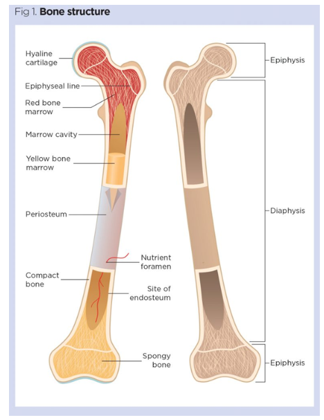
What is the diaphysis and what is its function?
The diaphysis is the shaft or long central portion of a long bone.
Characteristics:
Composed mainly of compact bone.
Surrounds the medullary cavity.
Provides strength and rigidity.
Functions:
Supports body weight.
Resists bending forces.
Provides attachment sites for muscles.
Examples:
The shaft of the femur.
The shaft of the humerus.
Because compact bone is very dense, the diaphysis is extremely strong and well suited for weight-bearing activities.
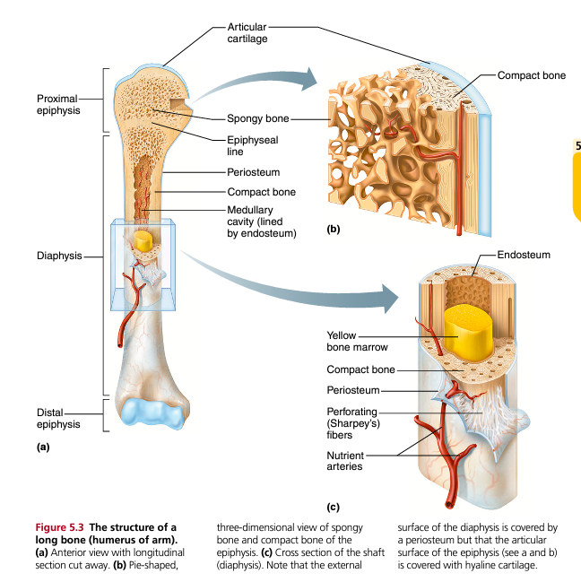
What are the epiphyses and what is their function?
The epiphyses are the enlarged ends of a long bone.
Characteristics:
Mostly composed of spongy bone.
Covered by a thin layer of compact bone.
Covered by articular cartilage where joints form.
Functions:
Form joints with neighboring bones.
Distribute forces across joints.
Reduce bone weight while maintaining strength.
The spongy bone inside the epiphyses contains trabeculae arranged along lines of stress, making the bone strong without being excessively heavy.
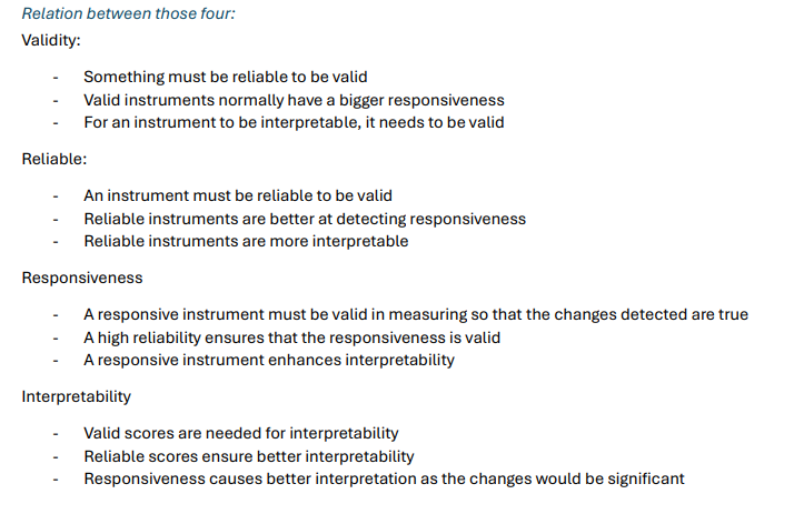
What is the metaphysis and why is it important?
The metaphysis is the region between the diaphysis and epiphysis.
In children and adolescents:
Contains the epiphyseal (growth) plate.
Bone length increases here.
In adults:
The growth plate closes.
It becomes the epiphyseal line.
Importance:
Responsible for longitudinal bone growth.
Site of active bone remodeling during development.
Without the metaphysis and growth plate, bones could not increase in length during childhood.
What is the epiphyseal plate and how does bone growth occur?
The epiphyseal plate, also called the growth plate, is a layer of hyaline cartilage found within the metaphysis of growing bones.
During childhood:
Cartilage cells divide rapidly.
New cartilage is produced.
Osteoblasts replace cartilage with bone tissue.
This process causes bones to become longer.
When growth ends:
Cartilage is completely replaced by bone.
The plate ossifies.
An epiphyseal line remains.
The closure of growth plates marks the end of height increase.
What is the periosteum and what are its functions?
The periosteum is a dense connective tissue membrane covering the external surface of bone, except where articular cartilage is present.
It contains:
Blood vessels
Nerves
Osteoblasts
Osteoclasts
Functions:
Protection
Protects underlying bone tissue.
Blood Supply
Provides nutrients and oxygen.
Muscle Attachment
Serves as an attachment point for tendons and ligaments.
Growth and Repair
Contains bone-forming cells important for healing fractures.
The periosteum is firmly attached to bone by Sharpey's fibers (perforating fibers).
What is red bone marrow and what does it do?
Red bone marrow is specialized tissue responsible for blood cell production.
Produces:
Red blood cells (erythrocytes)
White blood cells (leukocytes)
Platelets (thrombocytes)
This process is called hematopoiesis.
In adults, red marrow is found mainly in:
Vertebrae
Ribs
Sternum
Pelvis
Proximal ends of long bones
What is yellow bone marrow?
Yellow bone marrow consists mainly of adipose (fat) tissue.
Functions:
Energy storage.
Fat reserve.
In severe blood loss, yellow marrow can convert back into red marrow to increase blood cell production.
What is the endosteum?
The endosteum is a thin connective tissue membrane lining all internal bone surfaces.
It covers:
Medullary cavity
Trabeculae of spongy bone
Central canals
Functions:
Bone growth
Bone repair
Bone remodeling
Contains:
Osteoblasts
Osteoclasts
These cells constantly maintain bone tissue throughout life.
What is compact bone?
Compact bone is the dense outer layer of bone tissue.
Characteristics:
Strong
Heavy
Highly organized
Functions:
Provides strength.
Resists bending.
Protects internal bone structures.
Compact bone is especially abundant in the diaphysis of long bones.
What is spongy bone?
Spongy bone, also called cancellous bone, is a lightweight network of bony plates called trabeculae.
Characteristics:
Porous appearance.
Contains marrow spaces.
Lacks osteons.
Functions:
Reduces skeletal weight.
Absorbs stress.
Houses bone marrow.
Spongy bone is found mainly in:
Epiphyses
Vertebrae
Flat bones
What are trabeculae?
Trabeculae are thin bony plates that form the structural framework of spongy bone.
Characteristics:
Arranged along stress lines.
Contain osteocytes.
Surrounded by bone marrow.
Functions:
Provide strength with minimal weight.
Distribute forces efficiently.
Resist compression.
The arrangement of trabeculae changes according to mechanical stresses placed on the bone.
What are osteoblasts, osteocytes, and osteoclasts?
Osteoblasts
Bone-forming cells.
Functions:
Produce osteoid.
Deposit bone matrix.
Build new bone.
Osteocytes
Mature bone cells.
Functions:
Maintain bone tissue.
Monitor bone stress.
Coordinate remodeling.
Osteoclasts
Bone-resorbing cells.
Functions:
Break down bone matrix.
Release minerals into blood.
Participate in remodeling.
Bone health depends on a balance between osteoblast and osteoclast activity.
What is skeletal muscle, what are muscle fibers, and what are the main functions of skeletal muscle?
Skeletal muscle is one of the three muscle types in the body and is responsible for voluntary movement, meaning it is under conscious control. Skeletal muscles attach to bones through tendons and produce movement by pulling on the skeleton when they contract.
The basic cell of skeletal muscle is called a muscle fiber (or myocyte). Muscle fibers are very long, cylindrical cells that contain many nuclei. Unlike most cells, muscle fibers are specialized for contraction and contain numerous contractile structures called myofibrils.
The main functions of skeletal muscle are:
Producing body movement
Maintaining posture and body position
Stabilizing joints
Supporting soft tissues
Generating heat during contraction
Assisting breathing and circulation through muscle activity
Each muscle is made of thousands of muscle fibers organized into bundles called fascicles.
Describe the complete structure of skeletal muscle from the whole muscle down to the contractile proteins.
Skeletal muscle has several levels of organization:
Whole muscle
Surrounded by connective tissue called the epimysium
Fascicle
Bundle of muscle fibers
Surrounded by perimysium
Muscle fiber (muscle cell)
Surrounded by endomysium
Covered by the cell membrane called the sarcolemma
Filled with cytoplasm called the sarcoplasm
Inside each muscle fiber are:
Myofibrils
Long contractile structures running through the fiber
Each myofibril consists of repeating units called:
Sarcomeres
Basic contractile units of muscle
Within each sarcomere are:
Thin filaments
Made of actin
Thick filaments
Made of myosin
When actin and myosin interact, muscle contraction occurs.
What are the connective tissue layers of skeletal muscle and what are their functions?
Three connective tissue layers organize and support skeletal muscle:
Endomysium
Surrounds each individual muscle fiber
Contains small blood vessels and nerves
Helps support individual muscle cells
Perimysium
Surrounds bundles of fibers called fascicles
Provides structural organization
Contains larger blood vessels and nerves
Epimysium
Surrounds the entire muscle
Protects the muscle
Merges with tendons that attach muscle to bone
Together these layers help distribute force generated during contraction and provide protection and organization.
What is a sarcomere and what are its major parts?
The sarcomere is the basic contractile unit of skeletal muscle and is responsible for producing force and movement.
A sarcomere extends from one Z-line to the next.
Important structures include:
Z-line (Z-disc)
Marks the boundaries of a sarcomere
Anchors actin filaments
I-band
Light-colored region
Contains only actin filaments
A-band
Dark region
Contains the entire length of myosin filaments
Includes areas where actin overlaps with myosin
H-zone
Central region of the A-band
Contains only myosin filaments
These structures work together to allow muscle contraction through filament sliding.
Explain the sliding filament theory of muscle contraction and describe what happens to the sarcomere during contraction.
The sliding filament theory explains how muscles contract.
During contraction:
Calcium is released from the sarcoplasmic reticulum.
Myosin heads bind to actin filaments.
Myosin pulls actin toward the center of the sarcomere.
Actin slides over myosin.
The sarcomere shortens.
Importantly:
Actin filaments do not shorten.
Myosin filaments do not shorten.
Only the amount of overlap changes.
Changes during contraction:
Z-lines move closer together.
I-band becomes smaller.
H-zone becomes smaller or disappears.
A-band remains the same size.
The shortening of millions of sarcomeres produces whole-muscle contraction.
What are the different muscle fiber types and how do they differ?
Muscle fibers are classified according to contraction speed, fatigue resistance, and energy production.
Type I fibers (Slow-twitch)
Characteristics:
Slow contraction speed
Red appearance due to high myoglobin content
Many mitochondria
Aerobic metabolism
Highly fatigue resistant
Functions:
Standing
Walking
Posture maintenance
Long-distance running
Endurance activities
Additional feature:
Thicker Z-lines
Better structural integrity
Less likely to be damaged
Type IIa fibers
Characteristics:
Intermediate fibers
Mix of aerobic and anaerobic metabolism
Moderate fatigue resistance
Functions:
Middle-distance activities
Repeated moderate-intensity effort
Type IIx fibers
Characteristics:
Fastest contraction speed
White appearance
Few mitochondria
Anaerobic metabolism
Fatigue quickly
Functions:
Sprinting
Jumping
Heavy lifting
Explosive movements
Generate the greatest force and power.
What important supporting structures are found inside muscle fibers and what are their functions?
Several structures support muscle contraction and energy production.
Glycogen
Stores glucose
Provides energy for contraction
Myoglobin
Stores oxygen
Gives muscle a red color
Mitochondria
Produce ATP
Essential for muscle contraction
Sarcoplasmic reticulum
Stores calcium
Releases calcium to initiate contraction
T-tubules
Conduct electrical signals into the muscle fiber
Ensure synchronized contraction
Together these structures provide energy, oxygen, and signaling necessary for muscle function.
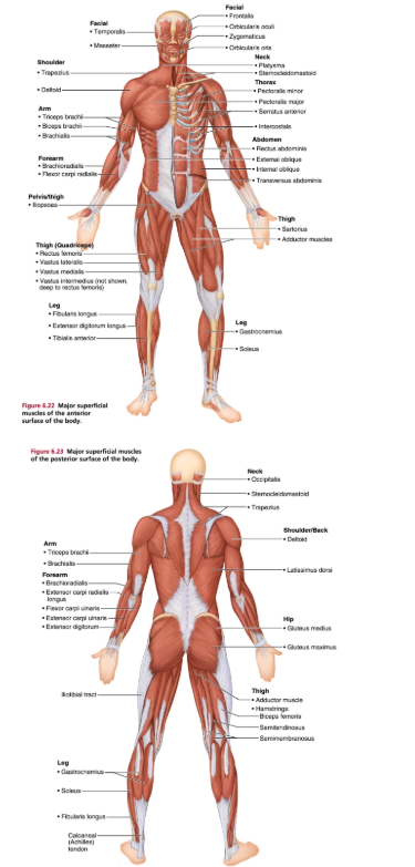
What are the names of the muscles in the body and where are they?
nog meer of niet? Alleen grote spiergroepen
What are fascicles and how does fascicle arrangement affect muscle function?
A fascicle is a bundle of muscle fibers surrounded by perimysium.
The arrangement of fascicles determines:
Force production
Range of motion
Direction of pull
Overall muscle function
Different muscles have different fascicle arrangements depending on the job they perform.
Parallel muscles favor movement.
Pennate muscles favor force production.
Convergent muscles allow versatile pulling directions.
Circular muscles control openings.
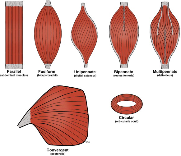
Describe all major fascicle arrangements, their characteristics, and examples.
Parallel
Characteristics:
Fibers run parallel to muscle length
Large range of motion
Lower force production
Example: sartorius
Fusiform
Characteristics:
Spindle-shaped
Thick middle and narrow ends
Good range of motion
Moderate force
Example:biceps brachii
Convergent
Characteristics:
Broad origin
Fibers converge into one tendon
Can pull in different directions
Example: pectoralis major
Unipennate
Characteristics:
Fibers attach to one side of tendon
High force production
Example: extensor digitorum longus
Bipennate
Characteristics:
Fibers attach on both sides of tendon
Example: rectus femoris
Multipennate
Characteristics:
Fibers attach from many directions
Greatest force production
Example: deltoid
Circular
Characteristics:
Fibers arranged in rings
Open and close body openings
Examples: orbicularis oculi
How are muscles attached to bones and how can muscle attachments help predict movement?
Skeletal muscles attach to bones through tendons.
Each muscle generally has:
Origin
Less movable attachment
Usually proximal
Acts as the fixed point
Insertion
More movable attachment
Usually distal
Moves toward the origin during contraction
A muscle's action can often be predicted by:
The bones it attaches to
The joint it crosses
Its position relative to the joint
General rules:
Crossing the front of a joint usually causes flexion.
Crossing the back of a joint usually causes extension.
Examples:
Biceps brachii
Origin: scapula
Insertion: radius
Action: elbow flexion and forearm supination
What are agonists, antagonists, synergists, and fixators?
Muscles rarely work alone.
Agonist (Prime mover)
The main muscle responsible for producing a movement.
Example:
Biceps during elbow flexion.
Antagonist
Produces the opposite action and relaxes while the agonist contracts.
Example:
Triceps during elbow flexion.
Synergist
Assists the agonist and helps produce smooth movement.
Example:
Brachialis assisting the biceps.
Fixator
Stabilizes the origin of the agonist.
Example:
Muscles stabilizing the scapula during arm movement.
These muscle relationships allow coordinated and efficient movement.
What is a joint (articulation) and what is its overall purpose in the body?
A joint, also called an articulation, is a location in the body where two or more bones meet. Joints are essential because they provide both stability and movement for the skeleton.
Their two main functions are:
To hold bones together securely
Joints maintain the structural integrity of the skeleton and keep bones aligned.
To allow movement
Depending on the joint type, they can allow no movement, slight movement, or free movement.
Joints range from completely immovable structures in the skull to highly mobile joints like the shoulder and hip. The type of joint determines how much motion is possible and in what directions.
What are the functional classifications of joints and what movements do they allow?
Joints are classified functionally based on how much movement they allow:
Synarthroses (immovable joints)
Allow no movement
Designed for protection and stability
Example: skull sutures
These joints are important for protecting delicate structures like the brain.
Amphiarthroses (slightly movable joints)
Allow limited movement
Provide both stability and flexibility
Example: intervertebral discs, pubic symphysis
These joints absorb shock and allow slight flexibility in the spine and pelvis.
Diarthroses (freely movable joints)
Allow wide range of movement
Found mainly in limbs
Example: shoulder, hip, elbow, knee
These are the most common functional joints for movement in daily life.
What are false and true joints?
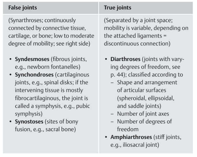
rue joints (movable joints)
These are joints where movement is possible
Bones are connected by cartilage and/or a joint capsule
They allow different types of motion (bend, rotate, slide)
Examples:
Knee joint
Shoulder joint
Elbow joint
Hip joint
These are the normal “functional” joints in the body.
False joints (immovable joints)
These joints allow no movement or very little movement
Bones are tightly connected by fibrous tissue
They are mostly found in the skull
Example:
Sutures between skull bones (like between frontal and parietal bones)
These are important for protection of the brain and keeping the skull rigid.
What are the structural classifications of joints and how do they differ in movement?
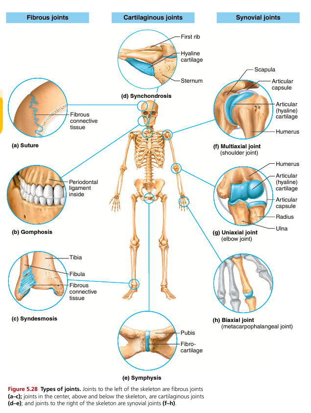
Joints are also classified structurally based on what connects the bones:
Fibrous joints
Bones connected by dense connective tissue
Usually immovable
Examples:
Skull sutures (no movement)
Gomphoses (teeth in sockets)
Syndesmoses (slight movement, e.g., tibia-fibula)
These joints prioritize stability over movement.
Cartilaginous joints
Bones connected by cartilage
Allow limited movement
Two types:
Synchondroses (hyaline cartilage, usually immovable)
Symphyses (fibrocartilage, slightly movable)
Examples:
Intervertebral discs (slight movement + shock absorption)
Pubic symphysis
These joints balance movement and shock absorption.
Synovial joints
Bones separated by a fluid-filled cavity
Freely movable joints
Most joints in limbs are synovial
These are the most important joints for body movement.
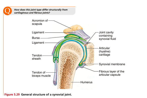
What is the structure of a synovial joint and how does it function?
Synovial joints are highly movable joints with a complex structure designed for smooth motion.
Key structures: Articular cartilage
Smooth hyaline cartilage covering bone ends
Reduces friction and absorbs shock
Has no direct blood supply
Synovial membrane
Inner lining of joint capsule
Rich in blood supply
Produces synovial fluid
Synovial fluid
Thick lubricating fluid inside joint cavity
Reduces friction during movement
Provides nutrients to articular cartilage
Joint cavity
Space between bones filled with synovial fluid
Fibrous capsule
Tough outer layer that encloses joint
Ligaments
Connect bone to bone
Provide stability and prevent dislocation
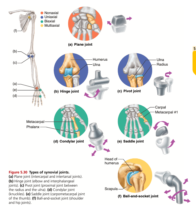
What are the types of synovial joints and what movements do they allow?
Plane joints
Flat surfaces slide over each other
Movement: gliding only (nonaxial)
Example: wrist bones, vertebrae
Hinge joints
One bone fits into a trough-like structure
Movement: flexion and extension only
Uniaxial
Examples: elbow, knee, fingers
Pivot joints
One bone rotates inside a ring
Movement: rotation only
Uniaxial
Examples: atlas-axis joint (neck), radioulnar joint
Condyloid (ellipsoid) joints
Oval-shaped surfaces
Movement: flexion, extension, abduction, adduction
Biaxial
Example: knuckles, wrist
Saddle joints
Both bones have concave and convex surfaces
Movement: biaxial, wide range
Example: thumb joint
Allows opposition (thumb movement)
Ball-and-socket joints
Spherical head fits into socket
Movement: all directions + rotation
Multiaxial
Examples: shoulder, hip
What are the main types of movement produced at joints?
Flexion
Decreases joint angle
Brings body parts closer together
Example: bending elbow or knee
Extension
Increases joint angle
Straightens body part
Example: straightening arm or leg
Abduction
Movement away from midline
Example: raising arms sideways
Adduction
Movement toward midline
Example: bringing arms back down
Rotation
Bone turns around its long axis
Example: turning head side to side
Circumduction
Combination of flexion, extension, abduction, adduction
Circular movement
Example: arm circles
What are special movements of the body and where do they occur?
Dorsiflexion
Lifting foot upward toward shin
Occurs at ankle
Plantar flexion
Pointing toes downward
Like pressing a gas pedal
Inversion
Sole turns inward (medially)
Eversion
Sole turns outward (laterally)
Supination
Palm faces upward/anteriorly
Radius and ulna parallel
Pronation
Palm faces downward/posteriorly
Radius crosses over ulna
Opposition
Thumb touches fingertips
Essential for grasping objects
Elevation / Depression
Elevation: movement upward (shoulders shrug)
Depression: movement downward
Protraction / Retraction
Protraction: forward movement (jaw, shoulders)
Retraction: backward movement
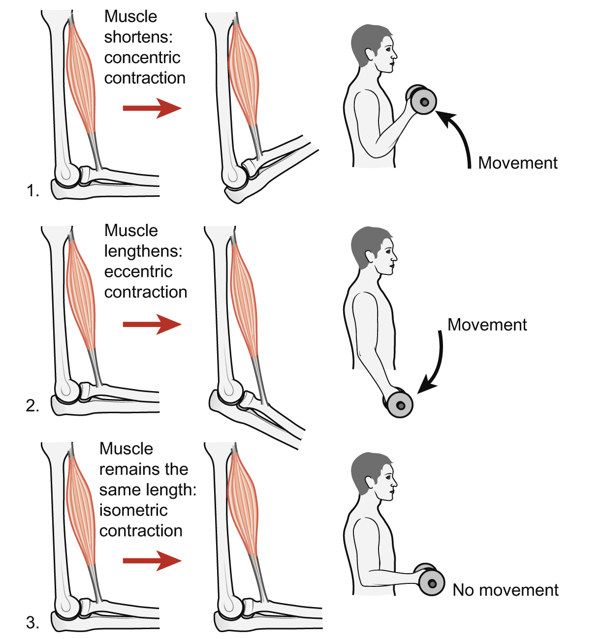
What are the movements of muscles?
Concentric contraction
Muscle shortens while generating force
Movement occurs because the muscle pulls the insertion toward the origin
Produces visible motion at the joint
Example:
lifting a dumbbell during a biceps curl (elbow flexion)
Eccentric contraction
Muscle lengthens while still producing force
Happens when the muscle is actively resisting a load
Controls or slows movement rather than creating it
Example:
lowering a dumbbell slowly during a biceps curl
Isometric contraction
Muscle produces force without changing length
No joint movement occurs, but tension is generated
Used for stabilization and posture
Example:
holding a weight still at 90° elbow flexion
maintaining posture while standing
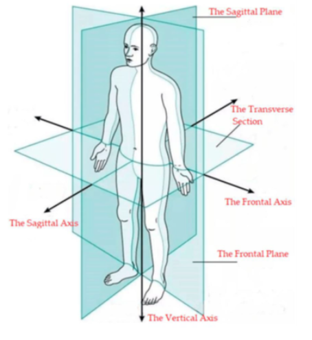
What are the axes and planes of movement?
Main axes of the body
Sagittal axis
Runs front to back
Perpendicular to frontal plane
Mainly involved in abduction/adduction movements
Transverse axis
Runs left to right
Perpendicular to sagittal plane
Mainly involved in flexion/extension movements
Longitudinal (vertical) axis
Runs top to bottom
Perpendicular to transverse plane
Mainly involved in rotation movements
Main anatomical planes
Median (mid-sagittal) plane
Divides body into equal left and right halves
Sagittal plane
Any plane parallel to median plane
Divides body into left and right portions
Frontal (coronal) plane
Divides body into anterior (front) and posterior (back) sections
Transverse (horizontal/axial) plane
Divides body into upper and lower parts
Key idea:
Movement happens in a plane and rotates around a perpendicular axis.
During elbow flexion, the biceps brachii acts as a primary agonist (along with the brachialis), while the triceps brachii functions as the main antagonist. Synergists that assist the biceps brachii include the brachialis and brachioradialis.
During knee flexion, the hamstrings act as the primary movers (agonists) to bend the knee. The quadriceps femoris act as the antagonists, relaxing to allow bending or resisting to control the speed. Synergists that assist with flexion include the gastrocnemius, popliteus, gracilis, and sartorius.