Pre-Clinic Radio: Final
1/34
There's no tags or description
Looks like no tags are added yet.
Name | Mastery | Learn | Test | Matching | Spaced | Call with Kai |
|---|
No analytics yet
Send a link to your students to track their progress
35 Terms
Where are x-rays produced in the x-ray tube?
anode, at the tungsten target
- electrons converted to x-rays here
Identify the part of the x-ray machine the removes long, lazy x-ray photons
the aluminum filter
Identify the part of the x-ray machine that restricts the size and shape of the beam
Lead collimator
1. Identify the error seen on the dental radiograph above
2. why did this error occur?
1. cone cut
2. misalignment of the receptor with the PID
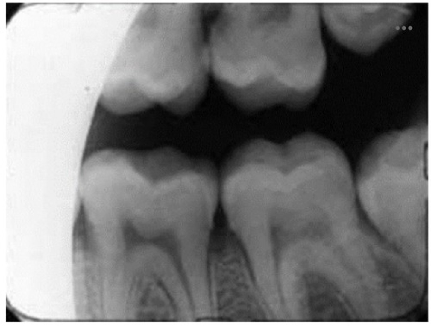
1. Identify the radiolucent area where the arrows are pointing.
2. Identify the radiopaque area where the arrows are pointing.
1. lingual foramen
2. genial tubercules
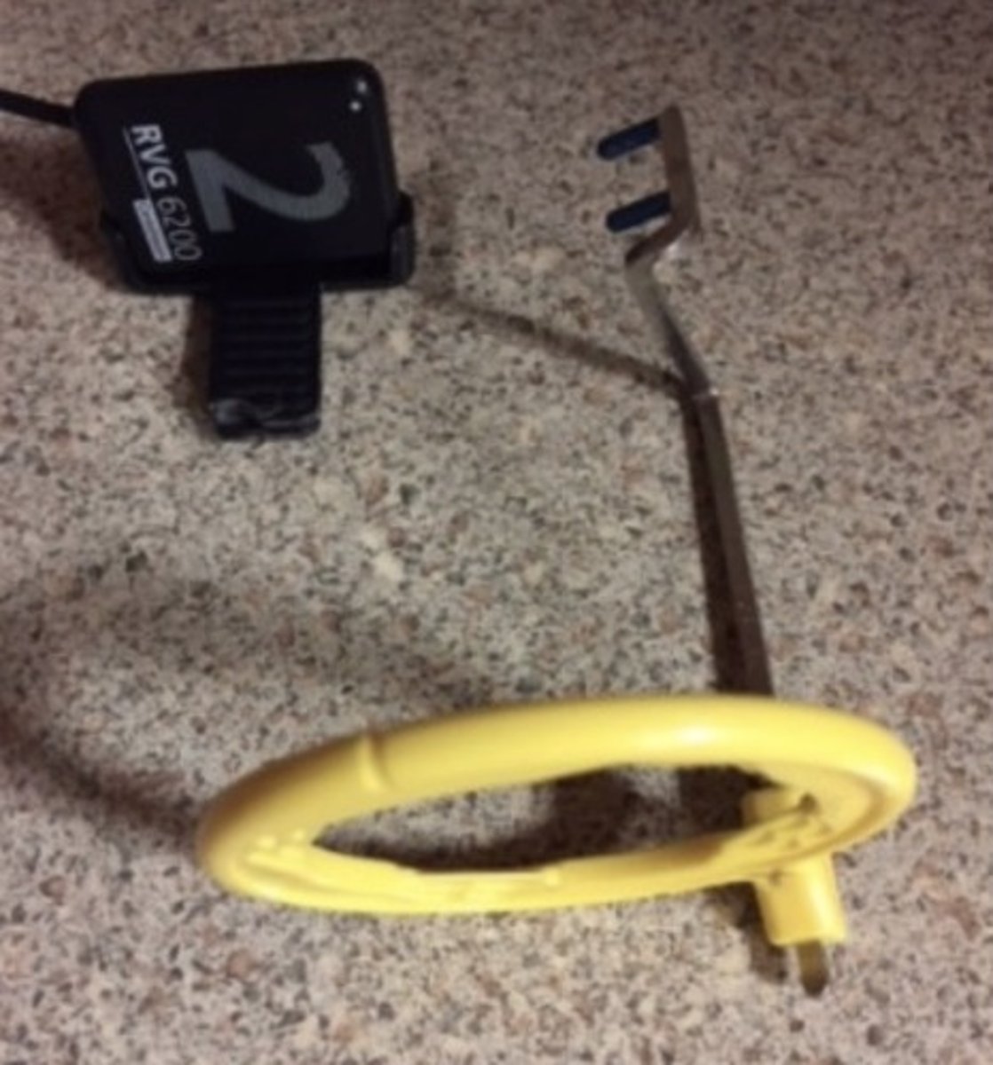
1. The beam alignment device above will be used to take a posterior molar radiograph. What is the error?
2. What size is the standard size receptor?
1. The wrong rod is being used. For posteriors, you should use yellow for PAs and red for bitewings. The blue is used for anterior radiographs.
2. Size 2 is the most standard, as it's used for all posterior radiographs + bitewings
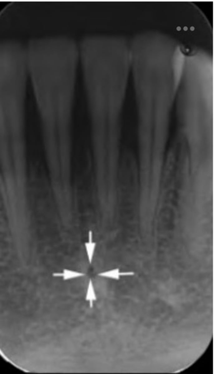
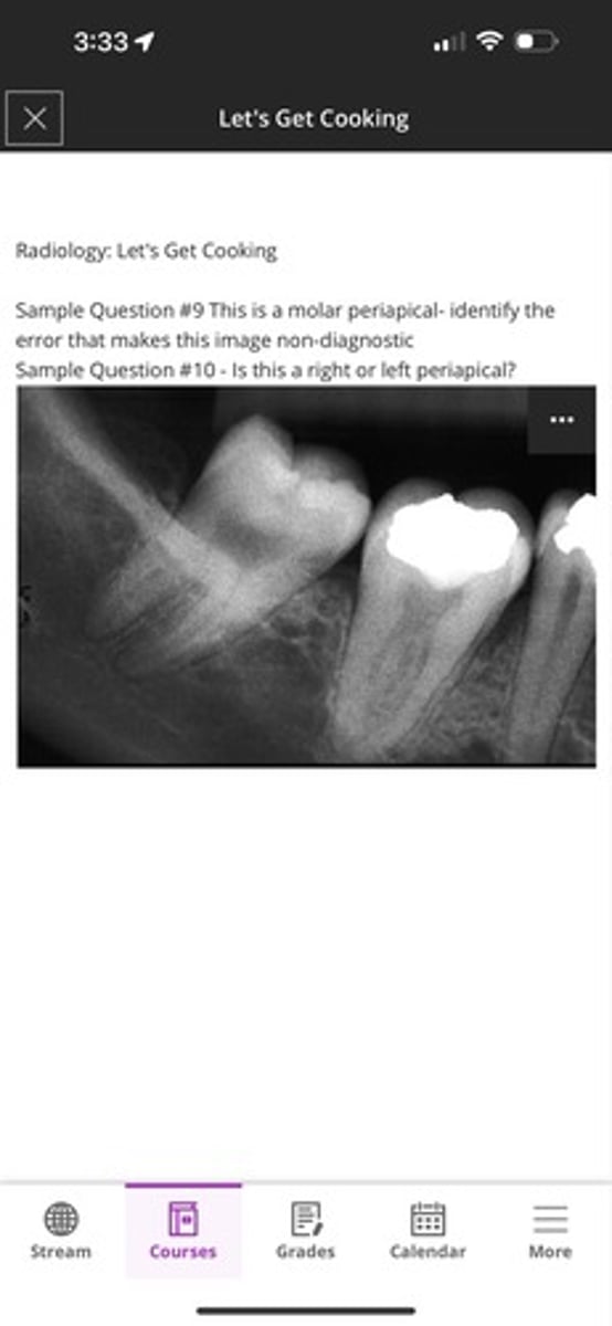
1. This is a molar periapical- identify the error that makes this image non-diagnostic
2. Is this a right or left periapical?
1. You cannot see the 2-3mm apical of both molars
2. right
Describe proper infection control procedures when taking radiographs
Wipe down all contact surfaces & put up barriers. Wear gloves and not touch surfaces with barriers
Describe how to foreshorten a maxillary canine
increased vertical angulation
Describe how to take an image of the distal Apex of a mandibular third molar
swing the alignment and PID distally, angle the sensor distally as well
Describe how to diminish airspace on a maxillary and mandibular premolar
Use proper receptor and tube angulation. Place the receptor over desired image location and line up the PID with the ring then angle to the tooth
What is the difference between paralleling and bisecting
paralleling: uses the rod & ring, angling the PID parallel to the tooth
bisecting: no rod or ring used, visualization techniques are used. This is better for patient with a strong reflex or abnormal anatomy
When do we use the bisecting angle technique
when paralleling does not provide a good image. It could be due to abnormalities or a patient's strong gag reflex. Not often used
Between paralleling and bisecting, which technique gives them more accurate image?
paralleling technique
Describe the inverse square law
The further the distance to the source, the more radiation intensity decreases
Define elongation and how to correct it
dimensional distortion making the object appear elongated. It can be fixed by increasing vertical angulation
describe foreshortening and how to correct it
dimensional distortion making the object appear shortened. It can be fixed by having decreasing the vertical angulation
if a bitewing has overlap of the key in approximate space how would you correct this issue?
you would correct this issue by lining up the rod with the key interproximal space
What color rods are used for the following:
- anterior PAs
- posterior PAs
- bitewings
- blue
- yellow
- red
What color rings (NOT RODS) are interchangeable?
blue and red
the total energy of the x-ray beam, it is the product of quantity (number of photons) and quality (energy of each photon)
intensity
What factors increase/decrease beam intensity?
increase: kVp, mA & exposure time
decrease: distance
What are the visual characteristics of diagnostic quality of a radiograph?
density & contrast
What are the geometric categories of the diagnostic quality of radiographs?
sharpness, magnification, & distortion
What is radiographic density?
the overall darkness of a radiograph
The darker the image, the (higher/lower) the density
higher
What can affect the overall radiographic density besides exposure factors?
patient's size, object density & film fog
What is the measurement of electrical force that causes the electrons to move from the negative cathode to the positive anode
voltage (kVp)
What exposure factors are concerned with the quantity/quality of the image?
quanitity: mA and exposure time
quality: kVp
- controls pentrating power
If you are trying to keep the same image density, what must be done when mA is increased?
exposure time must be decreased ; the two are inversely related
(T/F): decreased kVp results in a darker image
F; it results in a lighter image
Describe contrast
the variations in shades of grey
- higher contrast > less shades
- lower contrast > more shades
What is the only exposure factor has a direct influence on contrast?
kVp
What are considered the critical organs?
skin, thyroid gland, lens of the eye, & bone marrow
radiolucent artifact between the CEJ and alveolar bone, appears collar or wedge shaped. Most common in or root region
cervical burnout