DEVELOPMENT OF PHARYNGEAL ARCHES & THE FACE 1
1/48
There's no tags or description
Looks like no tags are added yet.
Name | Mastery | Learn | Test | Matching | Spaced | Call with Kai |
|---|
No analytics yet
Send a link to your students to track their progress
49 Terms
when does philtrum formation occur
between 1-3 months in utero
if the philtrum does not form between 1-3 weeks in utero, when will it form
if the philtrum does not form between 1-3 weeks in utero then it will never form
also any problems that occur during this stage will not resolve at a later stage unless surgery is done
diagram showing embryonic germ layers and their development
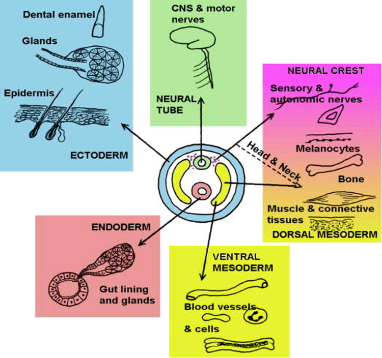
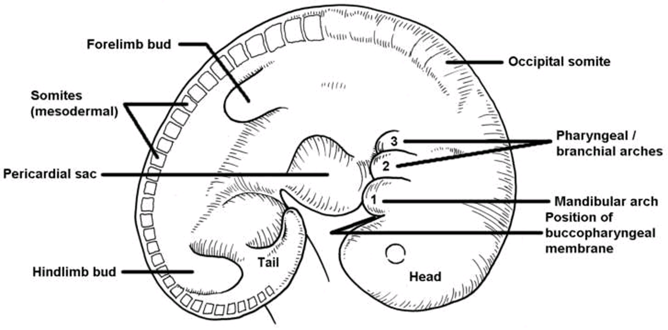
diagram of embryo at 3.5 weeks post conception
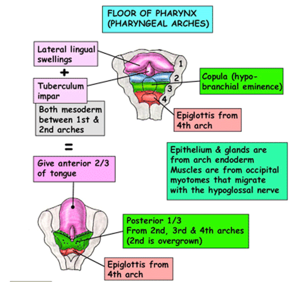
mesodermal blocks will develop into segmental structures like vertebrae, ribs, long bones, muscles
occipital somites will form close to the head and be involved in tongue development
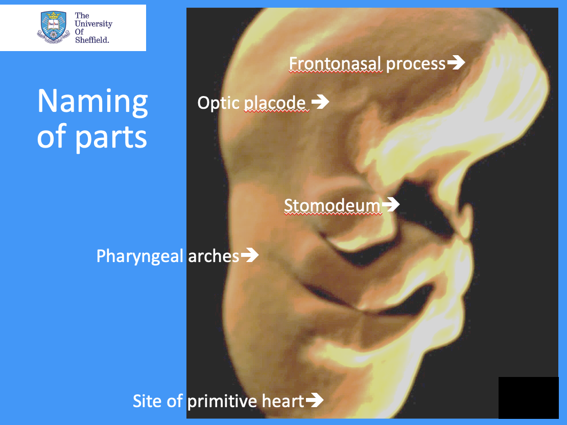
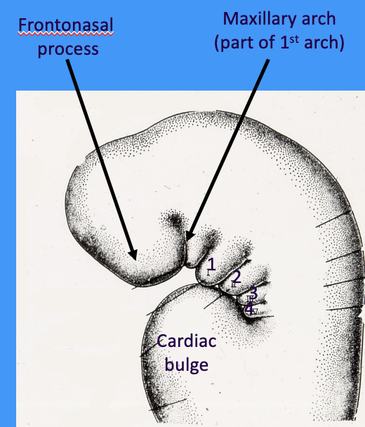
outline the stomodeum and optic placode
stomodeum = primitive developing mouth, represents where ectoderm layer folds in to meet endoderm layer
optic placode = future eye will develop as the optic placode interacts with underlying developing forebrain
placode = area of specialised thickened epithelium
how many pharyngeal arches are there
4 PAIRS (arches 4, 5 and 6 fuse to form a single arch)
what are the pharyngeal arches separated by externally and internally
externally arches are separated by clefts
internally arches are separated by pouches
what term is interchangeable with pharyngeal arch
branchial arch
in which direction do the pharyngeal arch pairs grow
pharyngeal arches grow towards the midline in pairs
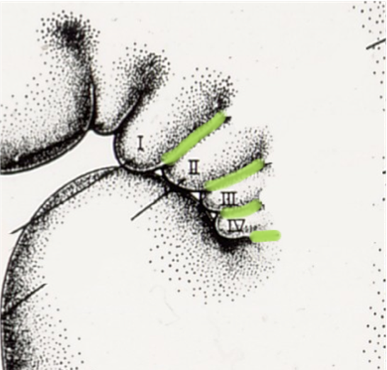
what do the green lines represent
clefts separating the pharyngeal arches externally
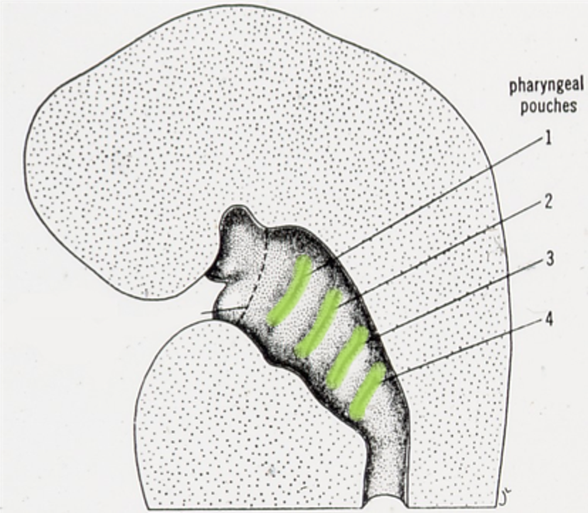
what do the green lines in this image represent
pouches separating the pharyngeal arches internally (these correspond with the clefts)
image shows embryo cut in half
what is the outer surface of pharyngeal arches covered by
ectoderm
what is the inner surface of pharyngeal arches lined with
endoderm
what is the space between the ectoderm and endoderm filled with (in the cranial region and elsewhere)
ectomesenchyme
towards head and neck = ectomesenchyme neural crest-derived cells
in lower part of embryo = mesoderm formed during gastrulation
each arch has a corresponding…
each arch has a corresponding
artery, vein and cranial nerve
skeletal element
muscle block
the migration of nerve fibres from the neural tube into the arches is…
consistent and predictable
they will always innervate specific muscles and regions of the skin and mucosa
which cranial nerve is associated with the first pharyngeal arch
1st arch = trigeminal (CN V)
which cranial nerve is associated with the second pharyngeal arch
2nd arch = facial (CN VII)
which cranial nerve is associated with the third pharyngeal arch
3rd arch = glossopharyngeal (CN IX)
which cranial nerve is associated with the fourth pharyngeal arch
4th arch = vagus (CN X)
what are hox genes
transcription factors
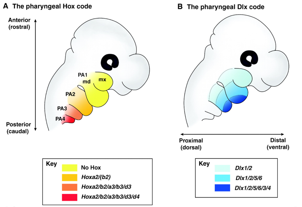
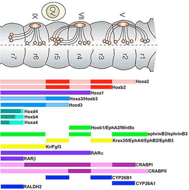
outline Hox genes and their function
patterns of Hox gene expression within the midbrain and hindbrain set up identity
as cells migrate from the neural tube they keep this pattern
Hox genes regulates movement and differentiation of cells
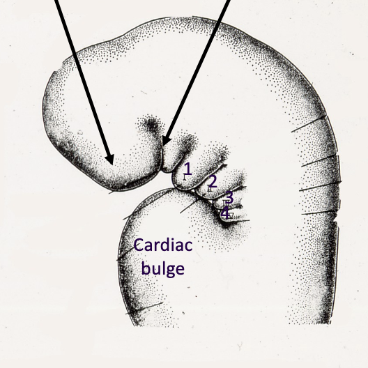
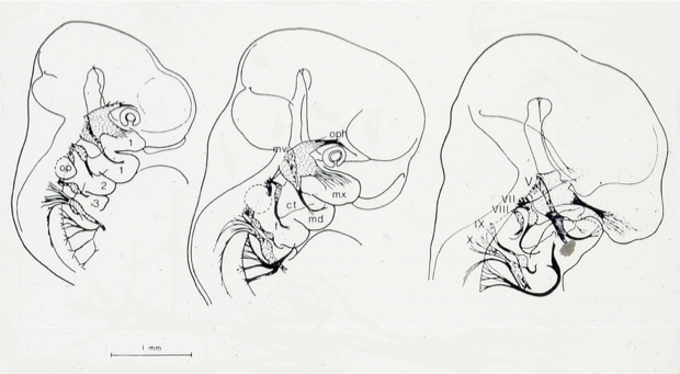
image showing growth of the arches
which muscles are associated with the 1st pharyngeal arch i.e. innervated by the trigeminal
muscles of mastication
some suprahyoids
tensor veli palatini
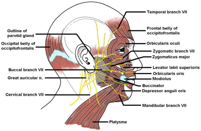
which muscles are associated with the 2nd pharyngeal arch i.e. innervated by the facial nerve
muscles of facial expression
some suprahyoids
stapedius
which muscles are associated with the 3rd pharyngeal arch i.e. innervated by the glossopharyngeal nerve
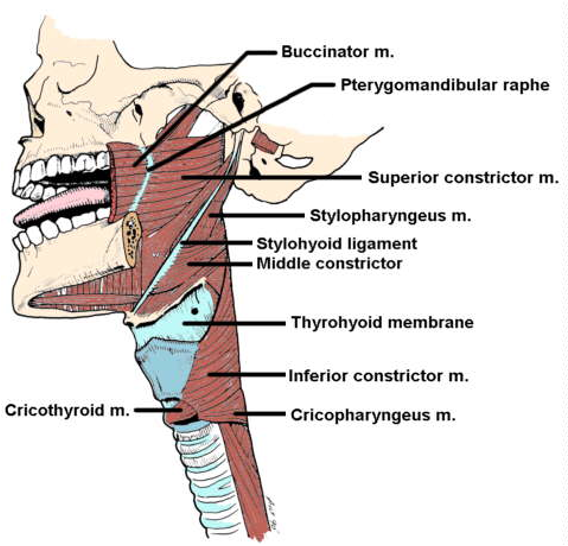
stylopharyngeus
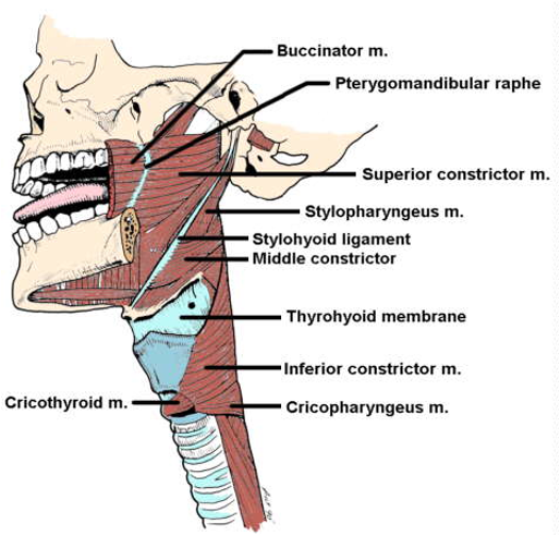
which muscles are associated with the 4th pharyngeal arch i.e. innervated by the vagus nerve
pharyngeal constrictors
muscles of soft palate and larynx

outline skeletal derivatives of the 1st pharyngeal arch
Meckel’s cartilage
this is a transient structure but develops into the incus and malleus

outline skeletal derivatives of the 2nd pharyngeal arch
stapes
styloid process
stylohyoid ligament
superior part of hyoid bone
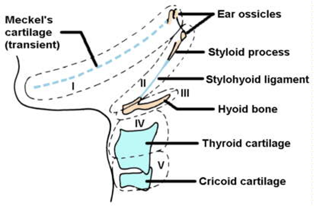
outline skeletal derivatives of the 3rd pharyngeal arch
majority of the hyoid bone

outline skeletal derivatives of the 4th pharyngeal arch
thyroid cartilage
cricoid cartilage
what does the 1st pharyngeal cleft form
the external auditory meatus
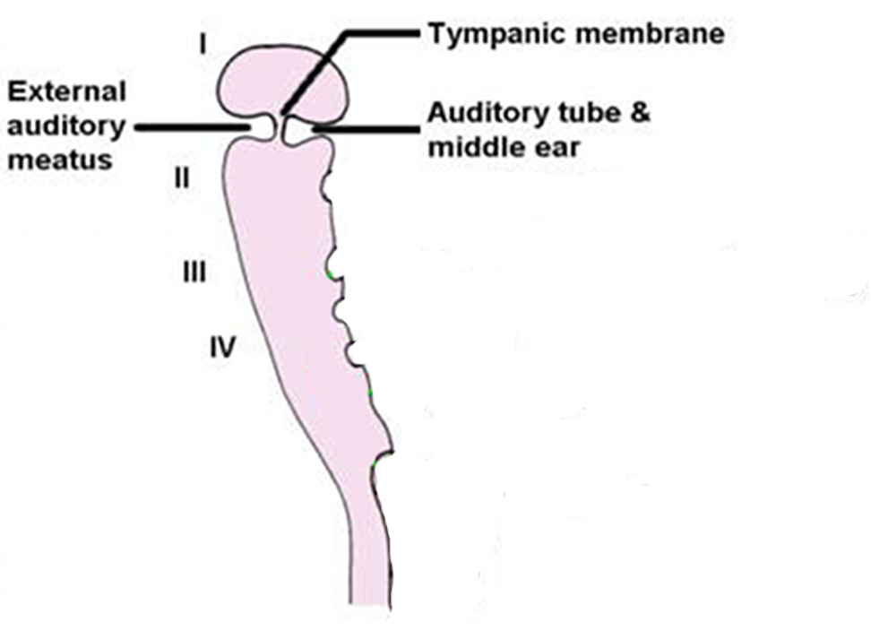
what does the 1st pharyngeal pouch form
the auditory tube and middle ear

what forms the tympanic membrane
a thin epithelial layer between the external cleft and internal pouch

what happens to the other pharyngeal clefts
the other clefts all grow over and become invisible
what does the 2nd pharyngeal pouch develop into
the palatine tonsils
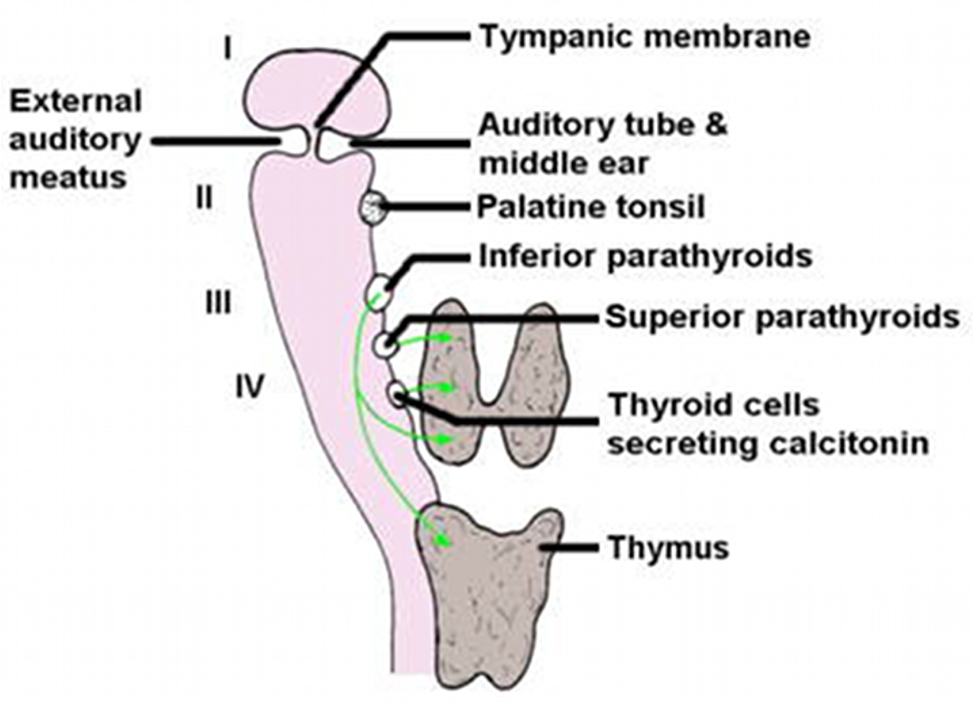
what does the 3rd pharyngeal pouch develop into
inferior parathyroids

what does the 4th pharyngeal pouch develop into
superior parathyroids

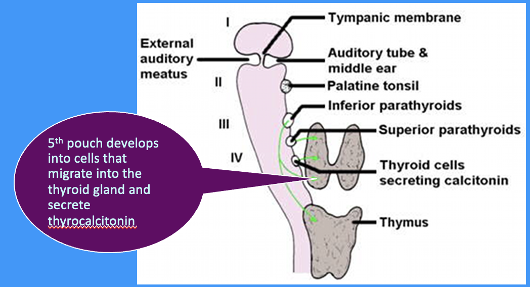
what does the 5th pharyngeal pouch develop into
cells that migrate into the thyroid gland and secrete thyrocalcitonin
(lowers blood calcium when levels are high by inhibiting osteoclasts which reduces calcium release from bone)
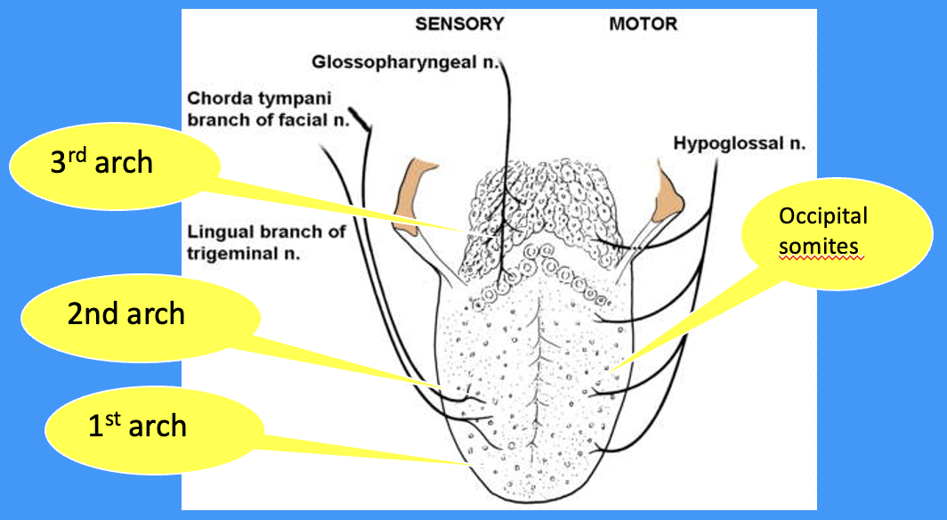
what does the sensory innervation of the tongue tell you about its pharyngeal arch derivation
mucosal components of the tongue are derived from the 1st, 2nd and 3rd pharyngeal arches
from which pharyngeal arch are taste buds formed
2nd pharyngeal arch

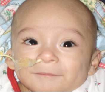
which arch do the majority of arch malformations affect
most arch malformations affect the 1st pharyngeal arch
which structure is the most impacted by arch malformations and give examples
skeletal structures:
hypotrophic mandible
conductive hearing loss
malformed external ear
(can be part of a syndrome or spontaneous/ isolate)
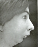
what skeletal defect is shown in this image
hypotrophic mandible
what skeletal defect is shown in this image
malformed (right) external ear
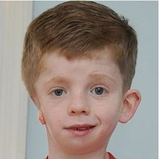
which skeletal defect is shown in this image
asymmetric hemifacial developmental disorder