bio unit 1: chapter 2
 Call Kai
 Learn
Learn Practice Test
 Spaced Repetition
 Match
Match Flashcards
 Knowt Play
1/86

There's no tags or description
Looks like no tags are added yet.
Name | Mastery | Learn | Test | Matching | Spaced | Call with Kai |
|---|
No analytics yet
Send a link to your students to track their progress
87 Terms
How do prokaryotic cells reproduce?
Binary Fission
the division of a cell into two without mitosis
Where is DNA found in a prokaryotic cell, and in what forms?
Nucleoid: A region containing one large, coiled chromosome.
Plasmids: Small rings of extra-chromosomal DNA found throughout the cytoplasm.
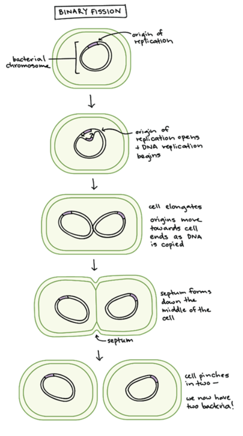
List the key steps of binary fission in order
1. Replication: DNA (chromosome and plasmids) and ribosomes replicate.
2. Elongation: The cell grows larger and longer.
3. Separation: Chromosomes move to opposite ends of the cell.
4. Cytokinesis: Proteins form a new cell wall in the middle, and the cell pinches in two.
Genes in the daughter vs parent cell
They are genetically identical.
During division, the replicated DNA is distributed evenly so each daughter cell receives the same genetic information.
Reproduction Rate of bacteria cells
Approximately every 20 minutes
Aside from DNA, what other cell components must be replicated or distributed during binary fission?
Ribosomes (replicated).
Plasmids (replicated).
Cytoplasm (distributed to both ends).
Peptidoglycan (used to build the new cell wall).
Parent cell
A cell before it divides by mitosis to produce two daughter cells
Daughter cells
A cell resulting from the mitotic division of a parent cell
Genes
Code for protein information
Origin of replication
The point at which the bacterial chromosome begins to replicate.
What is the Cell Cycle and why is it considered a "cycle"?
The series of phases a dividing eukaryotic cell passes through from one division to the next.
It is a cycle because the events repeat each time a cell divides.
While described in phases, it is a continuous process.
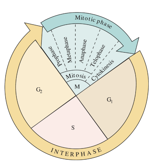
List the four main phases of the cell cycle in order.
G1 phase (Gap 1)
S phase (Synthesis)
G2 phase (Gap 2)
M phase (Mitosis & Cytokinesis)
What occurs during the G0 phase?
A non-proliferating (dormant) state where cells have withdrawn from the active cycle.
They perform normal functions but do not prepare for DNA replication.
E.g. Nerve cells
Define Interphase and its three constituent parts.
The stage between nuclear divisions characterized by active growth and DNA replication.
G1: Protein/organelle production and normal cell tasks.
S: DNA molecules are duplicated forming two chromatids per chromosome.
G2: Final growth and preparation before division.
Why are chromosomes not visible under a microscope during Interphase?
The DNA exists as chromatin—long, thin, unwound strands.
They only condense (thicken) just before mitosis begins.
Define Mitosis
A type of nuclear division that maintains the parental number of chromosomes for daughter cells
It is the basis of bodily growth and asexual reproduction in many eukaryotic species
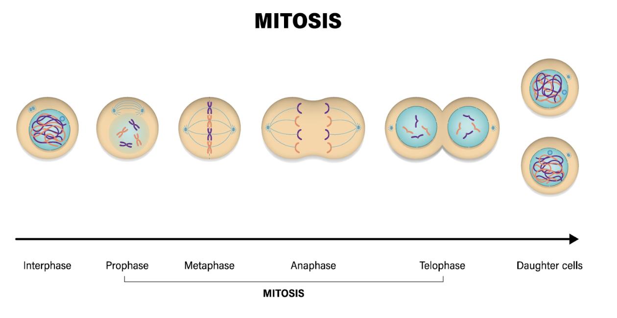
Stages of the cell cycle (IPMATC)
Interphase
Prophase
Metaphase
Anaphase
Telophase
Cytokinesis
Describe the key events of Prophase
Chromatin condenses into visible chromatids joined at a centromere.
Nucleolus disappears, and the nuclear membrane breaks down.
Centrioles move to poles and spindle fibres begin to form.
Describe the key events of Metaphase
Chromosomes line up along the equator (metaphase plate) and attach to spindle fibres.
Describe the key events of Anaphase
Spindle fibres contract
centromeres split
sister chromatids are pulled to opposite poles (now called chromosomes).
What are the key events of Telophase?
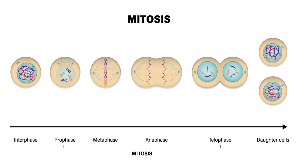
The two sets of separate chromosomes form tight clusters at each pole of the cell.
Chromosomes de-condense as the chromatin unwinds and becomes less visible.
A new nuclear envelope forms around each set of chromosomes.
Nucleoli reform within the new nuclei, and the spindle disassembles (breaks down).
Compare Cytokinesis in animal vs. plant cells.
Animal Cells: The plasma membrane draws together to form a cleavage furrow, pinching the cell in two.
Plant Cells: A cell plate forms in the centre; cellulose is deposited to create a new cell wall between daughter cells.
What is the purpose of Cell Cycle Checkpoints?
To stall the cycle if mistakes are detected, allowing for DNA repair.
This reduces the chance of mutations being passed to daughter cells.
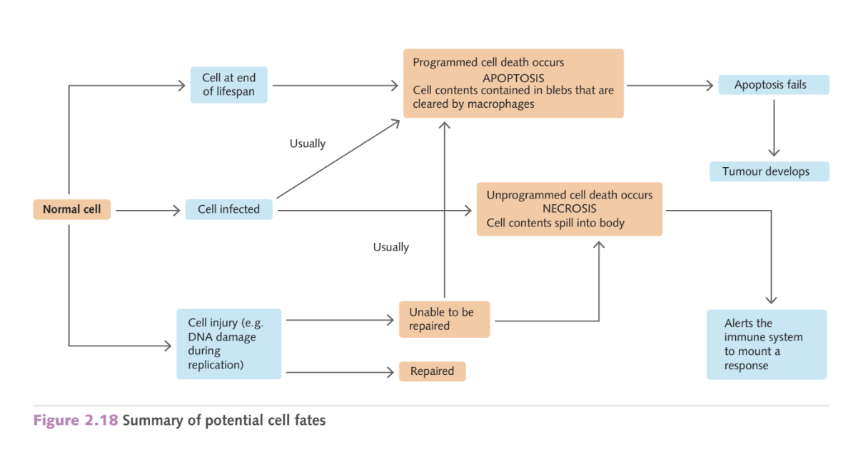
If damage is irreparable, apoptosis is triggered.
G1 checkpoint
Checks DNA damage, cell size, and nutrient levels before S phase.
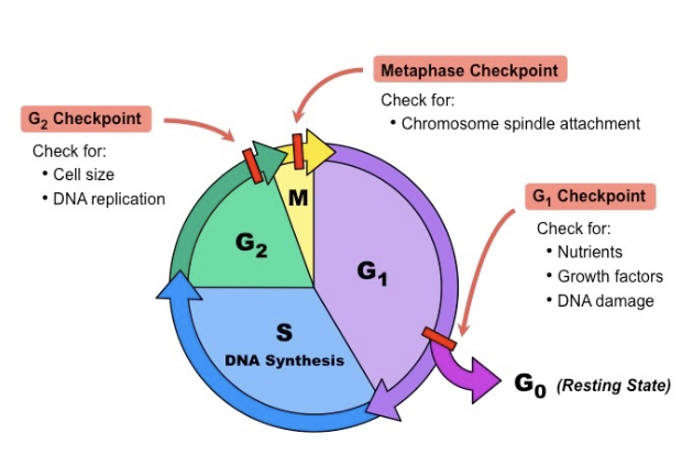
G2 Checkpoint
Screens for DNA replication errors and structural damage.
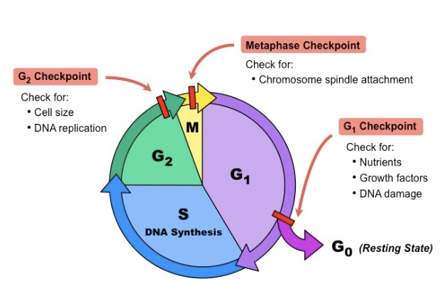
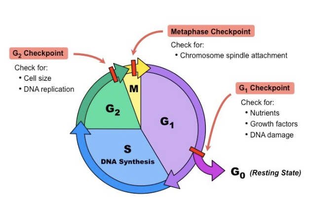
M Checkpoint
Ensures chromosomes are correctly attached to spindles before anaphase.
What molecules act as the "gatekeepers" of the cell cycle?
Cyclins and Cyclin-Dependent Kinases (CDKs).
They promote or inhibit progression depending on cellular signals.
Why do some cells (like skin or gut lining) divide more frequently than others?
They are in areas of high growth or wear.
Frequent division is required to replace cells that become useless due to mechanical disturbance or tissue damage.
Chromatin
A loose, unwound mass of DNA and associated proteins.
This is the "interphase" state of DNA, it looks like a bowl of spaghetti under a microscope and is not yet condensed into distinct shapes.
Chromosome
A structure composed of DNA and proteins that carries genetic information.
In the M phase, chromatin condenses into these tightly coiled, visible structures.
Chromatid
One of the two identical "arms" of a duplicated chromosome.
Sister Chromatids
The two identical copies formed during the S phase, held together by a centromere.
Once they separate during anaphase, they are each called an individual chromosome.
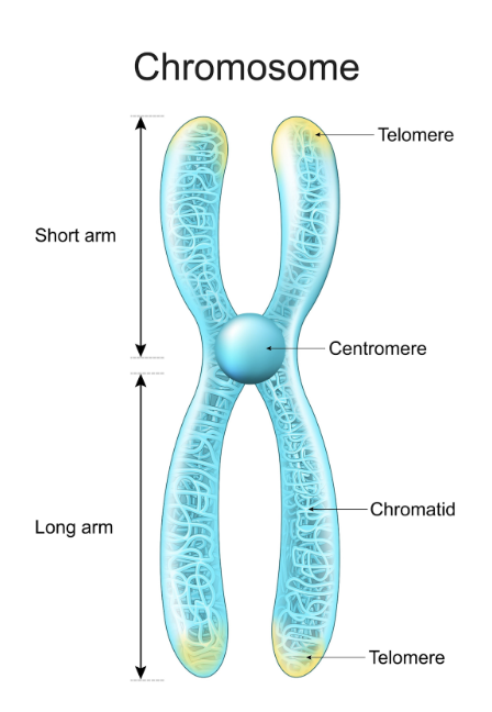
Centromere
The specialized DNA region that joins two sister chromatids together. It is also the attachment point for spindle fibres during cell division.
Centrioles
Cylindrical structures (found in pairs in many animal cells) that move to opposite poles of the cell during prophase to help organize the spindle fibres.
Spindle Fibres (or Spindle)
A network of microtubules that forms during mitosis.
These fibres attach to the centromeres and act like "cables" to pull the chromatids apart to opposite ends of the cell.
Equator (Metaphase Plate)
The imaginary line or plane in the middle of the cell where chromosomes line up during metaphase before being pulled apart.
Cytokinesis
The division of the cytoplasm.
This is distinct from mitosis (which is nuclear division) and results in the physical separation into two daughter cells.
Cleavage Furrow
The "pinch" or indentation that forms in the plasma membrane of an animal cell during cytokinesis as it pulls together to split the cell.
Cell Plate
The structure that forms in the center of a dividing plant cell.
Because plants have a rigid cell wall, they cannot "pinch" like animal cells; instead, they build this new wall from the inside out.
Why is mitosis important?
Allows for growth, repair, replacement and asexual reproduction.
What is Apoptosis?
A carefully regulated, active process of programmed cell death.
It is genetically programmed, meaning it is orchestrated by the regulated expression of dozens of genes.
Why is Apoptosis essential for development?
Shaping organs and tissues: e.g., removing webbing between a human embryo's fingers/toes or a tadpole losing its tail.
Removing unwanted cells: Getting rid of cells that are old, no longer needed, or have reached the end of their natural life span.
Removes cells with DNA damage
How is Apoptosis triggered?
Internal: Signals from within the cell (e.g., when DNA damage is detected).
External: Signals from other cells (external to the cell).
List the steps of the Apoptotic process in order.
1. Enzymes shred the cell's DNA into thousands of fragments.
2. The nucleus is gradually dismantled.
3. The plasma membrane forms blebs.
4. The cell breaks into apoptotic bodies.
5. Phagocytes (like macrophages) engulf and clear the remains.
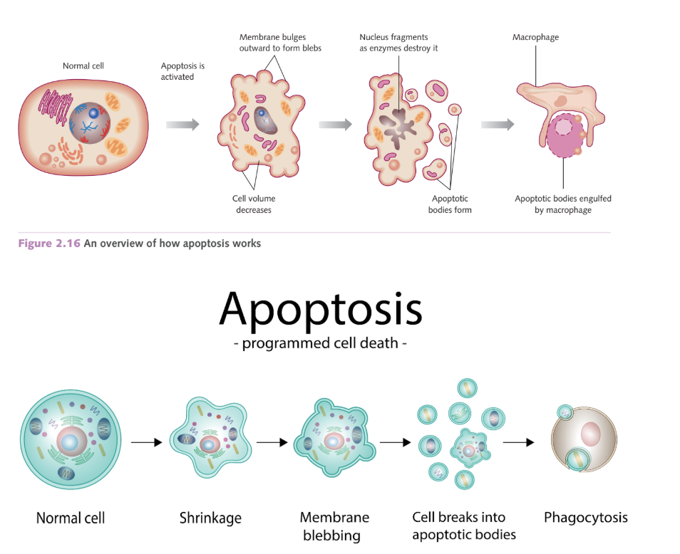
What is the difference between Apoptosis and Necrosis?
Apoptosis:
Programmed, orderly, and "clean" self-destruction.
The membrane stays intact (forming apoptotic bodies) so it doesn't harm neighbors.
Necrosis:
Accidental cell death caused by damage to the plasma membrane. It is not programmed or orderly.
How does a failure in Apoptosis lead to Cancer?
Cancer occurs when apoptosis is avoided.
If a cell has DNA damage that cannot be repaired and the apoptotic program fails to activate, the damaged cell persists and continues to divide, forming a cancerous tumour.
2 Apoptosis Pathways
Intrinsic or Mitochondrial Pathway
Extrinsic or Death Receptor Pathway
Intrinsic or Mitochondrial Pathway
Mitochondria determines how and when a damaged cell dies
Pores in mitochondria release cytochrome c in cytosol
Activates caspases
Caspase cleave cell contents
Apoptotic bodies are formed
Extrinsic pathway or Death Receptor Pathway
Cells are not stressed – healthy cells that are no longer needed.
Initiated when specific death ligands bind to transmembrane death receptors
Cleaves/cuts inactive pro-caspase 8 into active caspase 8
Then cleaves and activates caspase 3 6 7, dismantling the cell into apoptotic bodies.
Why do some tumour cells "ignore" apoptosis?
They may have changes (mutations) in their apoptosis genes, making them unable to activate the death program or making them unresponsive to the signals that tell them to die.
Blebs
Balloon-like outgrowths of the plasma membrane that occur when the membrane detaches from the underlying cytoskeleton.
Apoptotic Bodies
Small vesicles containing the contents of the dead cell, neatly encased by the plasma membrane to be cleared away.
Phagocytes (e.g., Macrophages)
"Scavenging cells" that find, engulf, and digest the apoptotic bodies to clean up the tissue.
Necrosis
Cell death resulting from external damage to the plasma membrane; unlike apoptosis, it is not a programmed or regulated process.
What are Proto-oncogenes?
Normal genes that code for proteins that stimulate cell division and cell growth, and prevent differentiation apoptosis.
If mutated or overactive, they lead to uncontrolled growth- link to cancer
What are Tumour Suppressor Genes?
Normal genes that slow down cell division, repair DNA mistakes, or tell cells when to die.
If mutated or "silenced", they can lead to a loss of control in cell growth and division, leading to cancer.
How is the "rate" of cell division maintained?
Through the balance between the activities of proto-oncogenes and tumour suppressor genes.
This ensures cells divide at a rate appropriate for their position and role in the body.
What is the p53 gene and why is it called the "guardian of the genome"?
It is a gene that activates when DNA damage or cell injury is detected. It controls two pathways:
Early stage (after M): Halts the cycle to allow for DNA repair.
Later stage (G2): Initiates apoptosis to prevent the damaged cell from replicating.
What happens if the p53 gene is mutated?
Loss of p53 protein results in:
Rapid, unchecked cell proliferation (growth).
Evasion of apoptosis (the cell won't die even when damaged).
Define "Genetic Predisposition."
An increased risk or susceptibility to cancer due to inherited mutations (like in BRCA1/2).
It does not guarantee a person will get cancer; other factors like viral infections, lifestyle (smoking), and environment play a role.
How does chemotherapy interact with apoptosis?
Many treatments focus on reinstating apoptosis (restarting the "self-destruct" program) in tumour cells.
However, cancer cells often develop resistance by producing "anti-apoptotic" proteins that act as a brake on death.
Malignant tumor (cancer)
Referring to cells that are cancerous and can spread or invade other tissues.
Benign tumor
Not harmful, slow growing
Anti-apoptotic Proteins
Proteins that prevent apoptosis; high levels of these lead to chemotherapy resistance.
What is a Mutagen?
An agent (chemical, physical, or biological) that increases the frequency of mutations in DNA.
What is a Carcinogen?
A specific type of mutagen that is known to cause cancer.
Oncogenes
Mutated proto-oncogens, which cause excessive, unregulated cell division, are "always on".
Chemical mutagens
Chemical substances that interfere with DNA (structure/sequence/replication)
Examples:
tobacco smoke, asbestos, mercury
Physical mutagens
Physical agents that cause DNA damage( all stages of the cell cycle)
Examples:
UV radiation, nuclear radiation, X-rays
Biological mutagens
Biologic agents that induce mutations
Examples:
viruses - they can physically insert their viral DNA into healthy cells, mutating the cell and disrupting its function
HIV
HPV
hepatitis B
Mutation
A change or mistake in the copying of a DNA sequence in chromosomes.
What is Differentiation?
The process by which unspecialised cells develop special characteristics and become specialised to perform specific functions.
What is a Stem Cell?
An undifferentiated cell that has the potential to both replicate (self-renew) and develop into many different kinds of cells.
Totipotent Stem Cells
Cells that can form any cell type, including the embryo itself and the supporting membranes (like the placenta).
E.g. the zygote
Pluripotent Stem Cells
Cells that can give rise to all cell types in the foetus, but cannot form the external support tissues like the placenta.
E.g. embryonic stem cells
Multipotent Stem Cells
Cells that are limited to giving rise to cells with a particular set of functions (e.g., blood stem cells making red/white cells).
The "Point of No Return"
Once a cell follows a specific developmental pathway and becomes fully differentiated,
there is usually "no turning back" to a stem cell state.
The Blastocyst
A hollow ball of cells formed ~5 days after fertilisation. It contains an inner cell mass (pluripotent) and an outer layer (forms the placenta).
Self-Renewal
The ability of a stem cell to divide and produce more stem cells of the same type
Maintains the "pool" of undifferentiated cells.
Specialisation
The structural and functional changes a cell undergoes to perform a specific job (e.g., a blood cell carrying oxygen).
Zygote (Fertilised Egg)
The initial totipotent cell formed by the fusion of sperm and egg.
Do all cells start off as stem cells after cell division?
No, not all cells start as stem cells after division.
Stem cells divide to produce more stem cells (self-renewal)
Specialized cells (like skin or muscle cells) usually divide to produce more of their own specific type
Why are stem cells important to the body?
Allows for growth and development- during fetal development these cells divide and differentiate to build every organ and system in the body
Replace cells lost through normal functioning e.g. stem cells in your bone marrow constantly produce new red blood cells to replace old ones.
Adult stem cell
A stem cell harvested from tissues such as bone marrow, that are not part of an embryo
Embryonic stem cell
A stem cell that is cultured from an embryo
Embryo
The early stage of development of an organism; in humans, from fertilisation to the end of the eighth week of pregnancy.
Foetus
The developing individual after the second month of pregnancy.