anatomy mid term
1/162
There's no tags or description
Looks like no tags are added yet.
Name | Mastery | Learn | Test | Matching | Spaced | Call with Kai |
|---|
No analytics yet
Send a link to your students to track their progress
163 Terms
Gross Anatomy
Study of large body structures
visible to naked eye
Ex. Heart, skull, foot
Systemic Anatomy
Study of an entire system within the body
organs working for a function
Ex. Cardiovascular system, Resp system
Microscopic anatomy
Study of very small structures that may not be seen by the unaided eye
Includes:
cytology: the study of individual cells
Histology: the study of tissues
Chemical Level
Most basic level
atoms (smallest units of matter) come together to form molecules
Ex. Water molecules (H2O) are formed when 2 hydrogen atoms (H) combine with one Oxygen atom (O)
Cellular Level
Individual cells
Smallest units of life
Basic unit of structure and function
Tissue Level
Cells of the same kind come together to form tissues
Defined as groups of similar cells coming together to perform a common function
Four main tissues types in the body:
1) Epithelial Tissue
2) Connective Tissue
3) Muscle Tissue
4) Nervous Tissue
Organ Level
Organs are composed of two or more tissues
Ex: bladder
organ composed of smooth muscle allows expansion in order to hold urine
Nervous tissue: to control urination
Epithelial Tissue:
Connective Tissue: reinforces the walls of the bladder
Organ system
A number of related organs working together to accomplish a common function
Ex.
Circulatory system: heart, arteries, veins all work together to circulate blood providing cells with oxygen and nutrients
Organism
All of the organ systems of the body come together performing necessary functions for life
ex. Respiratory system, cardiovascular system, digestive system, nervous system, reproductive system all come together to form a functional living human being
Matter
Anything that has mass and occupies space
Stuff the fills the universe
Composed of elements
Elements are made up of atoms
Atoms
The smallest unit of an element
atoms that make up a particular element are identical
Ex. All atoms that are found in the element oxygen are the exact same
Atomic structure
all atoms have protons, neutrons, and electrons
The protons (+) and the neutrons are found in the nucleus of the atom
The electrons (-) are found outside he nucleus
If the atom is neutral the number of protons=number of electrons
Total charge of the atom is equal to 0
An atom that gains or looses electrons is no longer neutral and is called an ion
Water
Most living cells are 60-80% water
Classified as a polar molecule
Polar molecules are defined as having a partial positive and a partial negative charge
Key functions of water include
Ability to act as a solvent
Ex. Salt or sugar can be dissolved in water
In chemical reactions
Maintaining body temperature
Lubricates joints and fills area around organs
Acids
Dissociate when placed in water
release H+ ions
Ex. HCl → H+ + Cl-
The more HCl that is added to water, the more H+ will be present following dissociation
The greater the amount (concentration) of H+ in the solution, the lower the pH of the solution
Lower pH means a greater acidity
Bases
Disssociate in water also
Release OH- ions
Ex. NaOH → Na+ + OH-
The OH- that is generated from the dissociation of NaOH binds to H+
The greater the amount (concentration) of OH- in the solution, the greater the pH of the solution
Higher pH means that the solution is more basic
Neutral pH
7
the amount of H+ in solution is equal to the OH- in solution
Acidic pH
0-6
0 is most ___ and 6 is the least
Basic pH
8-14
8 is the least ___ and 14 is the most
Carbohydrates
Sugars and starches
Contain carbon, hydrogen, and oxygen in a 1:2:1 ratio
Classified as either monosaccharides, disaccharides or polysaccharides
Monosaccharides
One sugar
Basic building blocks of carbs
Ex. Glucose, fructose
Disaccharides
Two monosaccharides covalently bound to one another
Ex. Sucrose
Sucrose is what we know as table sugar
Polysaccharides
Many monosaccharides bond together covalently
Ex. Glycogen (animals) and starch (plants), both long chains of glucose
Proteins
Made up of amino acids (20 different kinds of amino acids)
Contain carbon, oxygen, hydrogen and nitrogen
Have an enormous variety of different functions
Structural Proteins
Collagen (in skin) actin (muscles)
Cell function Proteins
Hemoglobin (oxygen transport), cytokines (cell to cell messengers)
Peptides
Amino acids bound together form ___
Dipeptides:
2 amino acids bound together
Polypeptides
Many amino acids bound together
One or more polypeptides folded into a characteristic shape
Lipids
Fancy name for fats
Most common lipids found in the human body are glycerides
obtained through diet
Glyceride is made of glycerol and fatty acids
Monoglycerides:
Glycerol and one fatty acid
Diglycerides:
Glycerol and two fatty acids
Triglycerides:
Glycerol and three fatty acids
Phospholipids
Composed of a diglyceride and a phosphate group
Two fatty acids Triglycerides tails (attached to the glycerol) are non-polar
non-polar is also called hydrophobic and means not water soluble
The phosphate ‘head’ group (attached to the other side of the glycerol) is polar
polar is also called hydrophilic and means ‘water loving’ or water soluble
Steroids
Produced from cholesterol
Ex. Includes the hormones estrogen and testosterone
Nucleic Acids
Composed of nucleotides
Made up of carbon, nitrogen, oxygen, hydrogen and phosphorous
Ex: DNA and RNA
Nucleotides
Basic building blocks of DNA and RNA composed of:
a phosphate group
A monosaccharide
Ribose sugar in RNA
Deoxyribose sugar in DNA
An organic base
DNA
Double stranded helix
Deoxyribose sugar In each nucleotide
Composed of Adenine, Guanine, Cytosine, and Thymine
Adenine base pairs with thymine forming 2 hydrogen bonds
Cytosine base pairs with guanine forming 3 hydrogen bonds
The main component of chromosomes:
Encodes genes
Used to produce RNA
RNA
A single stranded molecule
Ribose sugar in each nucleotide
Composed of Adenine, Guanine, Cytosine, and Uracil
adenine base pairs with uracil forming 2 hydrogen bonds
Cytosine base pairs with guanine forming 3 hydrogen bonds
Made from DNA
Used to produce protein
ATP
Adenosine tri-phosphate
Made of ribose sugar, adenine and three phosphate groups
Bonds between each of the phosphate groups are very high energy
Breaking these bonds releases energy so that it may be used to power processes within the cell
ATP becomes ADP when one of the bonds is broke removing a phosphate group
ADP becomes AMP when another phosphate is removed
Phospholipid Bilayer
A continuous layer: forms the bulk of the membrane structure
Consists of polar phosphate + head group that faces the inside of the cell and he outside of the cell → polar groups have favourable interactions with water
Also consists of two non-polar tails that are attached to the phosphate head group
point toward the interior of the plasma membrane
Protected from water because the are non-polar
Cholesterol is also present in the plasma membrane of humans
stabilizes the membrane especially as temperature increases/decreases
Membrane proteins
transmembrane proteins are embedded in the plasma membrane and pass all the way through the membrane
Peripheral membrane proteins are attached to either the cytoplasmic surface of the membrane or the extracellular surface
Membrane proteins function as channels, receptors, and enzymes among other things
Membrane Carbohydrates
Only located on the outer surface of the membrane
functions in cell to cell recognition
Attached to either:
Protein: called glycoprotein
Lipid: called glycolipid
Microvilli
Folds of the plasma membrane
Serve to increase surface area
Especially important in cells where nutrient absorption occurs
Cytosol
Semi-transparent, viscous fluid
Bathes the organelles inside of the cell
Water is the main component
Also contains:
Dissolved ions→Na+, Cl-,K+,Ca+
Suspended carbohydrates ad lipids
Melanin granules in certain cells
Ribosomes
Non-membranous
Composed of rRNA and protein
Responsible for protein synthesis
Can be located free in the cell or attached to the endoplasmic reticulum
Centrosomes
Are a region located near to the nucleus
Contains a granular looking matrix and 2 centrioles
Centrioles
Small, cylindrically shaped organelles
Composed of microtubules
Located perpendicular to one another
Function to direct the movement of chromosomes during cell division
Cytoskeleton
Determines/holds cell shape
Used to anchor organelles in place
Used to move materials throughout the cell
Composed of 3 types of protein rods located in the cytosol
Microfilments
Composed of actin
Thinnest component of the cytoskeleton
Important for muscle contraction, cell movement, maintenance of cell shape
Intermediate filaments
Composition differs based on tissue type
Works to support the cytoplasm
Microtubles
Composed of tubulin
Hollow tubes
Largest component of the cytoskeleton
Structural function to anchor and move organelles
Compromises centrioles, flagella, cilia, and the spindle apparatus used during cell division
Mitochondria
surrounded by a double membrane
Power house of the cell
Works to produce ATP → primary energy currency of the cell
Contains DNA, RNA, protein, and water
Rough Endoplasmic Reticulum
Appears rough because it has ribosomes attached to its surface
Ribosomes work to synthesize certain proteins here
Smooth Endoplasmic Reticulum
Appears smooth bc no ribosomes found
Functions to:
store calcium
Detoxify substances (drugs, alcohol)
Synthesizes lipids
Golgi Apparatus
Stacks of membranous disks
Function to modify newly synthesized proteins
Adds carbohydrate groups to these proteins
Form glycoproteins
Packages protein into vesicles that then carry protein to:
Cell membrane
Lysosomes
Be secreted to the extracellular environment
Nucleus
Control centre of the cell
surrounded by a double membrane that contains pores
The outermost membrane is continuous with the rough ER
Typically one nucleus per cell
Some cells contain multiple nuclei
The nucleus is located inside of the nucleus
Consists of DNA, RNA, and proteins
It is not separated from the nucleus by a membrane
Site of ribosome assembly
Chromosomes
Composed of DNA and Histone proteins
Found in 2 forms in the nucleus
thread-like and dispersed:
Will be found this way in cells that are not actively dividing
Coiled/Condensed:
Individually visible chromosomes
Thicker than thread-like chromosome structure
Will be found this way in a cell that is actively dividing
Interphase
Duplicates cell content to make enough for two cells
Mitotic Phase
Consists of:
Mitosis
Cytokinesis
G1 Phase
Lasts 8-10 hours
Period of intense growth and metabolism
At the end of G1 centrosomes replicate
Any cell that will not divide again will remain in G1 phase
Ex. Include neurons and muscle cells
Referred to as remaining in the G0 phase
Cells that are destined to divide will enter into S phase
S Phase
Lasts 6-8 hours
During this stage the DNA is replicated making identical copies of each chromosome
Replicates are called sister chromatids → attached to one another by the centromere
Ensures that each daughter cell will receive a complete set of chromosomes
Kinetochore protein attaches to each centromere
forms the kinetochore
A protein/DNA complex that is attached to the centromere of one chromosome
Occurs before mitosis and meiosis
G2 Phase
Lasts 4-6 hours
The final phase of interphase before the cells begins mitosis
Period of growth and metabolism
Enzymes and other proteins needed for cell division are produced
Each chromosome now has 2 sister chromatids attached to one another at then centromere
Prophase
Chromatin condenses and becomes visible
Nuclear membrane disappears
Nucleoli disappear
Centrosomes move to opposite poles of the cell
The spindle apparatus begins to form at the centromere
Kinetochore proteins attach to spindle microtubules → called kinetochore microtubules
The spindle apparatus moves the chromosomes to the equator of the cell
Metaphase
Each chromosome consists of 2 sister chromatids line up at the cell equator → called the metaphase plate
46 chromosomes in a straight line down the centre of the cell
Anaphase
The Kinetochores separate from one another
pulls the sister chromatids apart from one another
46 sister chromatids then migrate to each pole
Cytokinesis begins
Telophase
the spindle apparatus disassembles
Chromosomes uncoil forming chromatin once again
Nucleoli and the nuclear membrane reappear
Cytokinesis is completed
Mitosis ends and the cell enters G1 of interphase
There are now two identical daughter cells
Meiosis 1
Separates homologous pairs reduces the cell from diploid to haploid
1×46 duplicated chromosomes → 2×23 duplicated chromosomes
Meiosis 2
separates sister chromatids from one another
2 cells with 23 chromosomes each divide giving 4 cells with 23 chromosomes each
The stages are identical to the stages of mitosis but in a haploid cell
Prophase 1
Most complex phase in all of meiosis
Homologous pairs match up side → called synapsis
allows them to separate into two different daughter cells
Four chromosomes arranged in a line
2 sister chromatids from one member of the homologous pair and 2 sister chromatids from the other → called crossing over
Metaphase 1
Tetrads align on the metaphase plate
Sister chromatids remain attached at the centromere
Microtubules are attached to the kinetochore
Anaphase 1
Homologous pairs separate from one another
Each pair moves to opposite poles of the cell
sister chromatids still remain attached at the centromere
Telophase 1
Chromosomes arrive at the poles of the cell
Each pole of the cell now has haploid chromosome set
Sister chromatids still remain attached at the centromere
Cytokinesis 1
Overlaps with telophase 1
Forms two haploid daughter cells with two sister chromatids per chromosome
Fertilization
Haploid sperm and haploid oocyte come into contact with one another forming a diploid zygote
takes approximately 24 hours to complete
Pre-embryonic Development
Takes place in the first two weeks following fertilization
Series of developmental occurrences leading up to the zygote becoming an embryo
The diploid zygote begins as a single cell and divides by mitosis to produce many cells → cleavage divisions
these divisions increase the number of cells, producing a solid ball of 16-32 cell called a morula
Each cell is called a blastomere
The overall size of the morula is the same as the zygote but instead of one cell, there are many small cells
Gestation
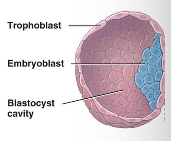
As the morula divides further, cells rearrange themselves and a blastocyst is produced
the blastocyst has a fluid filled cavity called a blastocoele (blastocyst cavity)
Forms ~5 days post-fertilization
Composed of:
Trophoblast cells that surround the blastocyst and eventually become the chorion
Provides nourishment to the developing embryo
An embryoblast which will become the embryo
Implantation
the attachment of the blastocyst to the endometrium of the uterus
occurs 5-7 days post-fertilization
The embryoblast develops into the embryonic disk following ____
The embryonic disk consists of the:
Epiblast layer
Hypoblast Layer
Epiblast Layer
Part if the embryonic disk
will give rise to the fetus
Hypoblast layer
Part of the embryonic disk
will give rise to the yolk sac
Ectoderm
Will go on to form the nervous system and the epidermal layer of the skin
Develops from Epiblast layer
Mesoderm
Will go on to form the muscle, bone, blood vessels and the dermis
Developed from the Epiblast layer
Endoderm
Will go on to form the epithelial lining of he digestive tract, respiratory tract, urinary tracts, reproductive tract and the associated glands
Embryonic Development
Occurs from week 3 to week 8 post fertilization
Epiblast layer develops into 3 germ layers
From week 4 to 8 post-fertilization
all major organ systems have completely developed
The heart begins to beat
The brain begins to develop
The limb buds begin to differentiate
Amnion
Embryonic Membrane
forms from Epiblast layer
A fluid filled cavity that acts to surround and cushion the developing embryo and fetus from bumps and other disturbances
Yolk Sac
Embryonic Membrane
forms from the Hypoblast layer
Produces the early blood cells and the germ cells
Chorion
Embryonic Membrane
forms from trophoblast cells
Becomes the fetal portion of the placenta
Surrounds all of the embryonic membranes
Allantois
Embryonic Membrane
an out pocketing of the yolk sac
Goes on to form the umbilical cord and the urinary bladder
Fetal Development
Occurs from the 9th week to the 40th week
referred to as the fetal period
period of growth and maturation of organs
Tight Junctions
Membrane junction
Protein molecules in the cell membrane fuse together
Serve to prevent substances from passing in between cells
Desmosomes
Membrane junction
Loose attachments
Use linker proteins to join adjacent cells
Gap Junction
Membrane junction
Protein channels that connect adjacent cells
Allow direct communication between cells
Allow substances to pass from the inside of one cell to the inside of another
Extremely important in smooth muscle and cardiac muscle cells
Epithelial Tissue
Found on all of the body surfaces and lines f the body cavities
An a vascular tissue → lacks blood vessels
Cells that form epithelial tissue have one free membrane
sit on top of a basement membrane
Held together by tight junctions
Reproduce via Mitotic division
Glands located here are formed from glandular epithelium
Ex. Salivary glands
The major tissue of any glands is epithelial tissue and the sub-type is glandular epithelium
Classified based on:
he number of cell layers and the shape of the epithelial cell
Simple Epithelium
A single cell layers with one free surface
Sits atop a basement membrane
Stratified epithelium
Several cell layers with one free surface
The basal/bottom-most layer sits atop a basement membrane
Pseudostratified epithelium
Appears as more than one layer but all cells really do sit on top of the same basement membrane Held together→ a single cell layer
Cuboidal
Cube-shaped cells
specialized for secretion and absorption
Columnar
Column-shaped cells
Specialized for secretion and absorption
Squamous
Irregularly shaped, scale-like cells
Specialized for secretion and absorption
Transitional
Stratified cell layer but the appearance varies with stretching
ex: cuboidal cells in the bladder appear columnar when the bladder is stretched
Exocrine glands
Secreted products onto a surface or into a cavity
Either single-celled or multicellular
single-celled
Ex. Goblet cells
Secrete mucous into a cavity
In digestive, urinary, reproductive and respiratory tracts
Multi-cellular
Have ducts→passageways
Secretions enter ducts
Sudorifierous glands produces sweat
Sebaceous glands produce sebum (oil)
Salivary glands produce saliva
Endocrine glands
Ductless
Secretions are called hormones
Release hormones directly into blood
Ex. Thyroid gland secretes thyroid hormone
Epithelial Function
Protection
stratified squamous epithelium
Skin provides a barrier that keeps microorganisms out of the body
Secretion
glandular epithelium
Secretion of lubricants, sweat, etc
Control of Permeability
simple epithelium
Found at sites where exchange of material occurs
Ex. Absorption of digested nutrients
Kidney, intestine, capillaries
Connective Tissue cells include
Blasts: form and secrete the matrix → example: osteoblasts
Cytes: maintain the matrix→ ex. Osteocytes
Clasts: breakdown the matrix → ex. Osteoclasts
*osteo indicates that these are cells in the bone
Matrix
Extracellular: surrrounds the cells of the connective tissue
Provides the connective tissue with its characteristics
The matrix has 3 primary components:
Protein fibers:
Collagen protein provides strength
Elastin protein provides stretch and recoil
Ground substances:
Unstructured material located between the cells
Contains fibers
Ex. Chondroitin sulfate And hyaluronic acid
Water
Primary Characteristics of connective tissue
Highly vascular with some exceptions
Cartilage is an avascular connective tissue
A lot of the extracellular matrix
Keeps the cells far apart from one another
Primary function is to support and connect the tissues in the body