L10- Protein Homeostasis 2
1/50
There's no tags or description
Looks like no tags are added yet.
Name | Mastery | Learn | Test | Matching | Spaced | Call with Kai |
|---|
No analytics yet
Send a link to your students to track their progress
51 Terms
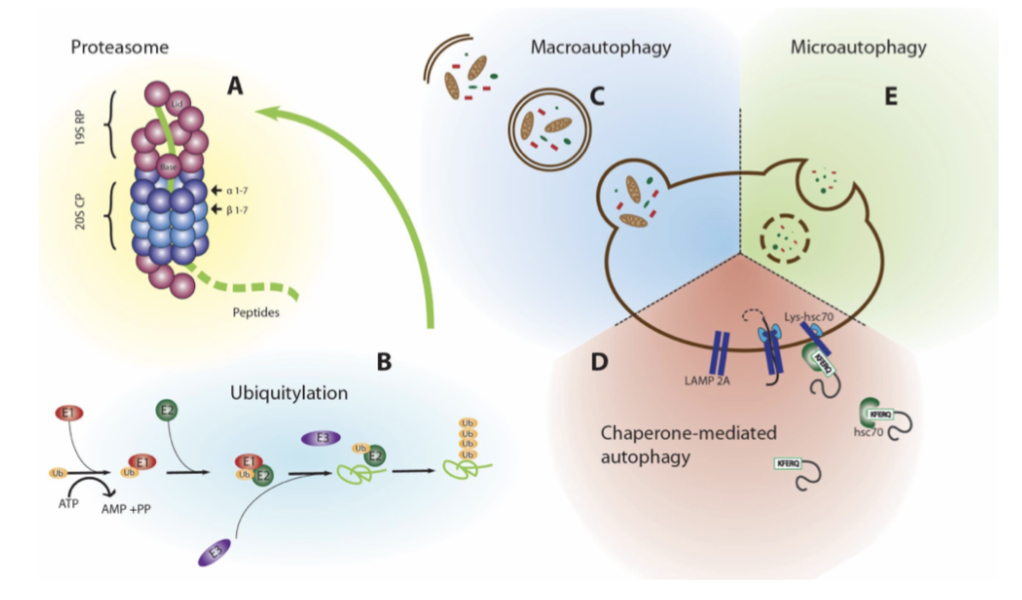
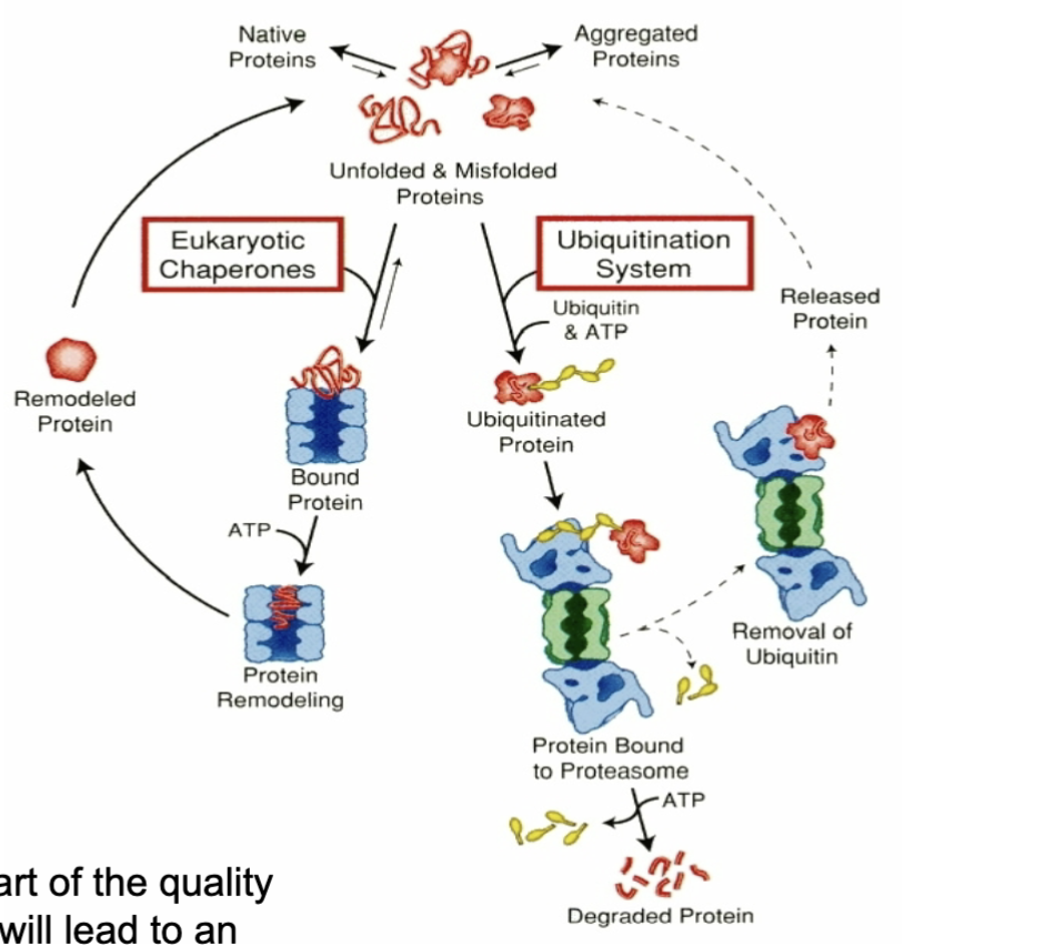
what are the 2 main protein degradation pathways
Proteasomal degradation and ubiquitination
lysosome
what are proteasomes
Proteasomes are large protein complexes- not organelles, as doesn't have a membrane
Located in the nucleus and cytoplasm- Possibly proteasomal subunits in the mitochondria
Work by proteolysis – a chemical reaction that breaks peptide bonds.
Yields peptides about 7-8 amino acids long, which are then further degraded into amino acids.
Regulates concentration of particular proteins and removes misfolded proteins.
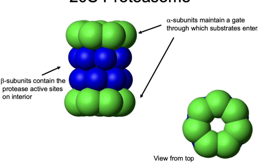
what is the structure of the 20S proteasome
Like a barrel with a pore
Enzymatic activity is localised in the centre
Protein disassembled, unfolded and fed through
alpha-subunits maintain a gate through which substrates enter
beta-subunits contain the protease active sites on interior
can proteasomes degrade aggregates
no
Cant degrade aggregated proteins but can prevent proteins from being aggregated by removing those with a tendency to be misfolded
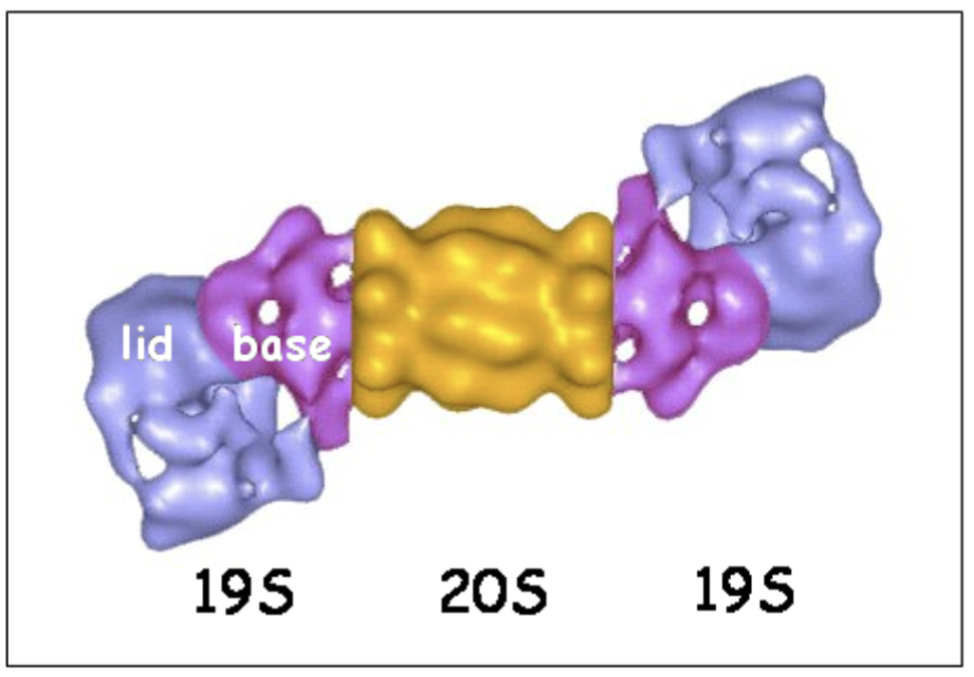
what is the structure of the 26S proteasome
alpha-subunits of 20S proteasome bind to the 19S regulatory cap.
The 19S cap contains ATPase active sites and ubiquitin binding sites.
Substrates must be tagged by ubiquitin to be recognised
what allows for the proteasome to detect ubiquitinated proteins
The Proteasome contains regulatory ends/subunits that provide specificity for recognising ubiquitinated proteins.
These subunits can use ATP to bind ubiquitin chains attached to target proteins.
The proteasome recognises ubiquitinated proteins, unfolds them using ATP, and feeds them through a central pore.
The protein is then degraded into peptides inside the proteasome.
what does S refer to (eg in 26S proteasome)
S refers to the Svedberg sedimentation coefficient which is used to characterise the behaviour of a particle type in ultracentrifugation.
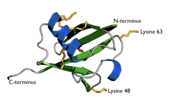
what is ubiquitin
Small regulatory protein (8.5kDa)
Single ubiquitin molecules or chains of ubiquitin are added to substrate proteins
Dependent in how chain is built- the signal will mean something different to the machinery
Ubiquitination (also called Ubiquitylation)
what is the effect of ubiquitination on the substrate
Degradation
Change in cellular location
Change in protein activity
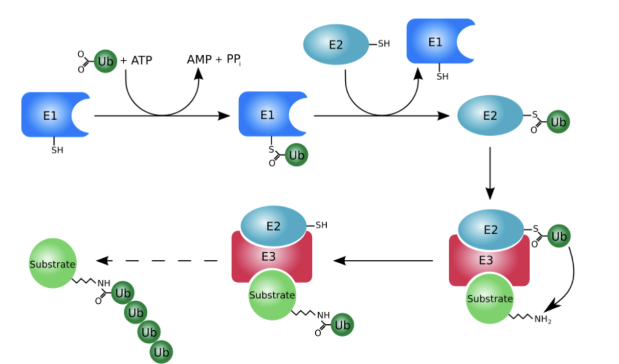
what are the 3 ubiquitin proteins
Ubiquitin-activating enzyme (E1 enzyme)
Ubiquitin-conjugating enzymes (E2 enzymes)
Ubiquitin ligases (E3 enzymes)
what is Ubiquitin-activating enzyme (E1 enzyme)
Only one type in mammalian cells
It activates ubiquitin (ATP-dependent reaction)
what are Ubiquitin-conjugating enzymes (E2 enzymes)
Several enzymes
They bind to activated ubiquitin
what are Ubiquitin ligases (E3 enzymes)
Many different enzymes e.g. Mdm2
Each enzyme has specific substrate proteins
They interact with E2 enzymes (E2 pick it up, relay it and pass it onto E3 )
how does ubiquitination occur
E1 uses energy from ATP to attach ubiquitin and make bond with sulphide group of cysteine
As it interacts with E2, it passes the Ub onto cysteine of E2, reaction continues to E3
Ub passed on via isopeptide bond- using lysine on protein to make initial single Ub chain
Can be done with single Ub, or straight away with chain of Ub
what are deubiquitinating enzymes (DUBs)
Large group of proteases that cleave ubiquitin molecules
• The majority are cysteine proteases e.g:
Ubiquitin-specific protease family (USPs)
Ubiquitin C-terminal hydrolases (UCHs)
what are the 2 functions of DUBs
Reverse the action of ubiquitination- remove chain of ubiquitin or chop chains into individual ubiquitin
Recycling ubiquitin back into cell
how does ubiquitination change with age
Ubiquitin levels and ubiquitin enzymes remain relatively stable with age.
However, ubiquitinated proteins accumulate with age, suggesting impaired proteasomal degradation.
This may reduce the pool of free available ubiquitin because ubiquitin becomes trapped on undegraded proteins.
how does the proteasome change with age
Some proteasomal components may decline with age
Proteasome activity decreases with age in many different tissues
Indirect reference to potentially impaired proteosome (with age)
If ubiquitin all attached to undegraded proteins- reduced ability to ubiquitinate other proteins
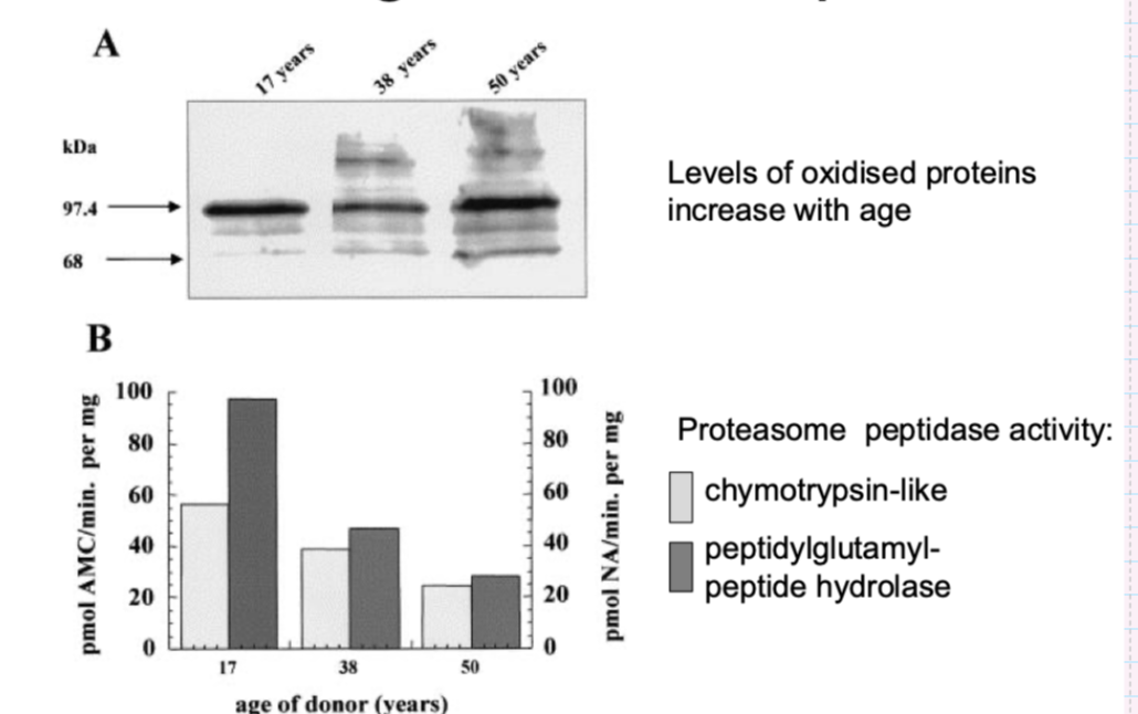
what is the evidence that proteasome activity declines with age in human epidermis
levels of oxidised proteins increased with age
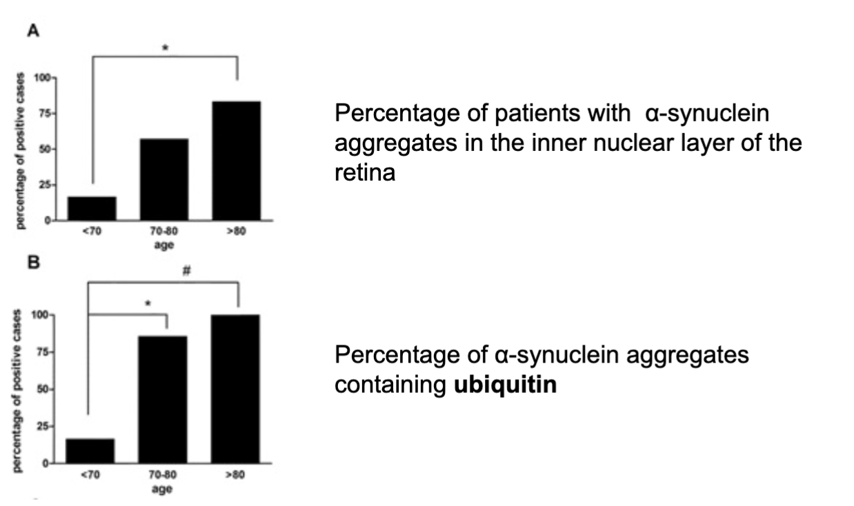
what happens to aggregated proteins containing ubiquitin in the ageing retina
increased % of patients with alpha-synuclein aggregates and those containing ubiquitin
Implies that proteasomal degradation is less efficient with age
Therefore see accumulation of proteins and protein aggregates
In peripheral tissues, skin and organs
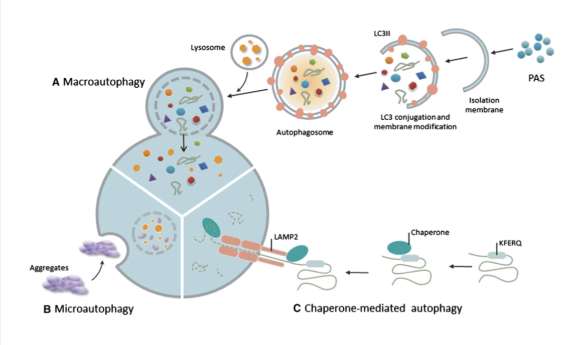
what are the 3 types of autophagy
macroautophagy
microautophagy
chaperone-mediated autophagy
what is macroautophagy
Intracellularly- Lysosome serves to degrade proteins by autophagy
Can degrade proteins carbs, lipids, nucleotides etc
The lysosome is surrounded by a single membrane.
Cellular material is enclosed within a double-membrane vesicle called an autophagosome.
The autophagosome fuses with the lysosome to deliver its cargo for degradation.
This allows cytoplasmic material to become accessible to lysosomal enzymes and be recycled by the cell.

what is microautophagy
Allows proteins to be taken in by invagination of membrane lysosome and then be degraded once budded in

what is chaperone-mediated autophagy
chaperone system- allows proteins to be translocated to lysosomal membrane
what is the role of macroautophagy and nutrient response pwathways
Nutrient sensing- everything in cell needs to be sensed- eg how much protein , amino acid etc to know if functions can occur like translation
Converge on mTOR complex – 2 key functions (autophagy or quality control)
autophagy-
Promote anabolic pathway- activated when enough nutrients
starvation- switched off, activate catabolic pathway
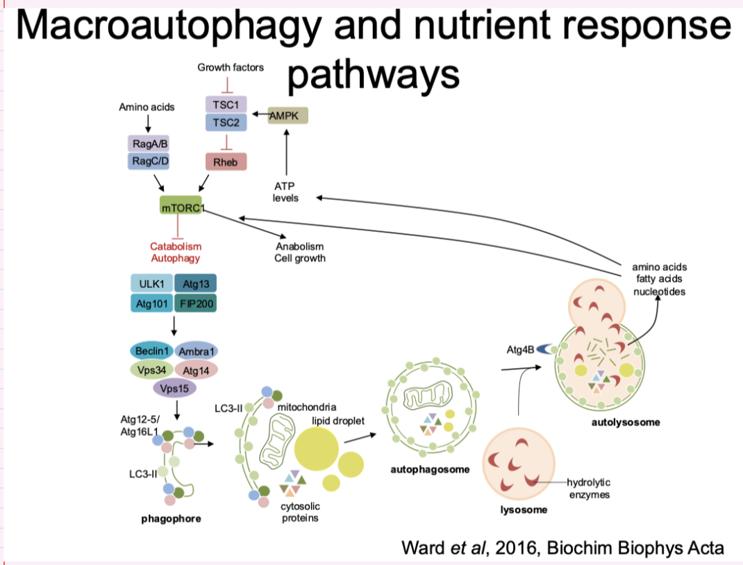
how is the autophagosome organelle unlike any other vesicle in the cell
Formed de novo , borrows lipiss from ER
like a flattened balloon that stretches- double membrane
When fuses with lysosome, second membrane can fuse and pop and lysozymes can degrade the contents
Discovered in budding yeast
what are the 2 key functions of macroautophagy
Converge on mTOR complex – 2 key functions
autophagy- to feed cell from inside- provide nutrients- cascade with many proteins in process
quality control
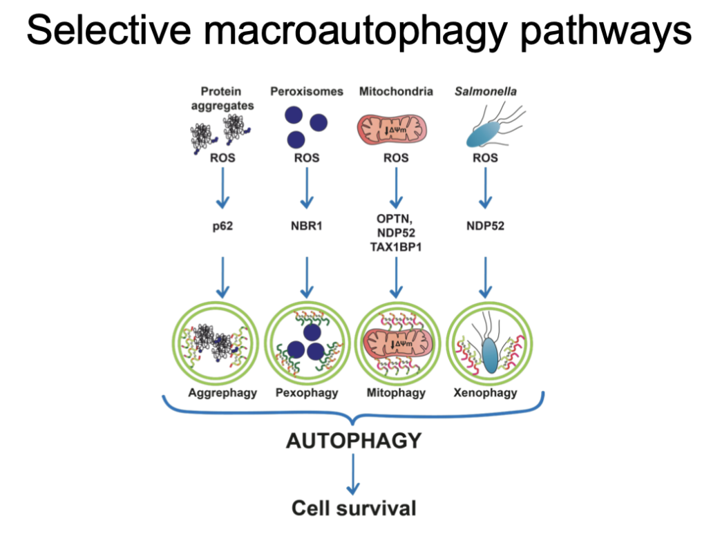
how does microautophagy ensure quality control
through selective macroautophagy pathways
Can pick up things that are undesirable
Broken or dangerous things- difunctional chromosomes and mitochondria etc
Can trigger apoptosis
what is the relationship between macroautophagy and ageing
Hallmark of ageing
Fluorescence- lysosomes with undegraded material- though to be mitochondrial components
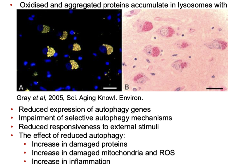
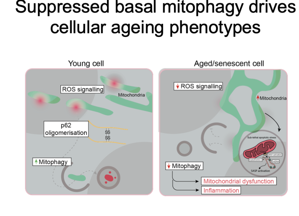
how does suppressed basal mitophagy drive cellular ageing phenotypes
Select autophagy pathways may be effected
Signal less with age so picked up less by autophagy machinery
See accumulation of mitochondria in ageing, but are less active
Don’t get moved when damage and start leaking intracellular mitochondrial components sending a signal that could lead to innate immune system response creating an inflammatory effect in cell and neighbouring cells
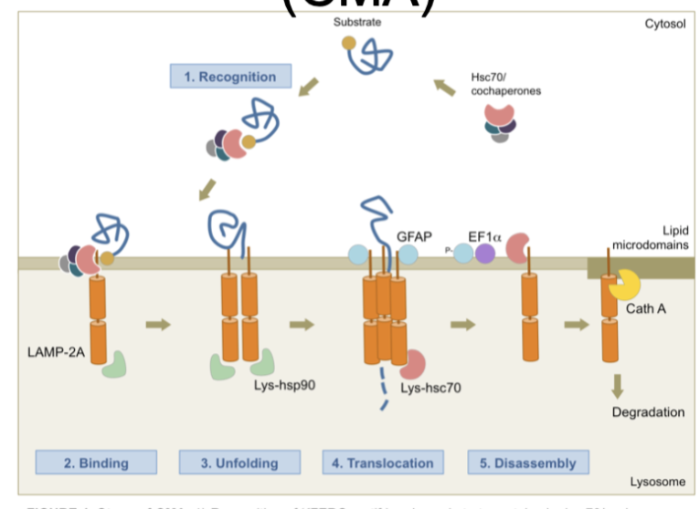
how does chaperone-mediated autophagy (CMA) change with ageing
Process that becomes less efficient with ageing
Uses chaperones, help assist degradation
Signal means exposure of specific motif, so when unfolded motif can be recognised at lysosomal surface
Protein passed to receptor lamp-2a
Pore formed and can pass through
Chaperones help form pore and disassemble it
Reduced in ageing due to reduction of lamp-2a itself
what happens to LAMP-2A in CMA with age
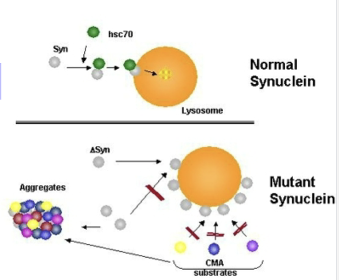
Decline in LAMP-2A receptors with age (due to increase in its degradation)- System also susceptible to clogging - Misfolded proteins can block the pore
Mutant proteins bind to LAMP-2A receptors and block them e.g.
α-synuclein, UCHL1 (Parkinson’s disease)
Tau (Alzheimer’s disease)
Insufficient degradation can be caused directly by the misfolded proteins
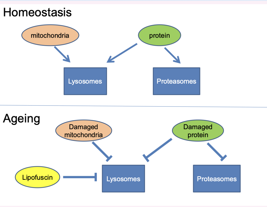
what happens regardless of if lysosomal or proteasome pathways become inhibited first
Both lysosomal and proteasomal pathways decline with age
• Regardless of which system becomes inhibited first:
Lysosomes (from their burden of defective mitochondria and lipofuscin) or
Proteasomes (from their burden of damaged proteins)
Both systems may become overwhelmed leading to a loss in protein homeostasis.
how is protein homeostasis altered with age
normal- Proteins degraded by both systems, mitochondria only by lysosomes
aged- Not degraded efficiently- causing impairment of homeostasis
what are some theories of disturbed protein homeostasis in ageing
Chaperone overload
Quality control of proteins depends on cross-talk between chaperones and proteolytic pathways. Failure of any part of the system leads to aggregation
Garbage catastrophe theory of ageing
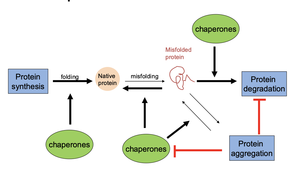
what is the protein triage model for quality control
Failure in any part of the quality control system will lead to an increase in aggregation
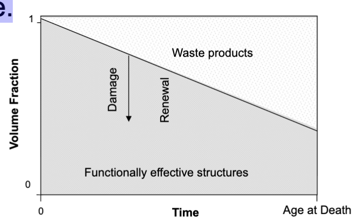
what is the garbage catastrophe theory of ageing
More generic take on the theory that ageing is a build up of damage
Imbalance between oxidative damage and renewal of biological structures may lead to a progressive loss of functionally effective elements and accumulation of waste products with age.
but- Don’t know if it’s a result of ageing or a cause of ageing still
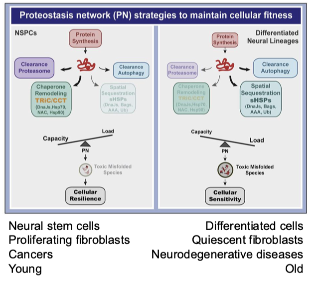
How does cellular plasticity and protein homeostasis change with ageing or cell state?
Young/healthy cells:
High capacity > load
Rely on fast, flexible mechanisms (e.g. chaperones)
Less need for last-minute/emergency responses
Older or altered-state cells (e.g. differentiated, quiescent):
Reduced plasticity and efficiency
Loss of rapid-response mechanisms
Greater reliance on slower systems (e.g. autophagy)
More “last-ditch” responses to maintain homeostasis
what is the effect of protein aggregation on the proteostasis network
If not degraded/folded
Potentially may be assembled into aggregates
Happens increasingly in age related diseases
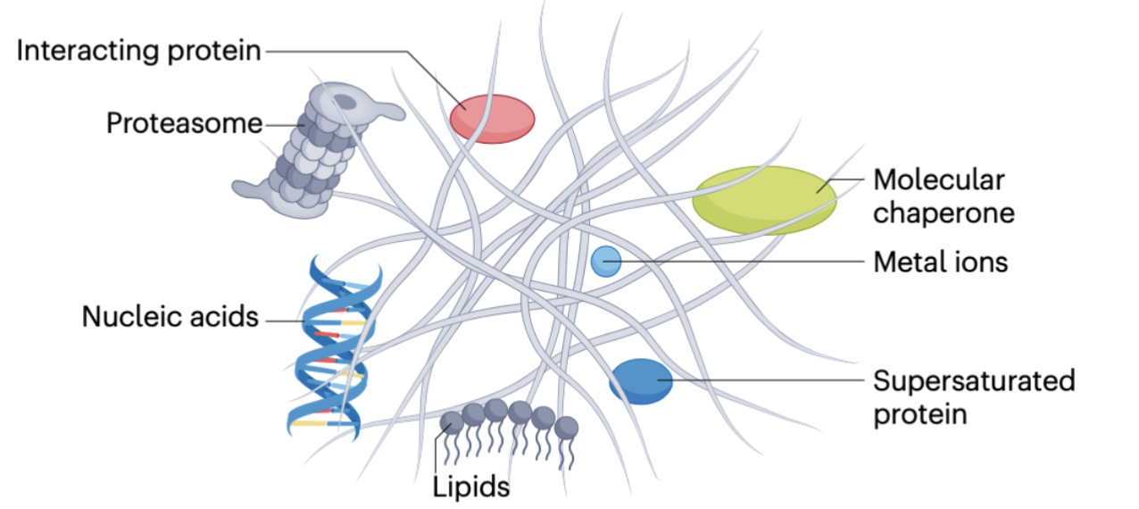
what is the definition of a protein aggregate
Change in secondary/tertiary structure
• Poor solubility in aqueous or detergent solvents
• Aberrant sub-cellular or extracellular localisation
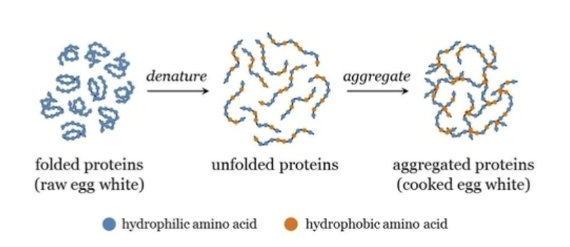
what are the features of a protein aggregate
Protein misfolds and becomes clumped together
Preceded by change in structure of protein
Can be inside or outtside cell
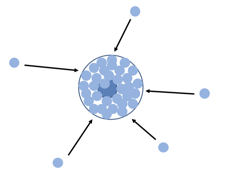
Triggered by hydrophobic residue- proteins likely to stick to each other
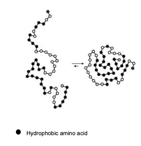
what happens to hydrophobic regions of aggregates
they are internalised
can all proteins aggregate
Not all proteins aggregate prone and some more likely than others
Some basically pre destined to become aggregates
what does the propensity of a protein to aggregate depend on
The secondary structure
Stability of the tertiary structure
Degree of disorder (intrinsically disordered proteins)
how are aggregates formed
Once something is misfolded, causes aggregation of other things
what is chaperones cannot remove aggregates
If process is inefficient, cause inclusion- hallmark of various diseases
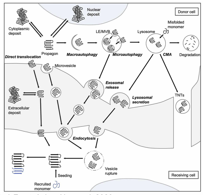
how are misfolded proteins propagated
aggregates can be transported around
Can toxify cell form within but can also spread
what is the aggresome
Coalescence of inclusion bodies by active retrograde transport of misfolded proteins along microtubules
Protective vs damaging function
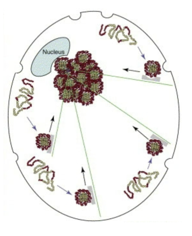
what is the function of the aggresome
Formation of aggresome within the cell is a protective mechanism
Try to contain aggregates together
Near nucleus as aggregates travel along microtubules
Forms a depot of unwanted things
Aggresomes are something that ultimately kills the cells
Can become so big that they trap organelles, can disrupt processes of the cell
Maybe suffocate the cell from inside
describe the sequestration of cellular components with aggregates
both loss-of-function phenotype and gain-of-function toxicity can contribute to the detrimental effect of protein aggregates on cell function
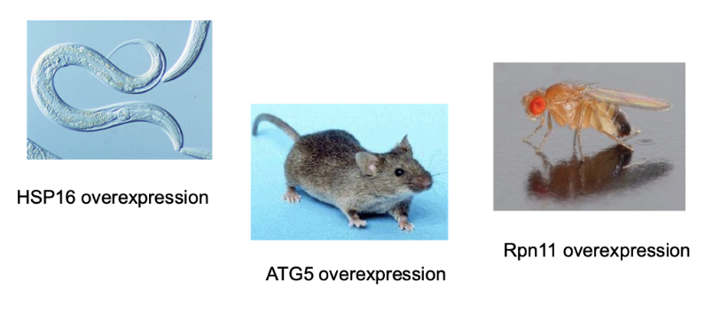
what are some interventions that improve proteostasis and extend lifespan
If we upregulating selectively and meaningly
Could be selective to the degradation fo age related things
HSP16 overexpression in c. elegans
ATG5 overexpression in mice
Rpn11 overexpression in drosophila