07/08 Anatomy and Physiology of the Respiratory System Review
1/35
There's no tags or description
Looks like no tags are added yet.
Name | Mastery | Learn | Test | Matching | Spaced | Call with Kai |
|---|
No analytics yet
Send a link to your students to track their progress
36 Terms
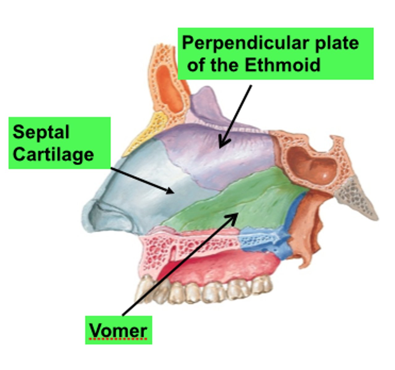
Bones of the Nasal cavity
Nasal bones, vomer, ethmoid bone with perpendicular plate, sphenoid, maxilla hard palate, and palatine bone.
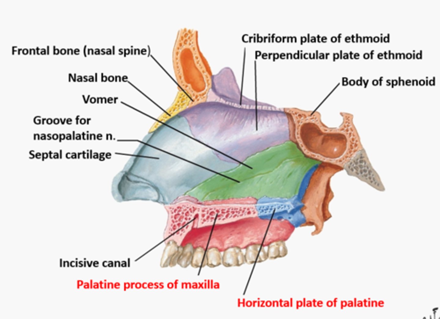
Nasal Septum
is made up of Hyaline cartilage
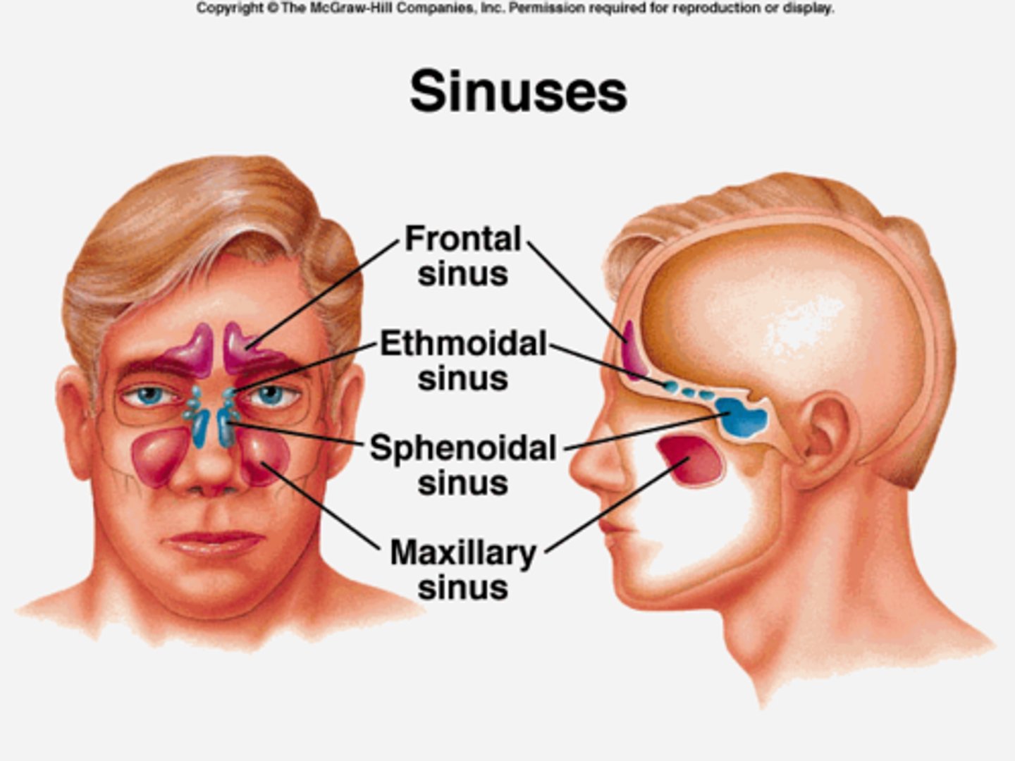
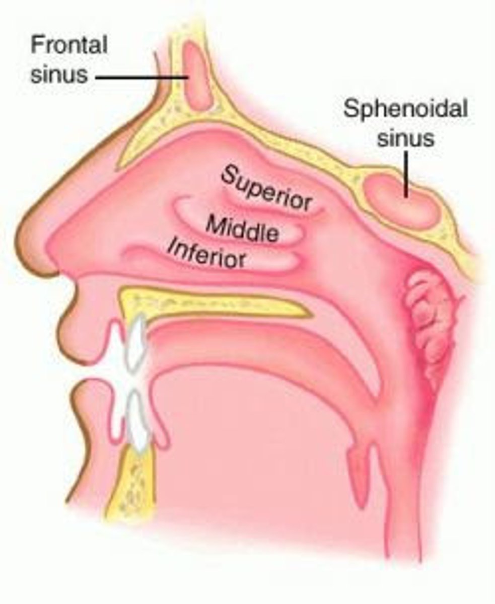
Paranasal sinuses
Frontal, Sphenoid, Ethmoid, Maxillary Sinus
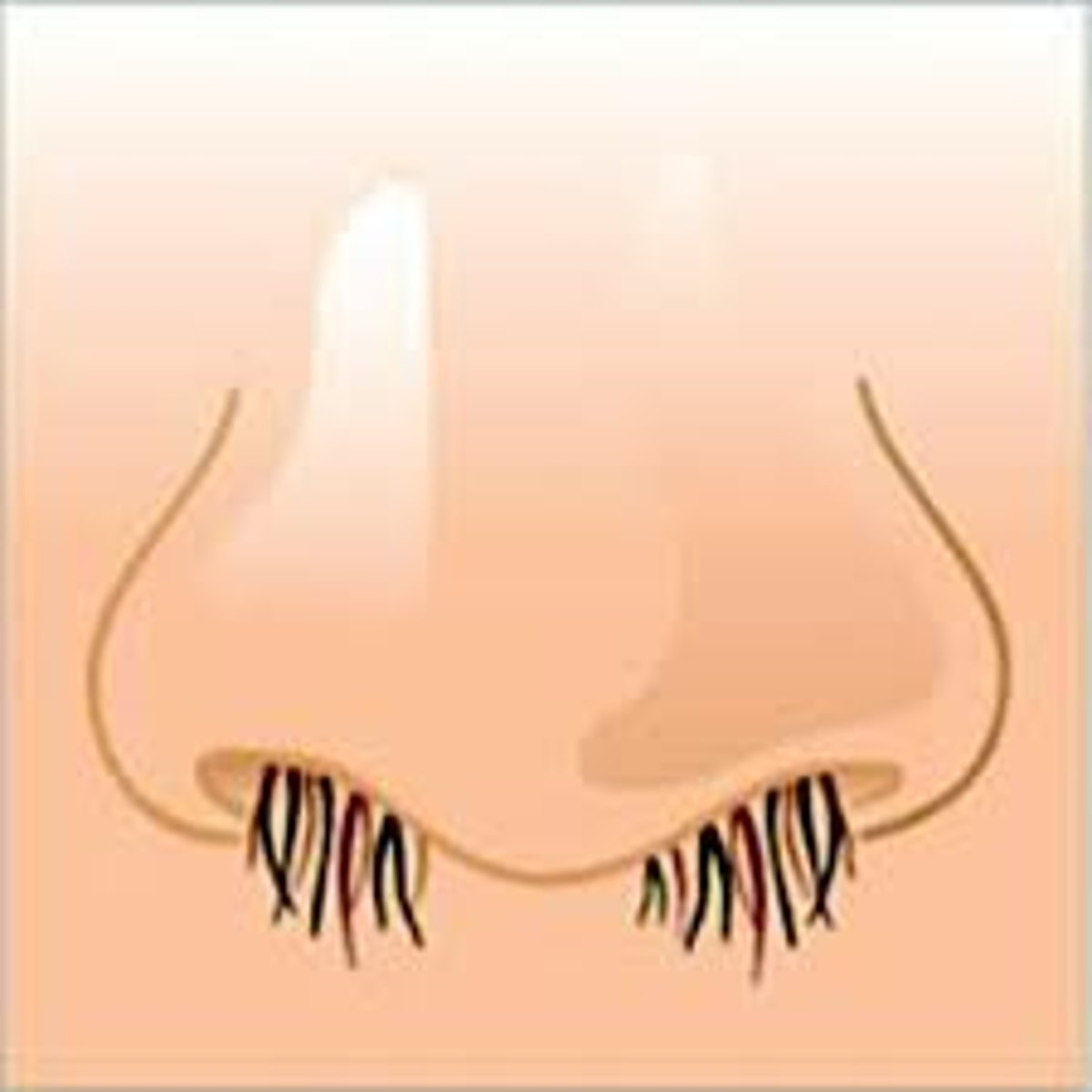
Naris (e)
nostril
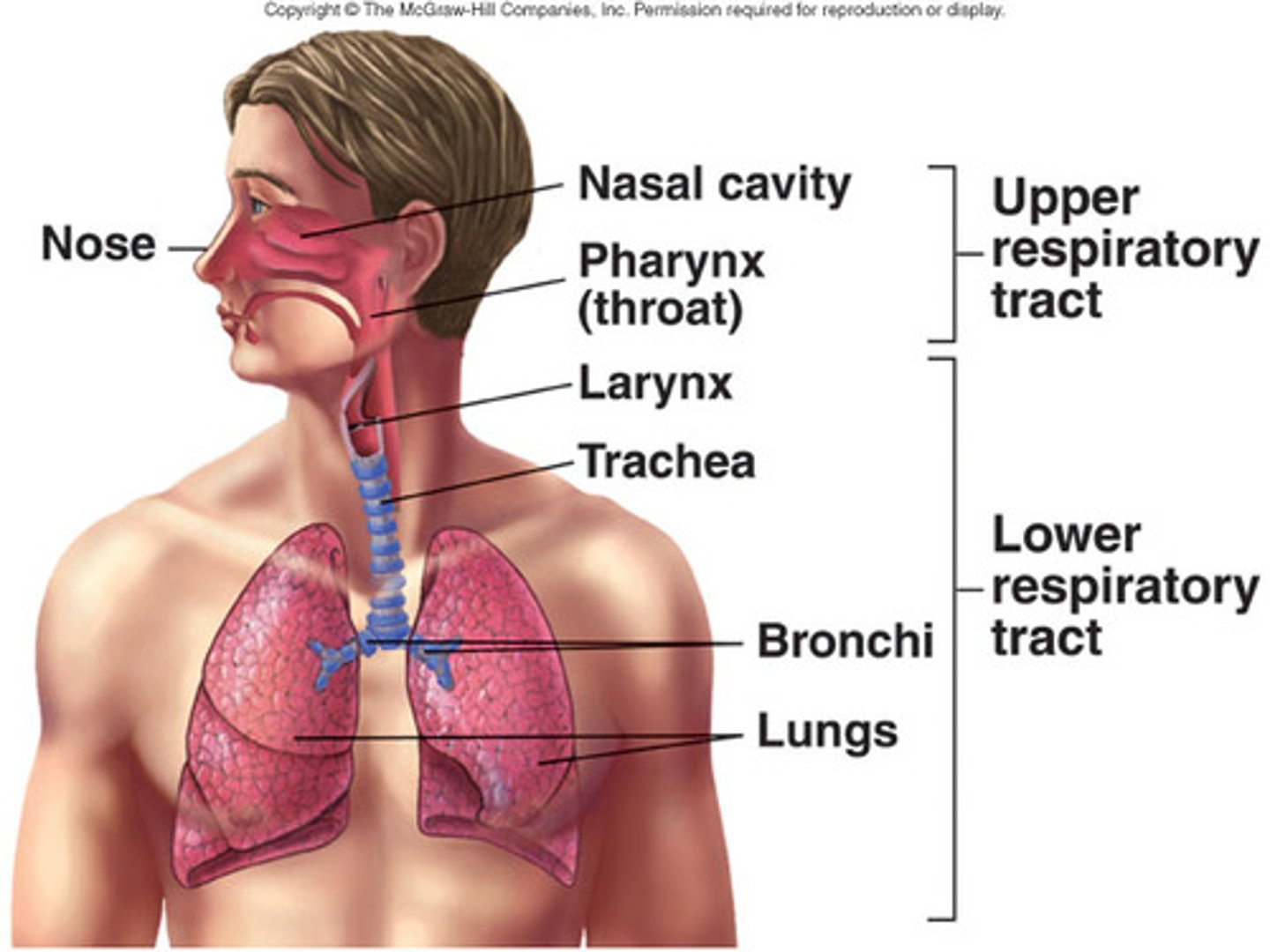
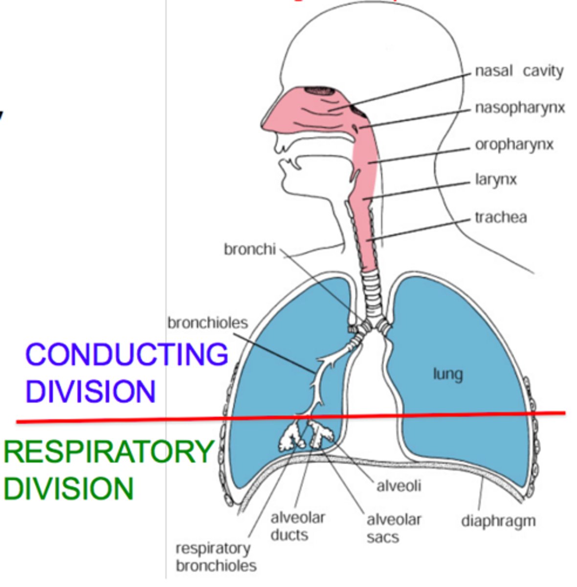
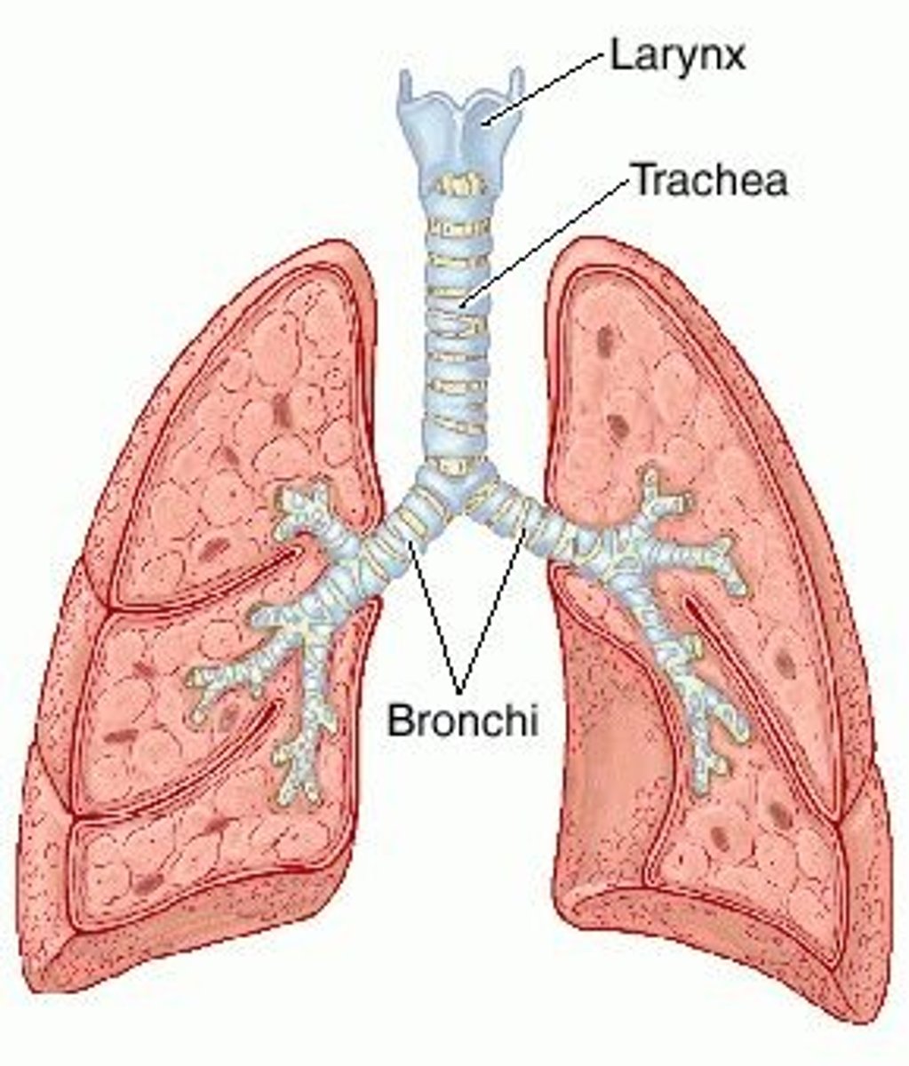
upper respiratory tract
external nose, nasal cavity, pharynx and superior margin of the larynx

lower respiratory tract
larynx, bronchi, lungs
respiratory division
alveoli and other gas-exchange regions such as the bloodstream through the alveolar wall
Chonchae
Superior, Middle and Inferior nasal conchae
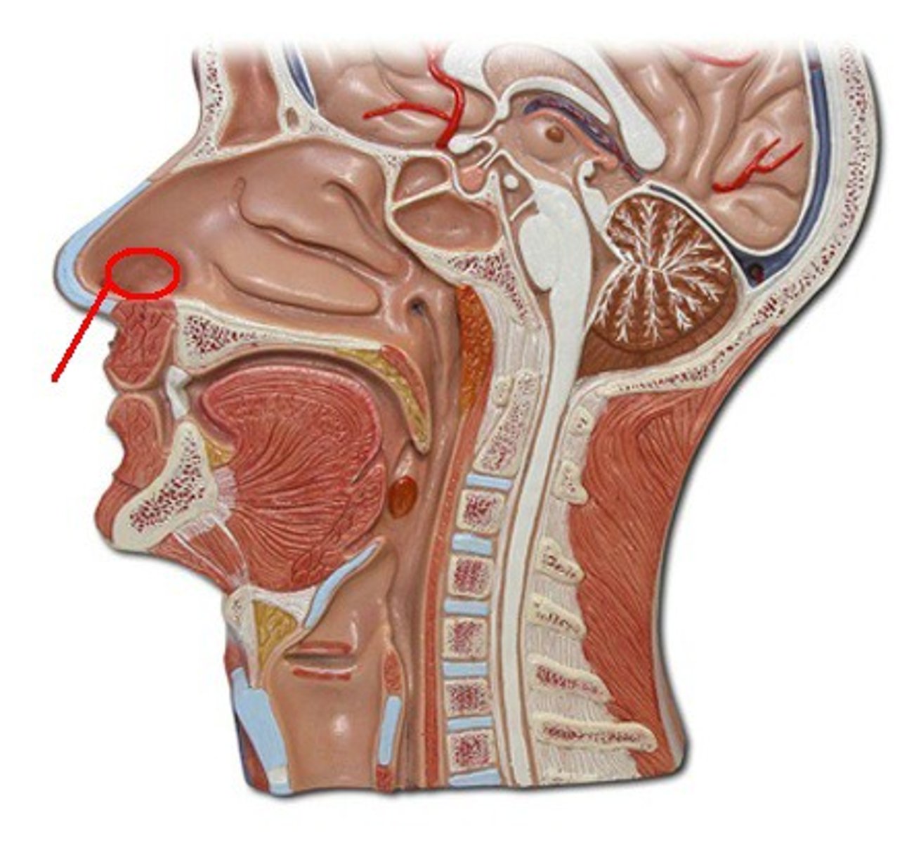
Vestibule
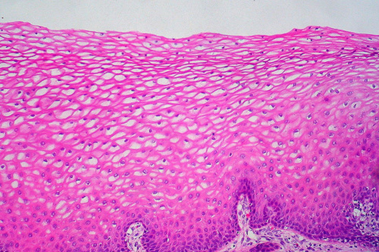
just inside the nostril, lined with stratified squamous epithelium
Vibrissae
hairs in the nose that block some of the inhaled debris
Nasal cavity and nasopharynx epithelium
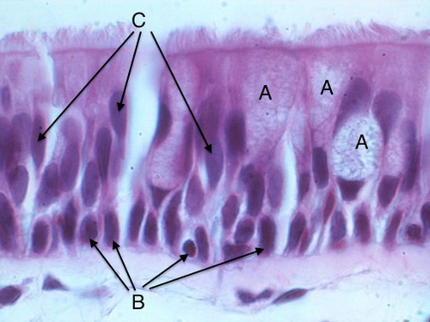
Most of the nasal cavity is lined with ciliated pseudostratified columnar epithelium
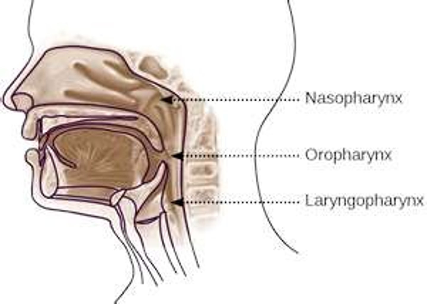
Oropharynx and Laryngopharynx epithelium
Lined with stratified squamous epithelium, passageway for food, fluid and air.
Goblet cells (A)
produce protective mucus
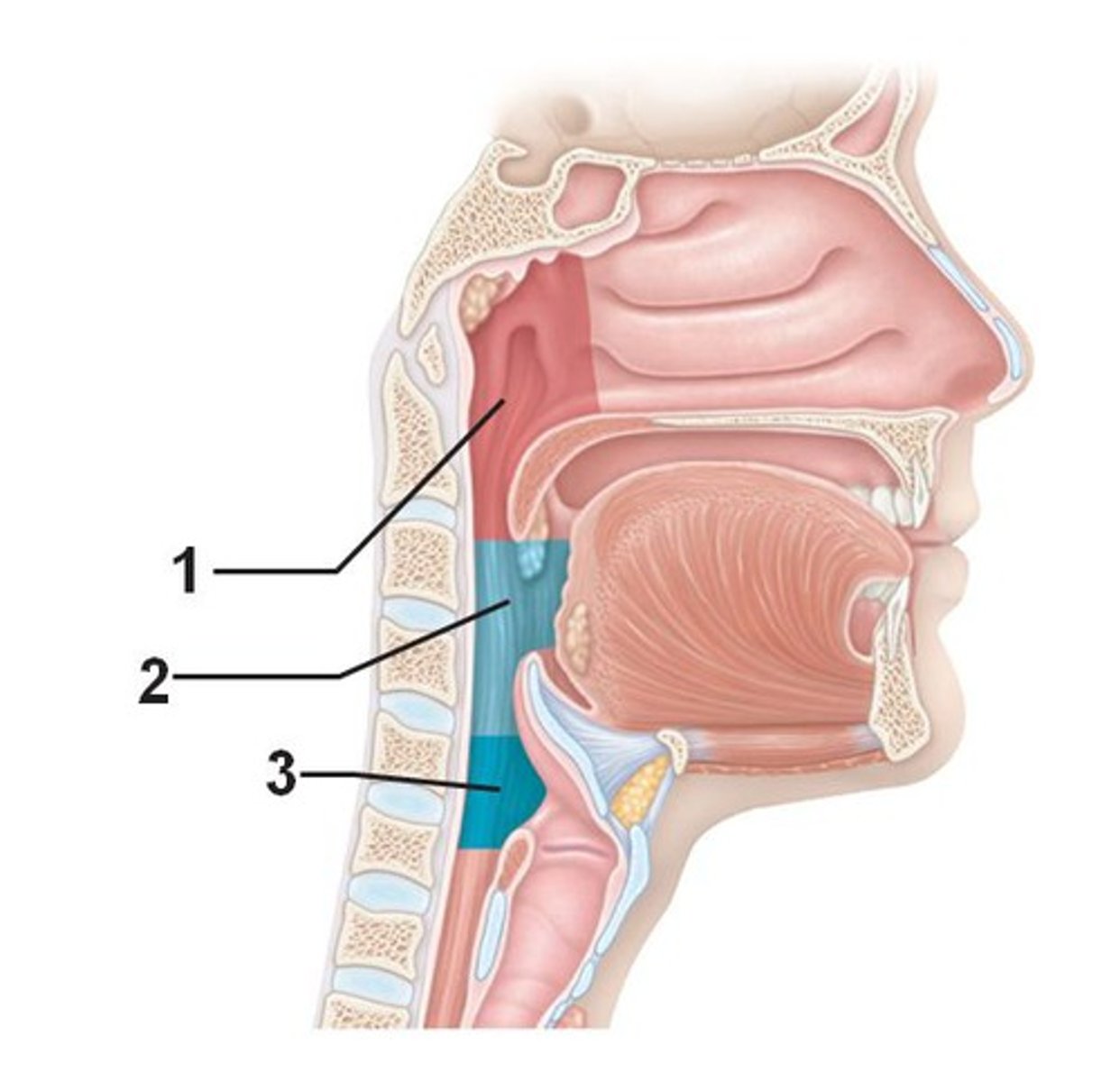
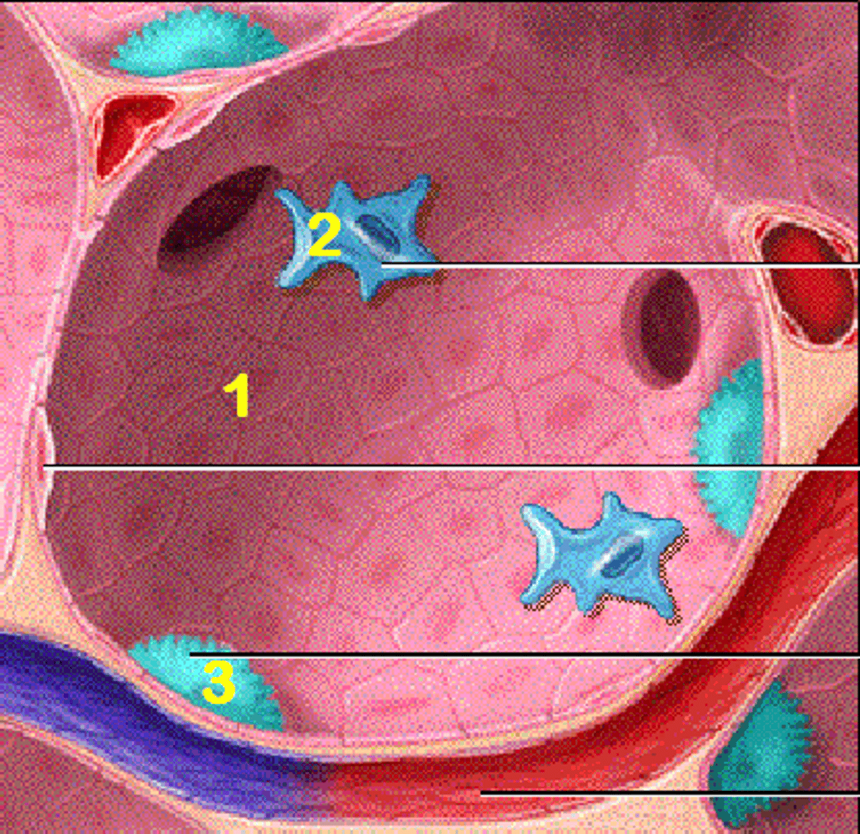
oropharynx (2)
from the soft palate to the epiglottis

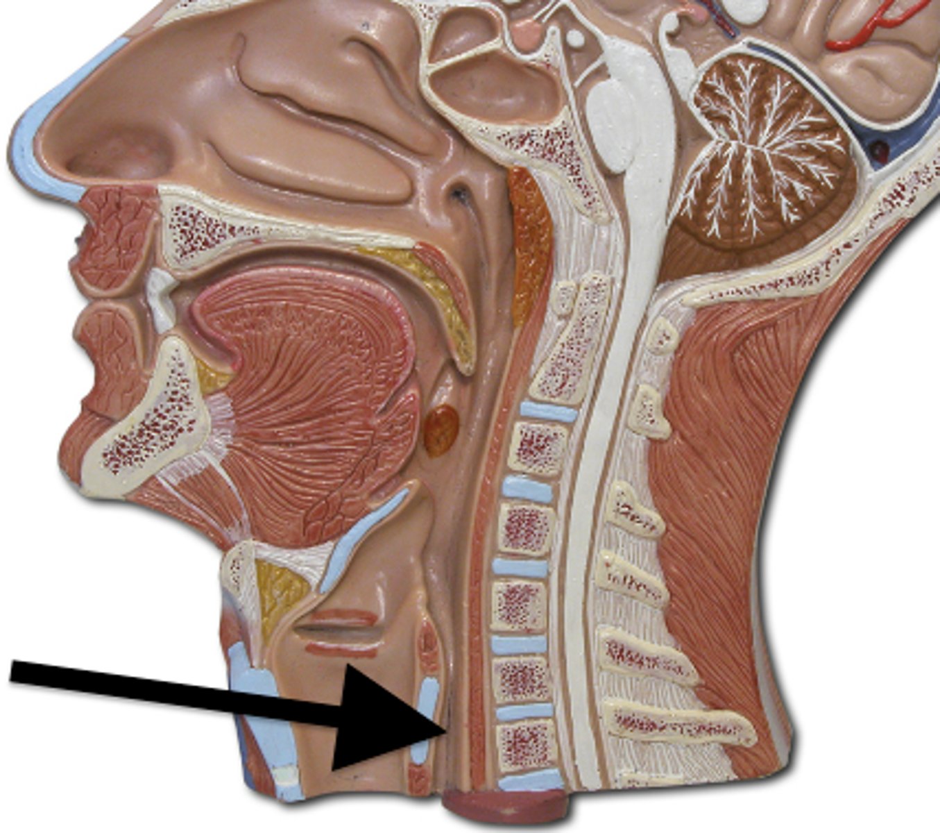
laryngopharynx (3)
from the superior margin of the epiglottis to the inferior margin of the cricoid cartilage
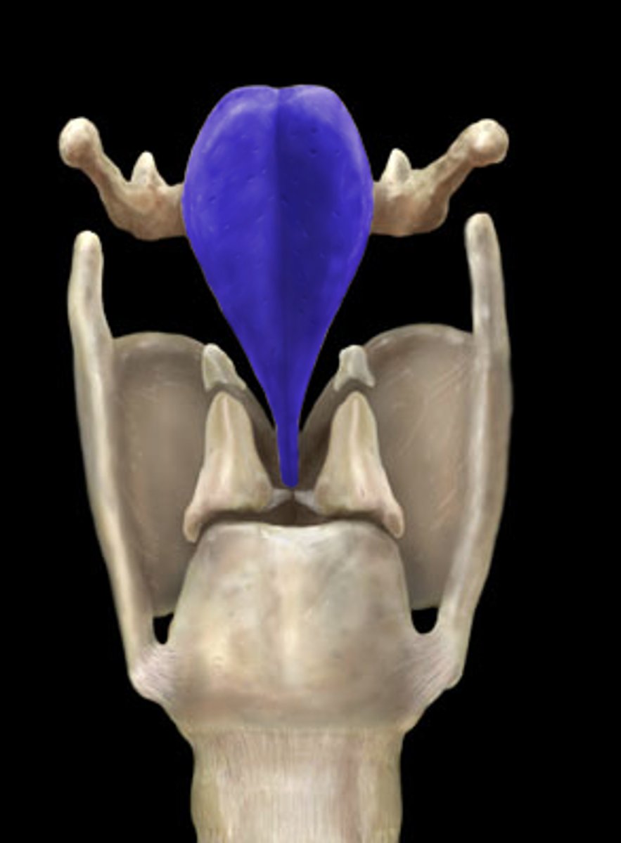
epiglottis
closes the airway and directs food and drink into the posterior esophagus
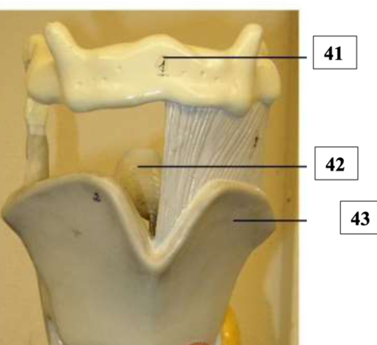
thyroid cartilage (Adam's apple)
called the laryngeal prominence
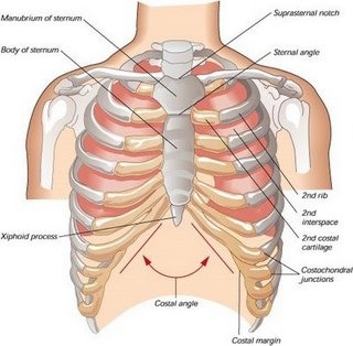
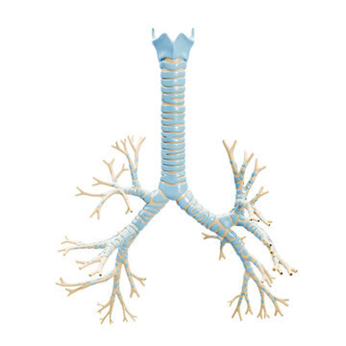
Trachea
located posterior to the sternum
Esophagus
located posterior to the trachea
sternum
located anterior to the trachea and esophagus
C-shaped cartilaginous rings
made up of hyaline cartilage
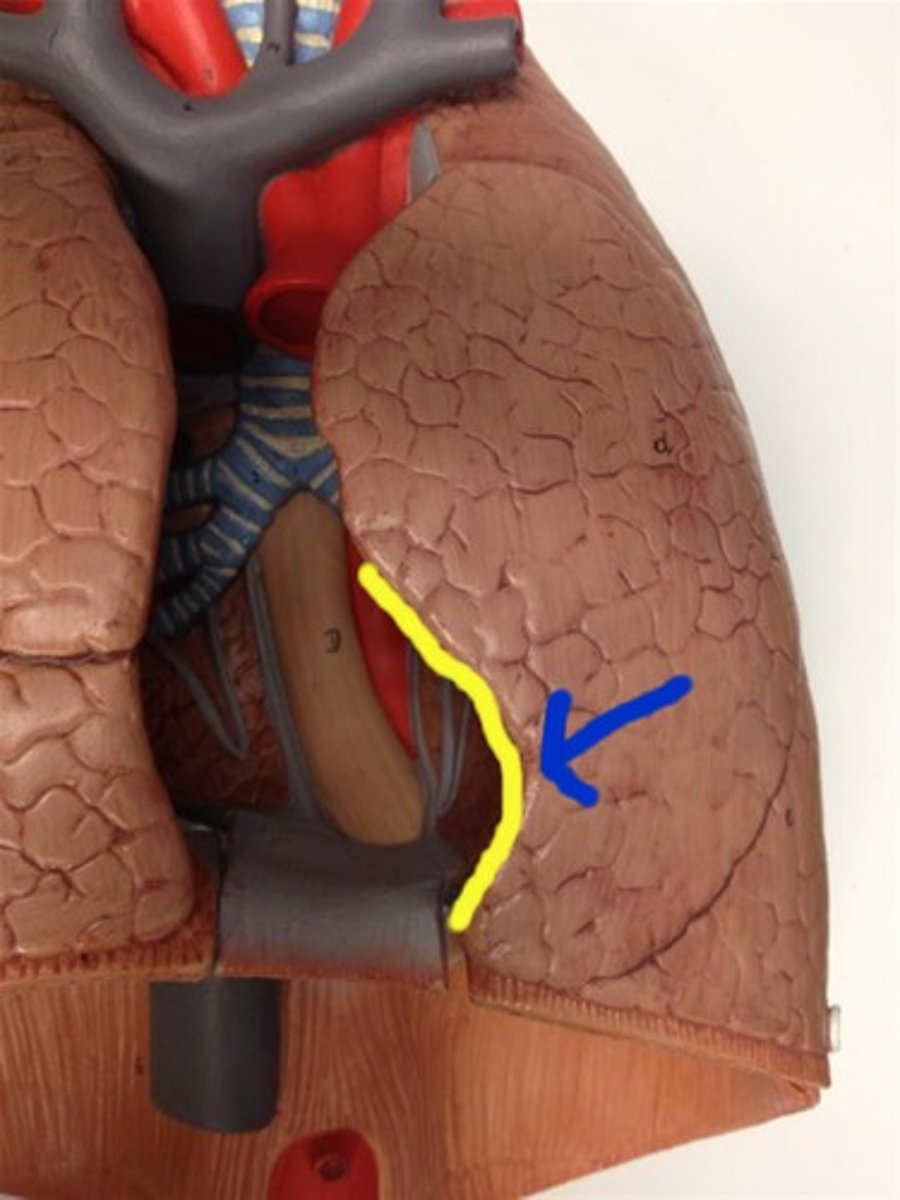
Cardiac notch
indentation on the surface of the left lung that allows space for the heart
Right lung
Has three lobes: superior, middle, inferior. Horizontal and Oblique fissure
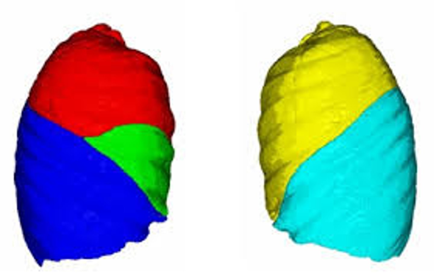
Left lung
has 2 lobes: superior and inferior. Only oblique fissure

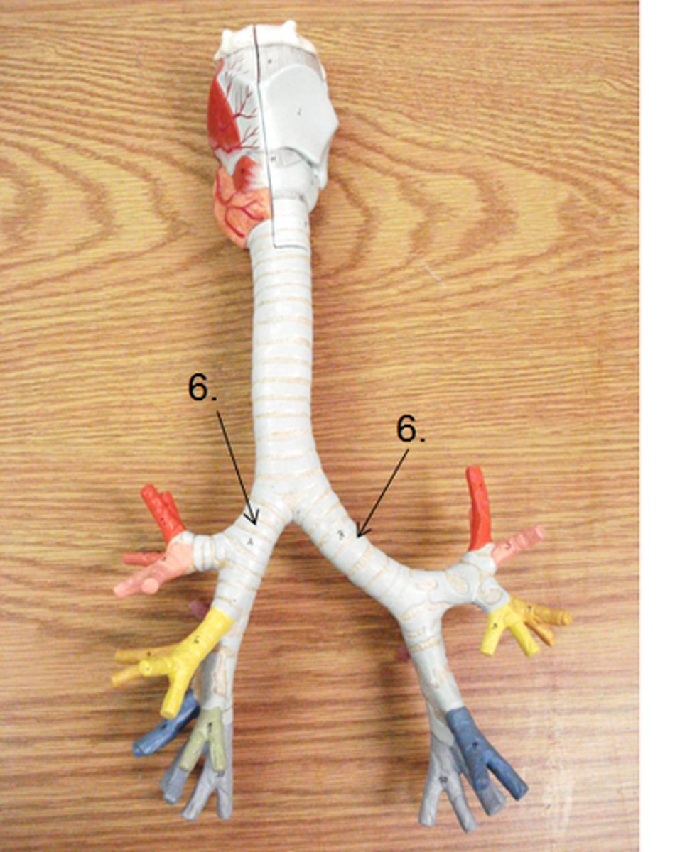
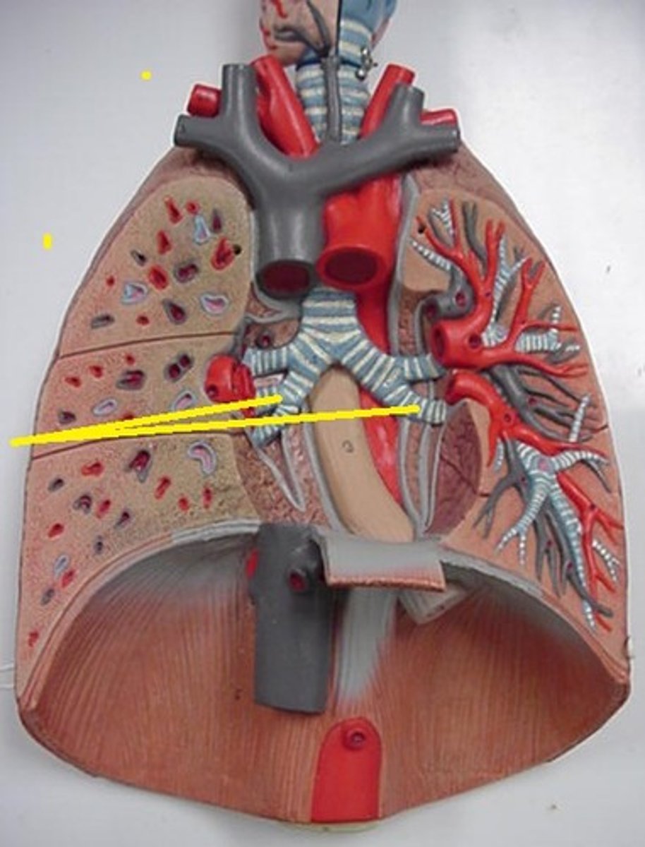
primary bronchus (6)
Main bronchus leading to each lung.
secondary (lobar) bronchus
Paired branches of main bronchus: Left one has two, and right lung has three
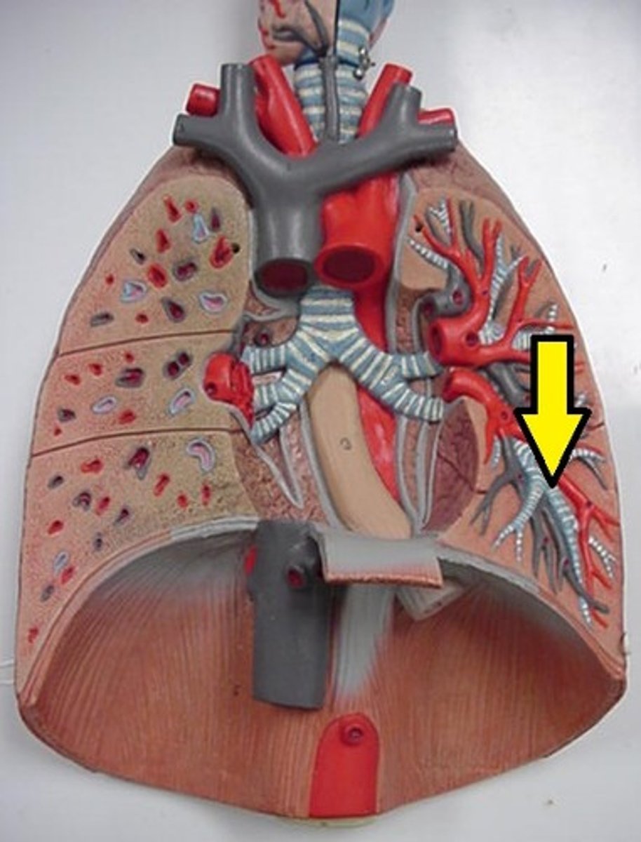
tertiary (segmental) bronchus
Last segment of the bronchioles
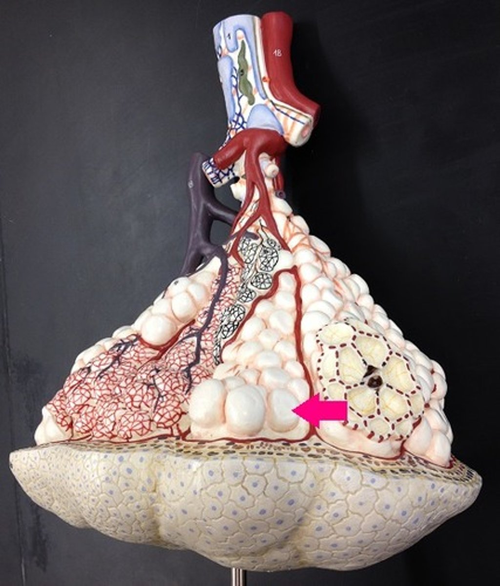
Alveoli sac
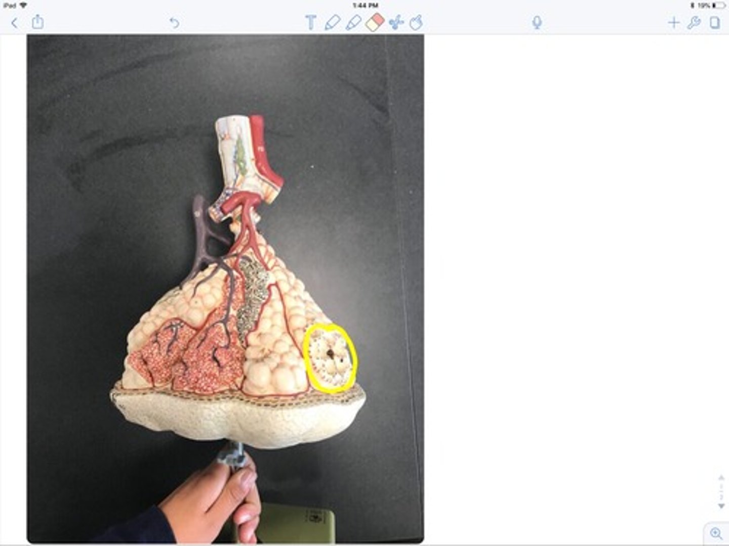
alveoli
Terminal air sacs that constitute the gas exchange surface of the lungs.
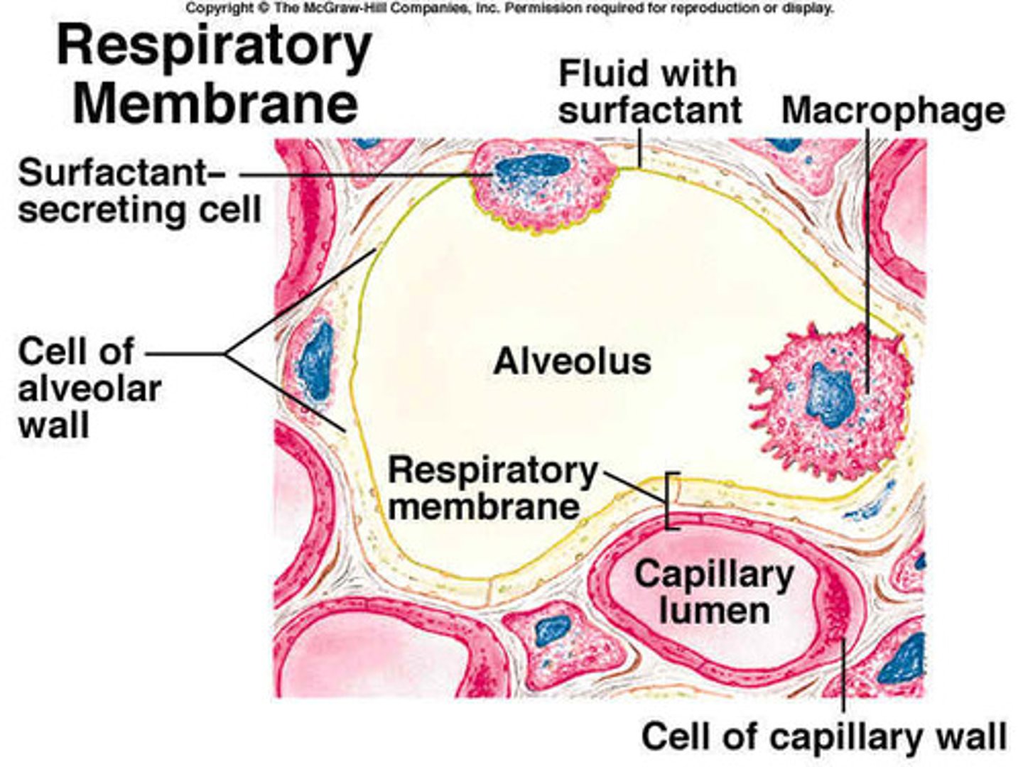
Squamous (type I) alveolar type
simple squamous epithelial cell that forms the walls of the alveoli of the lungs
cuboidal great (type II) alveolar cells
5%, function to repair type I cells and secrete surfactant
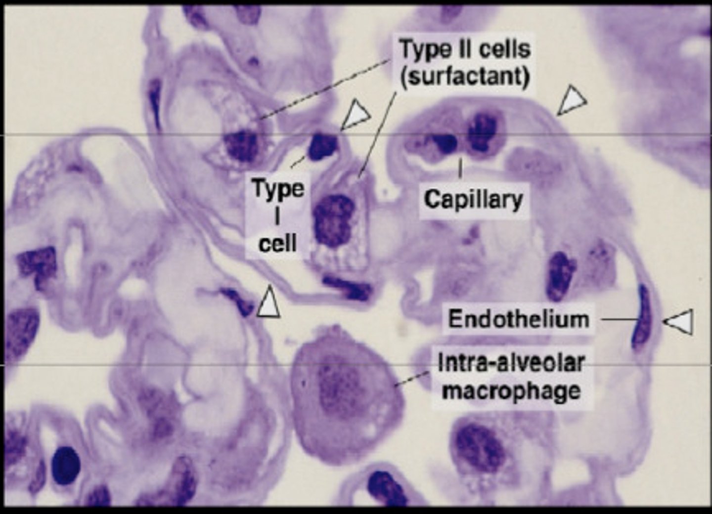
surfactant
prevents alveoli from collapsing when one exhales
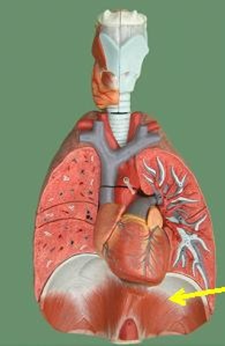
Diaphram
a dome-shaped muscle that separates the thoracic cavity from the abdominopelvic cavity
inhalation
diaphragm and external intercostal muscles contract, thoracic cavity increases volume thus decreasing pressure. Air flows in, raises the ribs and pushes the sternum out. Air moves into the lungs when pressure inside of the lungs is lower than the pressure of the atmosphere.
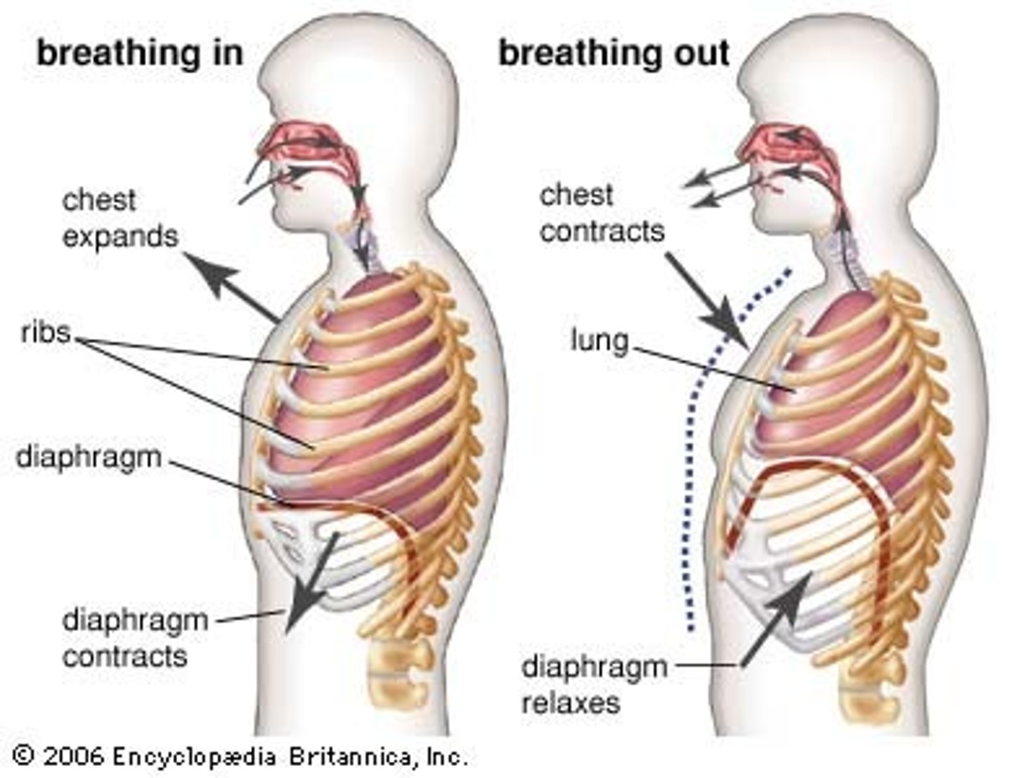
exhalation
diaphragm and external intercostal muscles relax, thoracic cavity decreases volume thus increasing pressure. air flows out, sternum and ribs move inward and up. Air moves out of the lungs when pressure inside of the lungs is greater than the pressure in the atmosphere
Boyle's Law
A principle that describes the relationship between the pressure and volume of a gas at constant temperature. Air always moves from an area of high pressure to an area of low pressure.