UGA CBIO 2200 Lab Practical 1
1/136
There's no tags or description
Looks like no tags are added yet.
Name | Mastery | Learn | Test | Matching | Spaced | Call with Kai |
|---|
No analytics yet
Send a link to your students to track their progress
137 Terms
Anterior/Ventral
front
Posterior/Dorsal
back
Superior/Cranial
higher
Inferior/Caudal
Lower
Lateral
closer to the side
Medial
closer to the middle
Proximal
a position in a limb that is closer to the point of attachment (trunk)
Distal
a position in a limb that is farther from the point of attachment (the trunk)
Superficial
shallow (closer to the surface)
Deep
farther from the surface of the body
Contralateral
structures found on different sides of the body (right vs. left)
Ipsilateral
structures on the same side of the body (right and right; left and left)
Sagittal plane
divides the body/organ into left and right segments
Midsagittal
divides right down the middle (left and right)
Paragagittal
Divides the half into halves again; adjacent/parallel to the midsagittal region
Frontal (Coronal) plane
divides the body/organ into a front and back portion (anterior and posterior portion)
Transverse plane
divides the body/organ into a top and bottom half
Oblique plane
diagonal cut (uneven distribution; i.e. right eye visible, left eye is not)
Cranial cavity
brain (protection: bones, cerebrospinal fluid)
vertebral cavity
contains the spinal cord
thoracic cavity
contains heart and lungs
pleural cavity
contains the lungs
pericardial cavity
contains the heart
abdominal cavity
Contains stomach, intestines, spleen, and liver, and other organs
pelvic cavity
Contains urinary bladder, reproductive organs, and rectum
Thoracic cavity
protected by ribcage; contains pericardial cavity (heart), pleural cavities (lungs); floor: diaphragm
Abdominopelvic cavity
Contains the abdominal cavity (digestive organs) and the pelvic cavity (reproductive organs)
- largest cavity in the body
- no membrane physically divides abdomen and pelvis
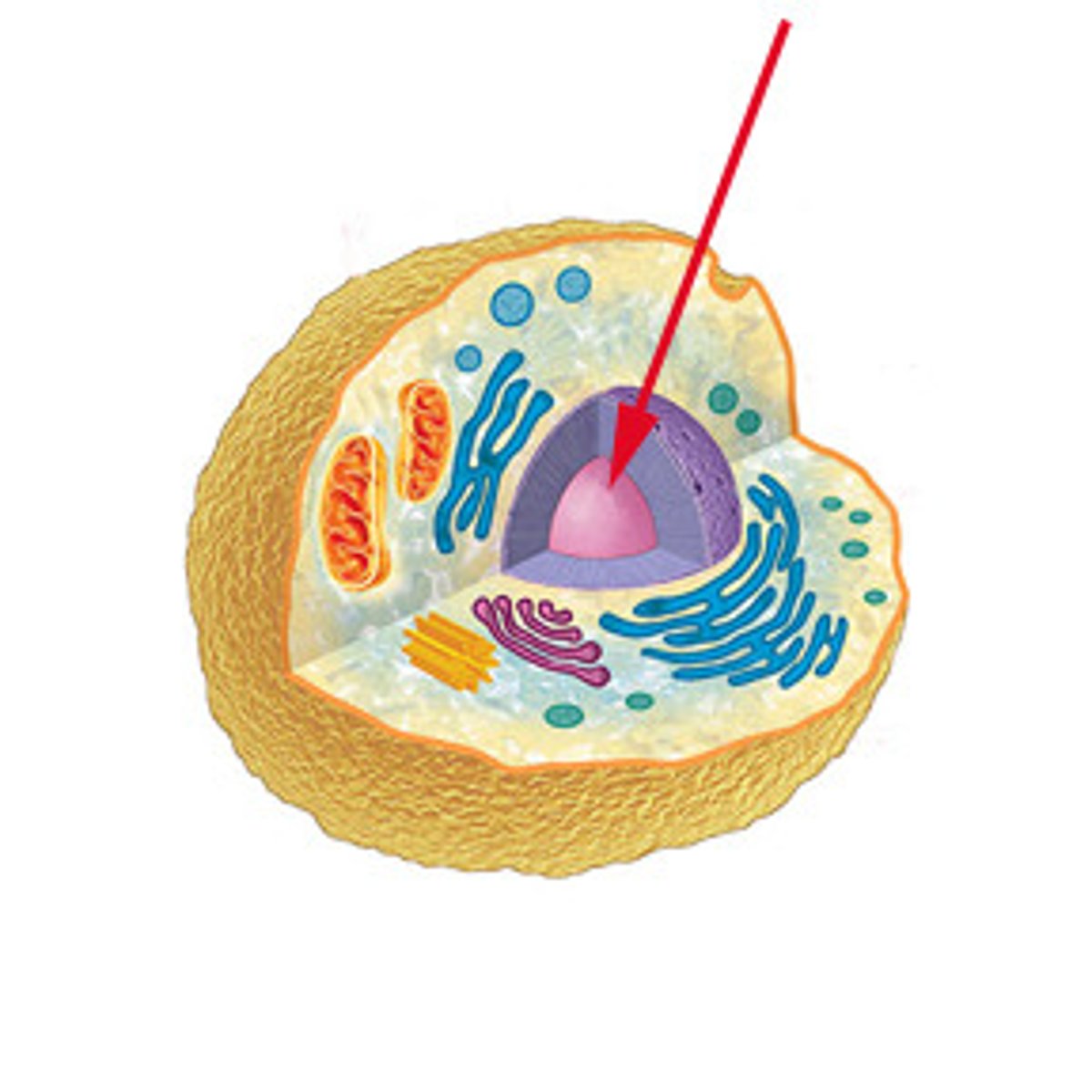
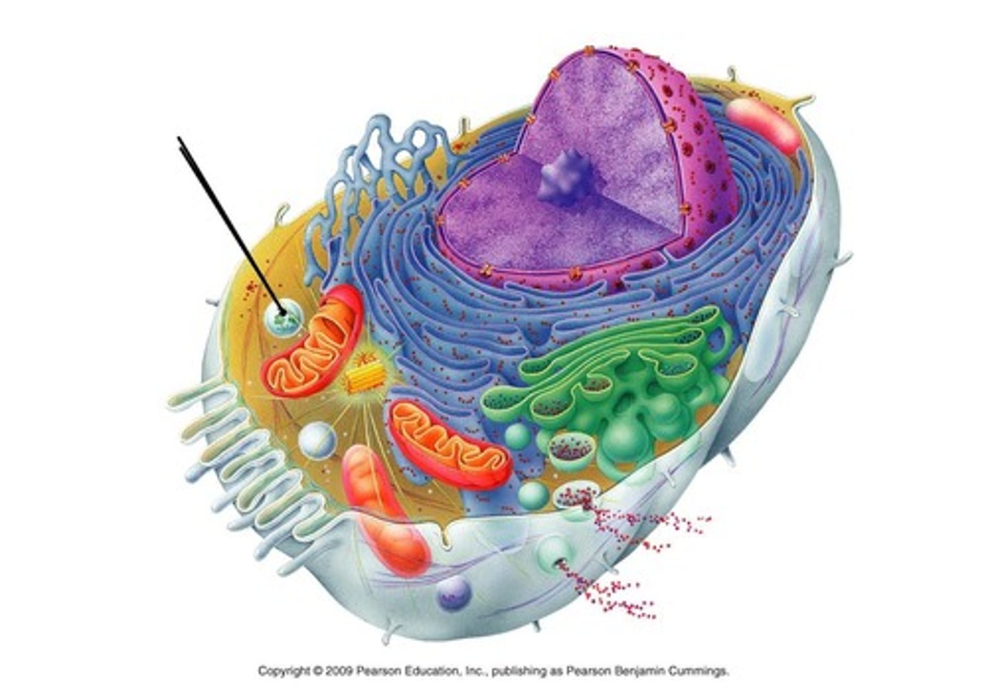
Nucleus
contains cellular DNA; "control center" of the cell (directs cellular functions)
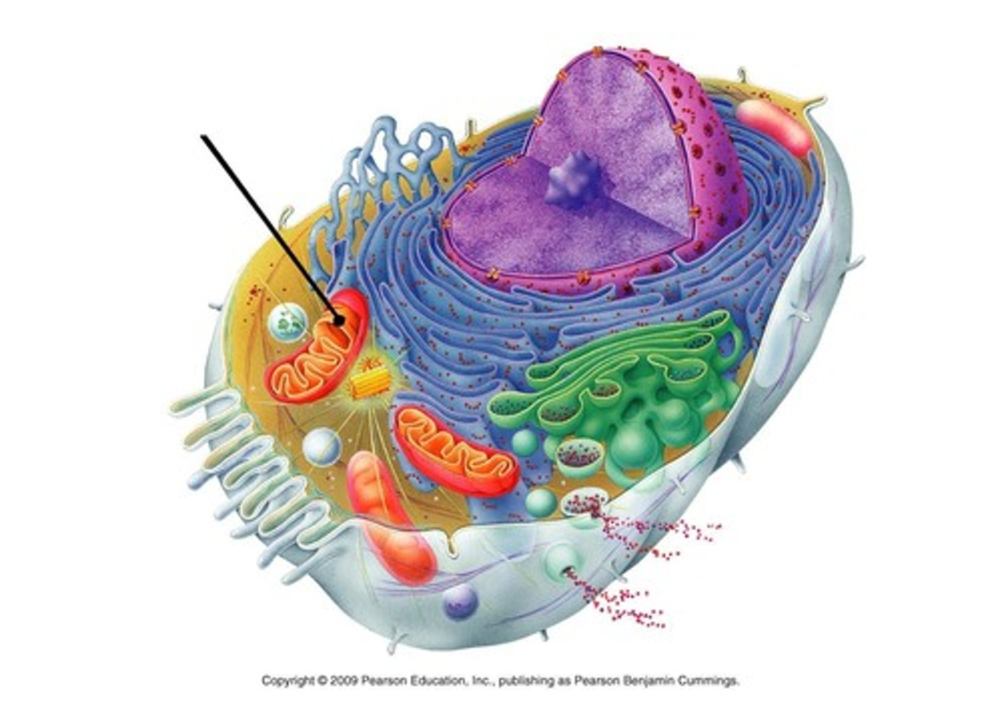
Mitochondrion
powerhouse of the cell; produces ATP by converting energy storage into usable energy (ATP) to power cellular function
Ribosomes
protein synthesis; found free in cytosol, found on rough ER, made in Nucleolus
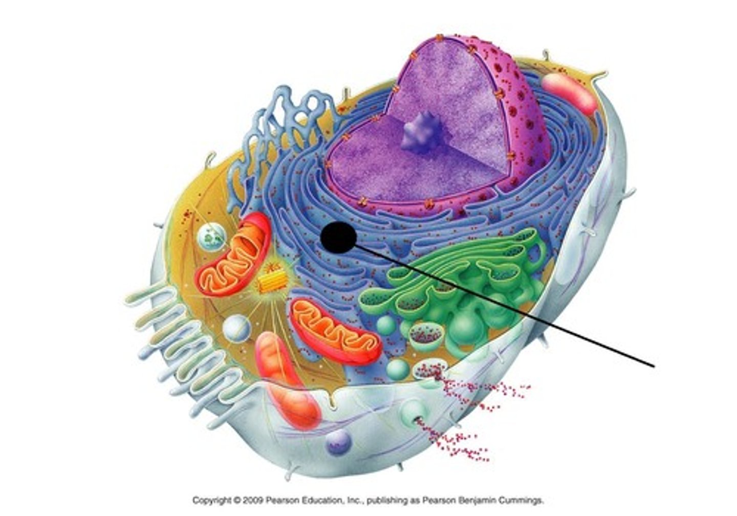
Rough Endoplasmic Reticulum
surrounds the nucleus; has ribosomes so it can function to synthesize and modify protein
Smooth Endoplasmic Reticulum
synthesizes lipids

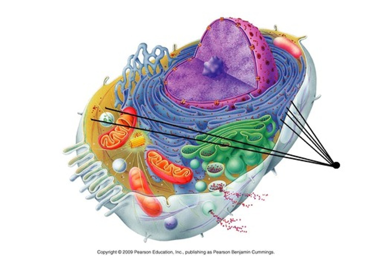
Golgi apparatus
packages things and send it out to different parts of the cell; sorts, modifies, and SHIPS products of the ER

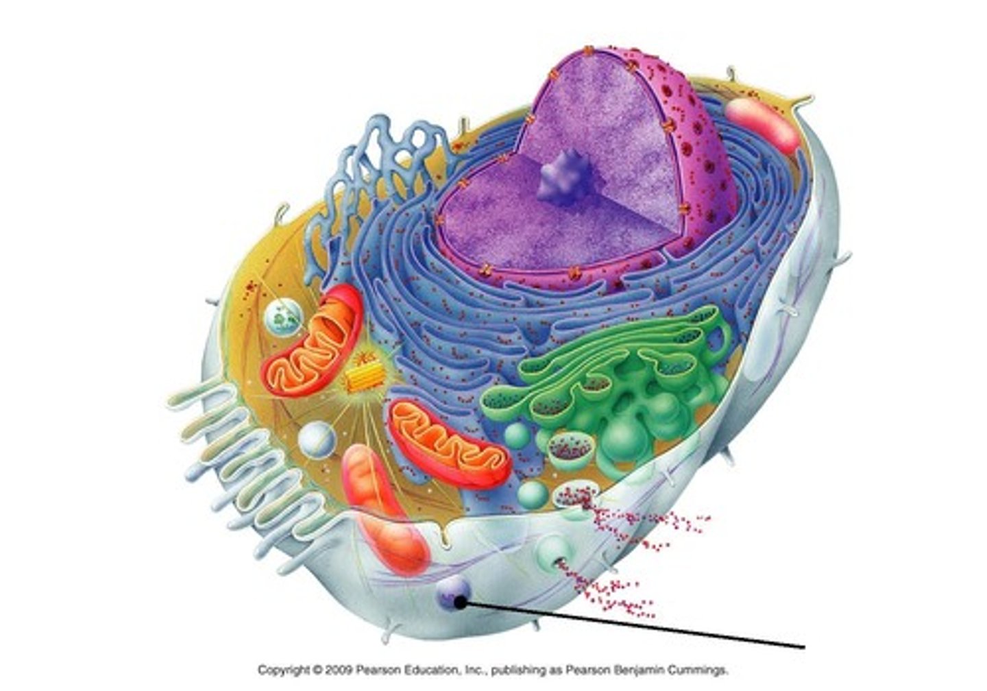
Lysosome
breaks down waste sent to it from the golgi apparatus; uses digestive enzymes to break down materials
Peroxisome
organelle that contains enzymes for metabolzing lipids; chemically detoxifies
Interphase
growth phase; cells not dividing
G1: metabolic functions carried out (growth); could last days
S: DNA replication
G2: grows, prepares for mitosis
G0: resting phase before prepare to divide again (or just permanent rest)
Mitosis
division of genetic material between two nuclei
PMAT
- Prophase
- Metaphase
- Anaphase
- Telophase
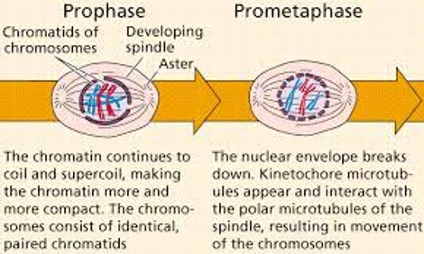
Prophase
1st mitotic phase; chromosomes are condensing (become tightly packed and observable), spindle fibers emerge from centrosomes, nuclear envelope breaks down, centrosomes more toward opposite poles
Prometaphase
in-between phase of mitotic division; kinetochores appear at centromeres, mitotic spindle microtubules attach to kinetochores
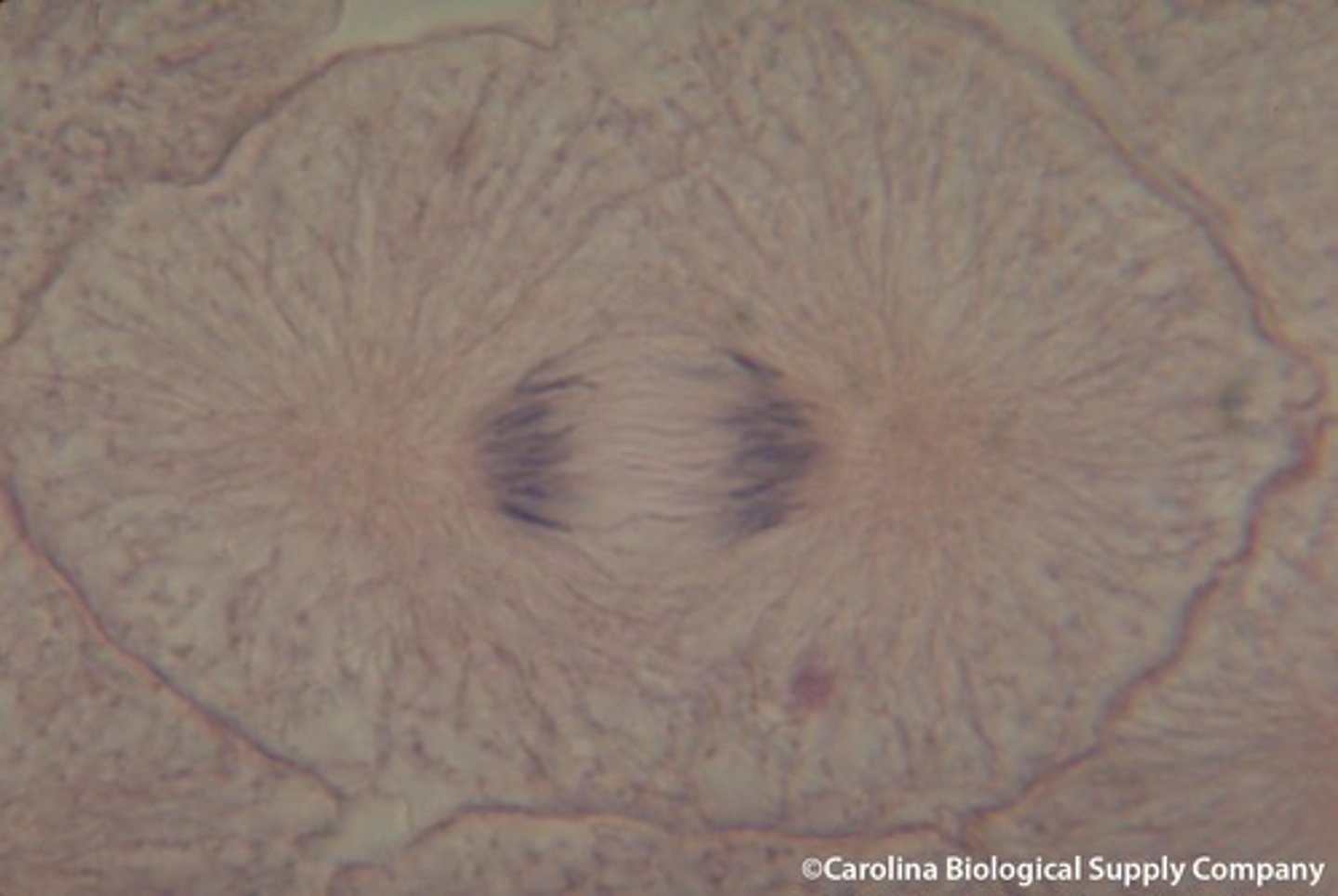
Metaphase
2nd phase of mitotic division; chromosomes are fully condensed and lined up in the center (METAPHASE PLATE); each sister chromatid is attached
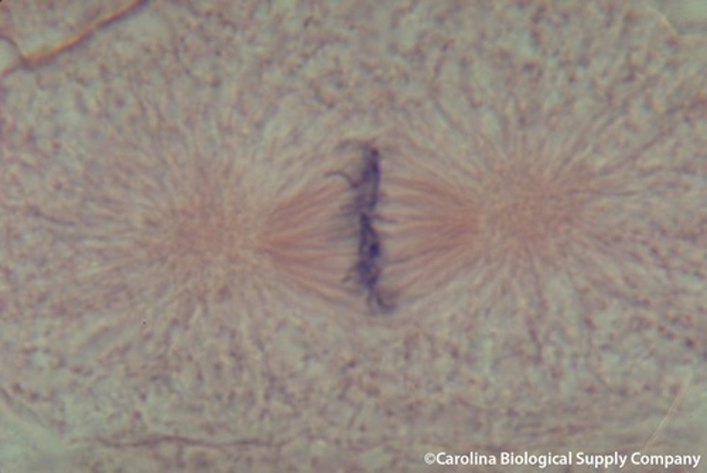
Anaphase
3rd phase of mitotic division; sister chromatids are pulled AWAY from each other (now separate chromosomes); centromeres split in two; spindle fibers begin to elongate the cell
Telophase
4th phase of mitotic division; chromosomes arrive at opposite poles; decondense; nuclear envelope begins to surround these new chromosomes; mitotic spindle breaks down; spindle fibers continue to push poles apart

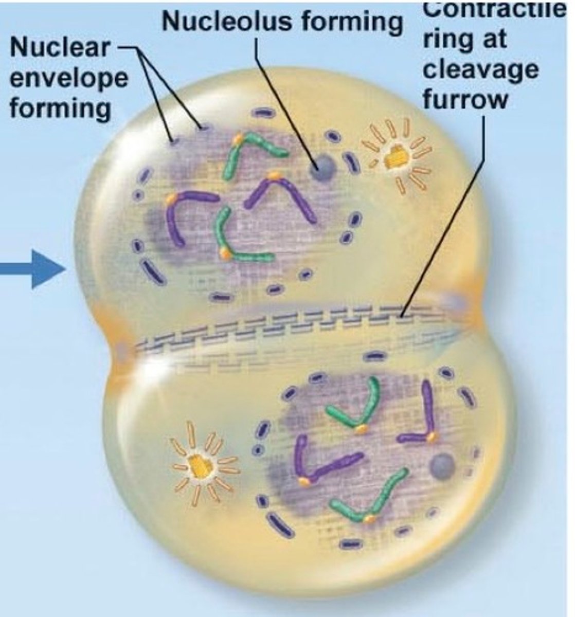
Cytokinesis
division of two separate nuclei formed during mitosis into two separate cells
- a cleavage furrow separates the daughter cells
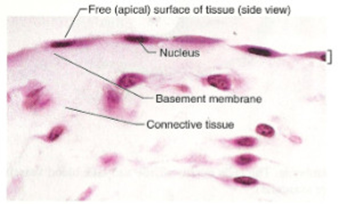
Epithelial Tissue
tissue that lines outer surfaces of the body, internal cavities, and forms glands
- LARGE SHEETS of cells (lining outside of body/outside of organs)
- CELLULAR (no extracellular present between cells)
Found in: lining of GI tract organs (other hollow organs), skin surface (epidermis)
- GLANDULAR TISSUE
- Skin, digestive tract, airways, urinary/repro systems)
Functional Features of Epithelial Tissue?
- POLARITY (apical/basal surfaces)
- Needs CONNECTIVE TISSUE (so skin doesn't fall apart)
- AVASCULAR (no blood vessels, so everything either diffuses or absorbs from other tissue)
- INNERVATED (nervous tissue allows interaction with external envt)
- REGENERATION (rapid replacement of dead cells)
What is the goal of Epithelial tissue?
first layer of protection from the external envt
- permeability (selectivity)
- temp regulation?
- secrete mucous (i.e. digestive enzymes from small intestine)
Epithelial Tissue Classification
- Simple squamous
- Simple cuboidal
- Simple columnar
- Stratified squamous
- Stratified cuboidal
- Pseudostratified columnar
- Transitional
Simple squamous
- simple: one layer
- squamous: thin, flat
- look like: scales
- rapid passage of chemical compounds
- i.e. alveoli of lungs, kidney tubules, capillary lining
FUNCTION: diffusion and filtration of materials; secretes lubricating substance
Simple cuboidal
- simple: one layer
- cuboidal: cube shape (squarish), round nucleus
- secretion, absorption of molecules
- lining of kidney tubules, ducts of glands
FUNCTION: secretes/absorbs
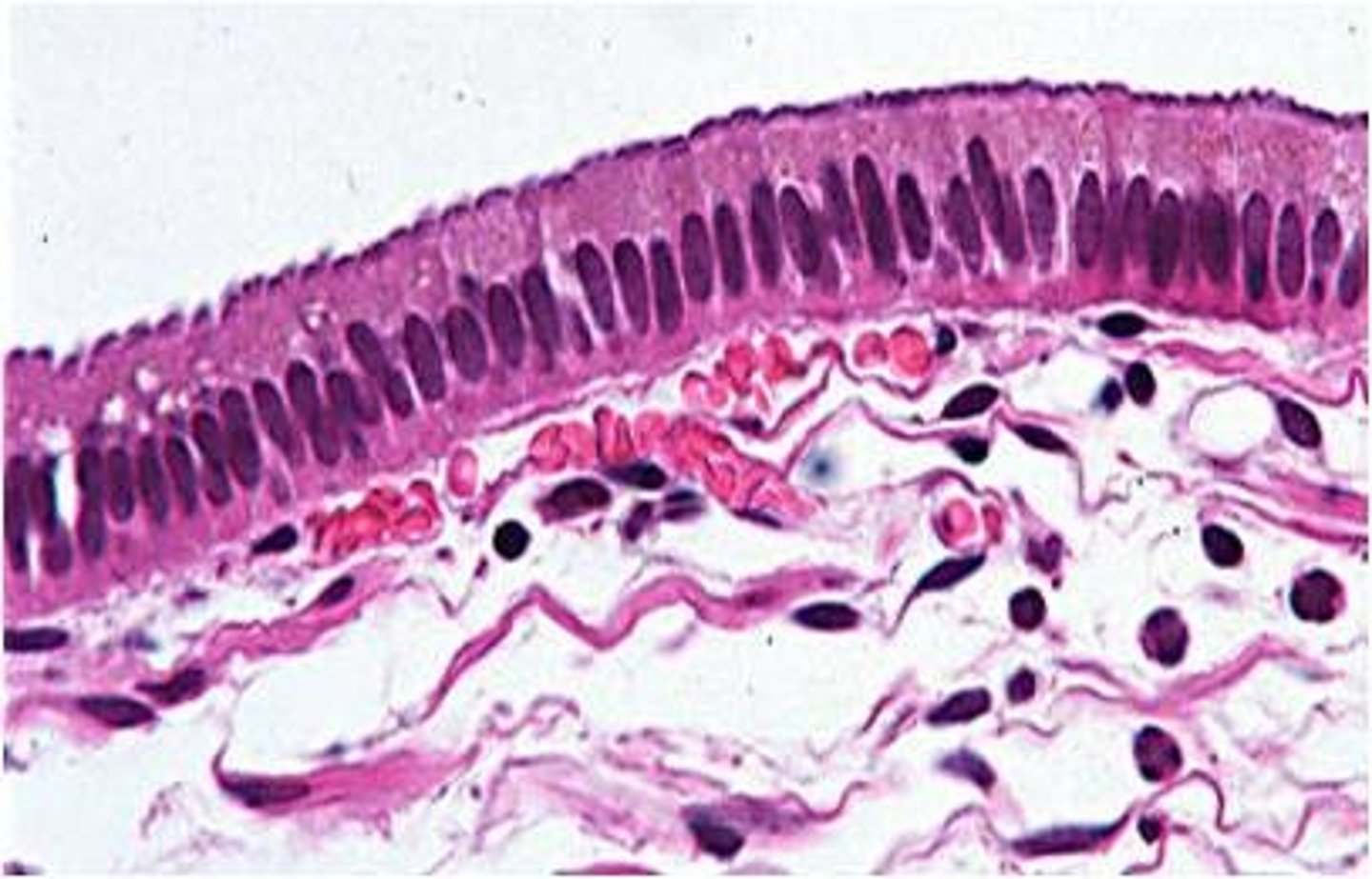
Simple columnar
- simple: one layer
- columnar: tall
- nucleus = tall, located on basal membrane side
- secretion/absorption of molecules
- digestive system, female repro tract
- ciliated = cilia located on apical surfaces (i.e. uterine tube, respiratory system)
-include interspersed cells of other kinds?
-FUNCTION: absorbs; secretes mucous and enzymes
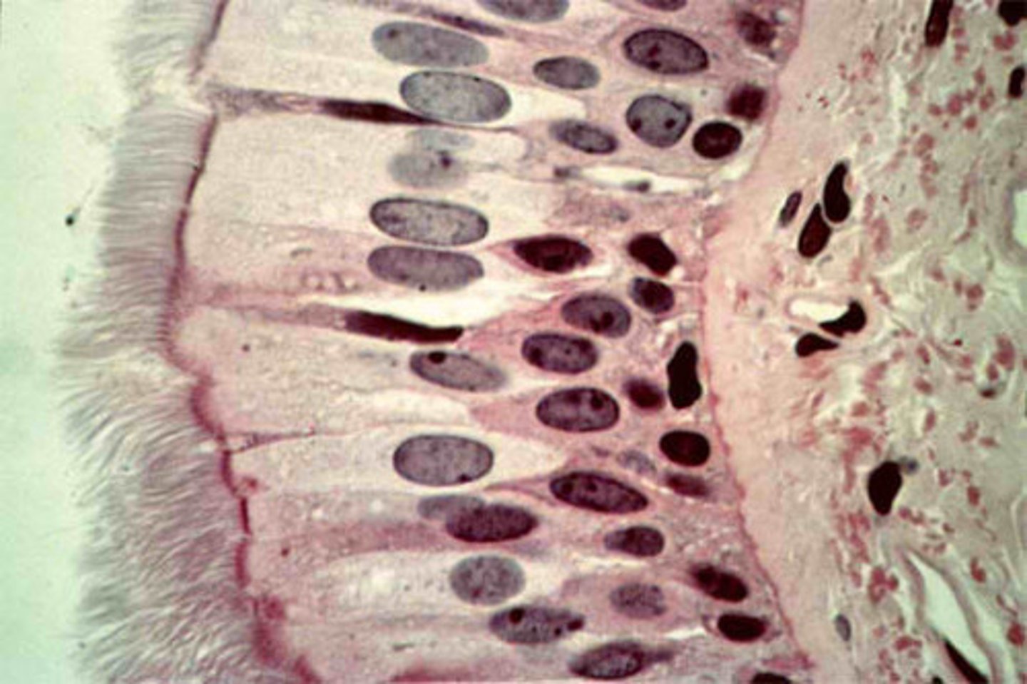
Pseudostratified columnar
pseudostratified: appears to have many layers but only has one layer (irregularly shaped columnar cells)
- nuclei are at differing levels
- all cells in contact with basal lamina despite appearance
- respiratory tract (cilia)
- include interspersed cells of other kinds?
FUNCTION: secretes mucous; ciliated tissue moves the mucous
Stratified
for protection against chemical and physical wear and tear
- many layers
- named for MOST APICAL layer
- Strat squamous = most common
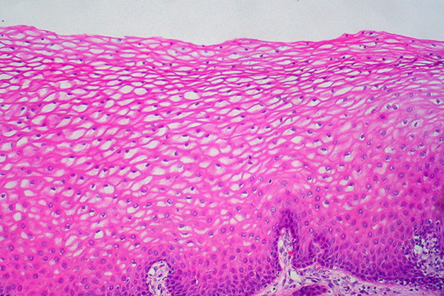
Keratinized Stratified squamous
epithelial tissue with dead cells filled with keratin as the TOP LAYER (most apical)
- i.e. Mammalian skin
FUNCTION: protects against abrasion
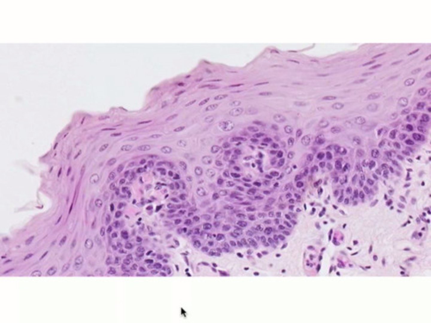
non-keratinized stratified squamous
no layer of dead cells filled with keratin on top
- i.e. lining of the oral cavity
FUNCTION: protects against abrasion
Stratified cuboidal/stratified columnar
UNCOMMON
- can be found in glands and ducts
-Strat cuboidal: protective tissue
- Strat columnar: secretes/protects
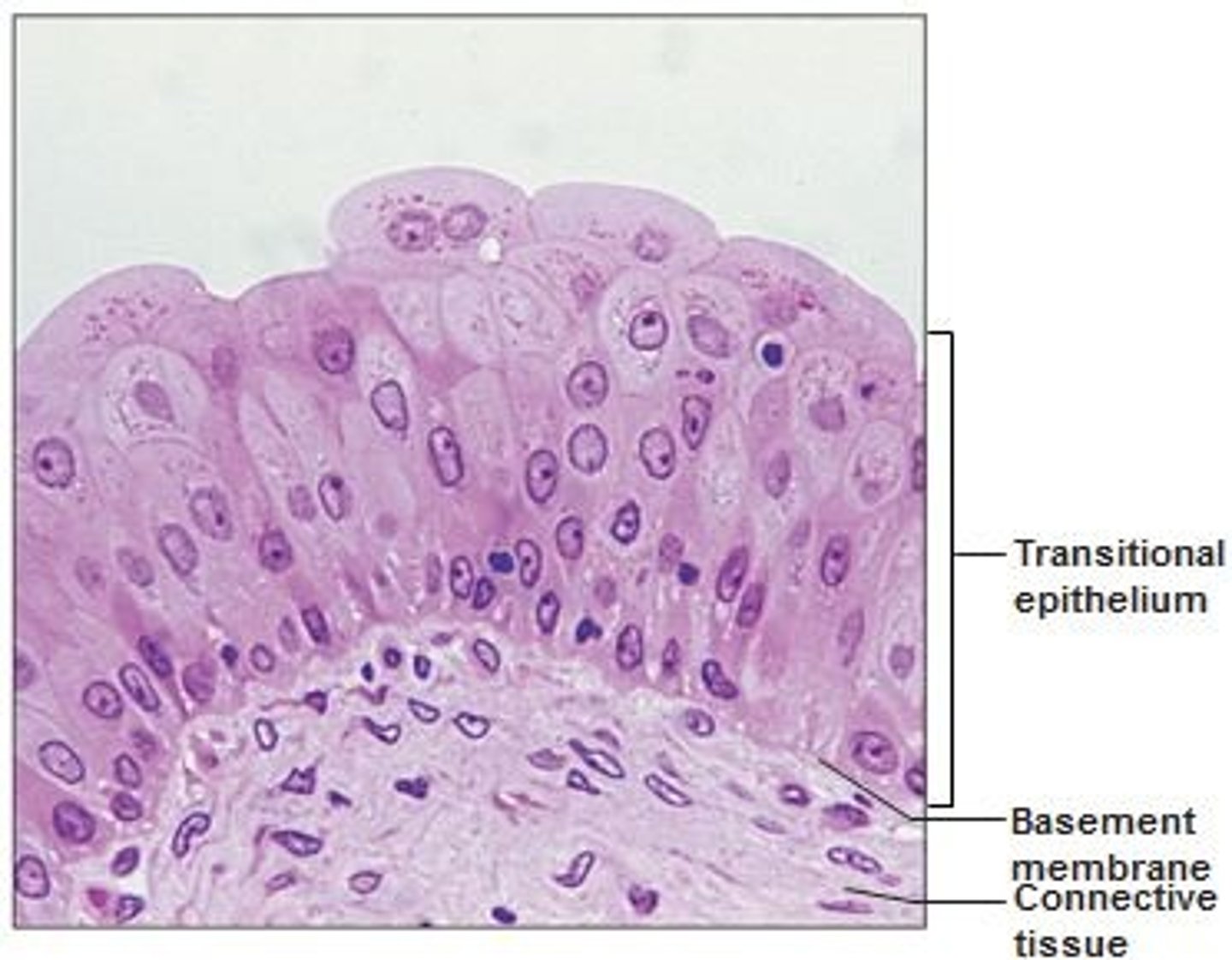
Transitional stratified
apical cells can change shape
- ONLY in urinary system (ureters/urinary bladder)
- umbrella shaped, apical surface
- appears thicker and more layered when bladder is EMPTY
- stretched out, less stratified when bladder is full
FUNCTION: allows urinary organs to expand/stretch
Connective Tissue
binds cells and organs together and helps protect, support, and integrate all parts of the body
Found in: Fat (soft padding), bone, tendon
FUNCTION: support and connect tissues and organs; protect organs (bones), protect from microorganisms; adipose tissues (store thermal energy in fat) = insulation
- specialized fluid CT - blood/lymph (transport fluid, nutrients, waste, chem messengers)
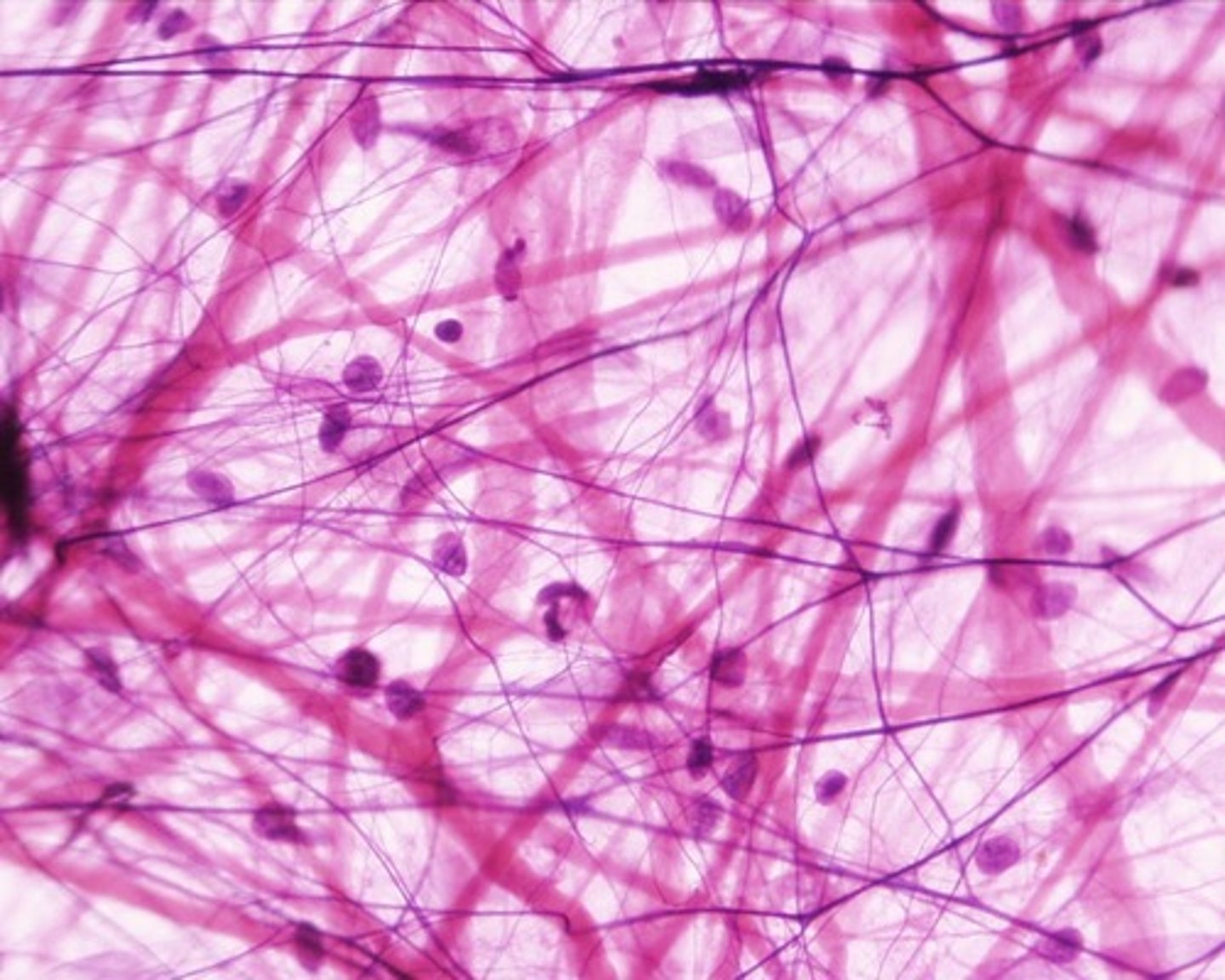
Areolar Tissue
Loose Connective Tissue:
- little specialization (all cell/fiber types in giant web)
- protein fibers: collagen, elastic, reticular
- cell types: mesenchymal, fibroblasts, fibrocytes, adipocytes, macrophages, lymphocytes, mast cells
- fills space between muscle fibers
- surrounds blood/lymph vessels
- supports abdominal organs
- connective tissue of EPITHELIAL MEMBRANES
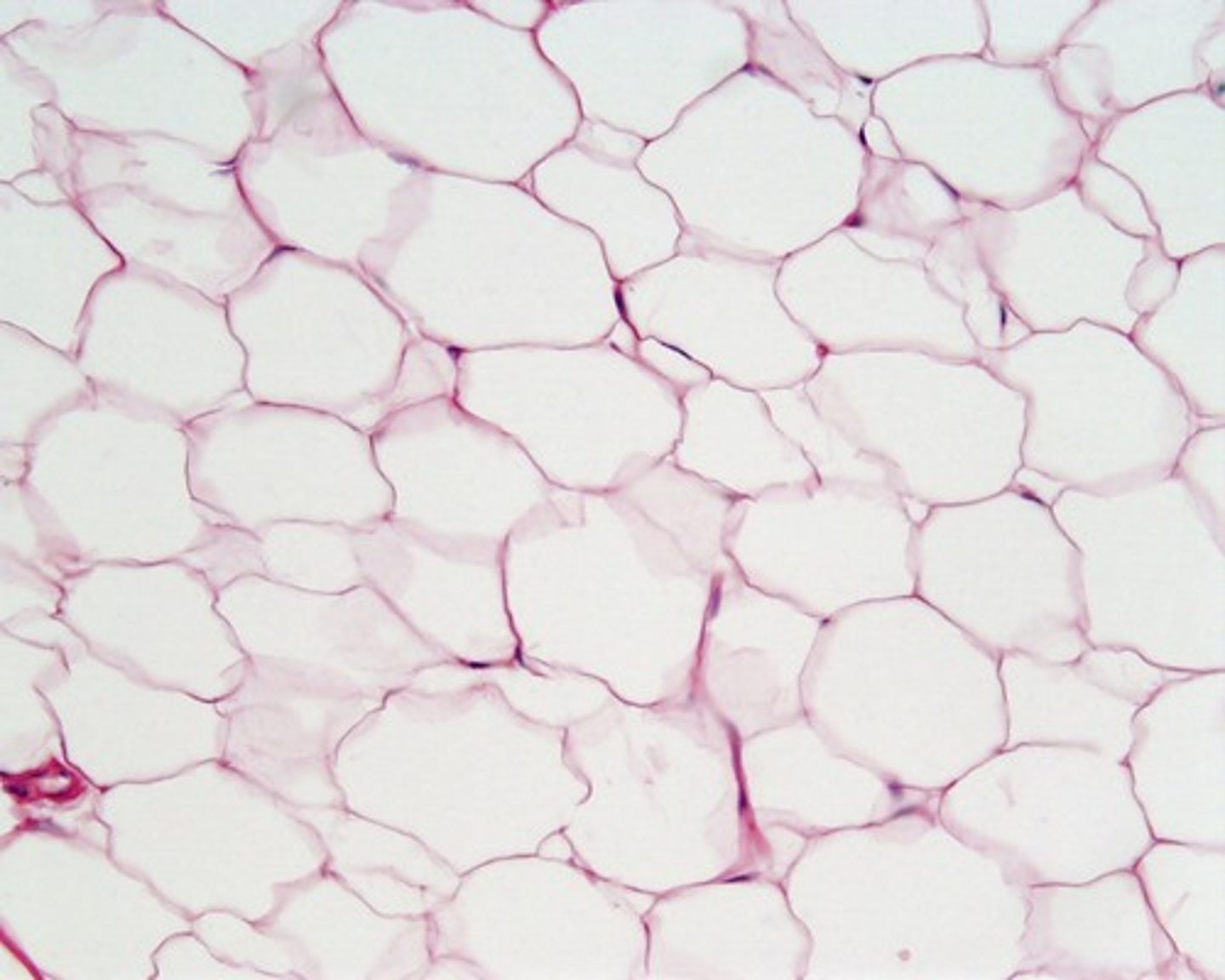
Adipose Tissue
Loose Connective Tissue:
- ADIPOCYTES, stored lipids, nucleus
- fat storage cells (little matrix)
- capillaries = storage and mobilization of lipid molecules
- White adipose = most abundant
- insulation, protecting kidneys, cushioning back of eye
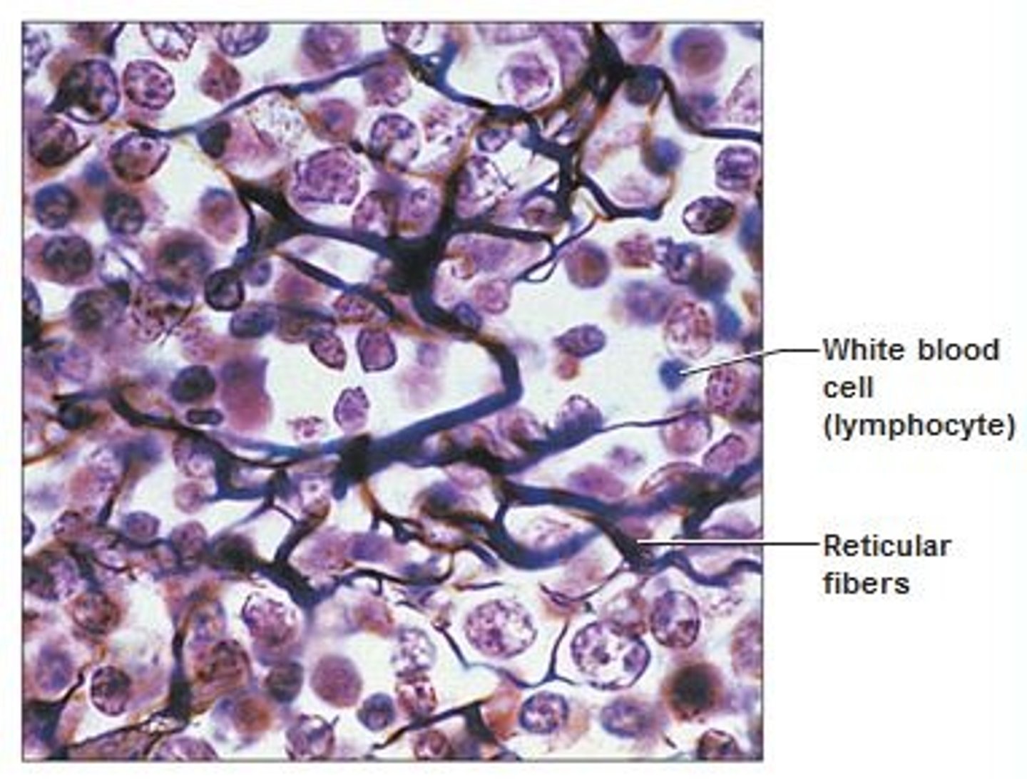
Reticular Tissue
Loose Connective Tissue:
- mesh, supportive framework for soft organs (spleen, liver)
-The cells produce reticular fibers where the cells attach
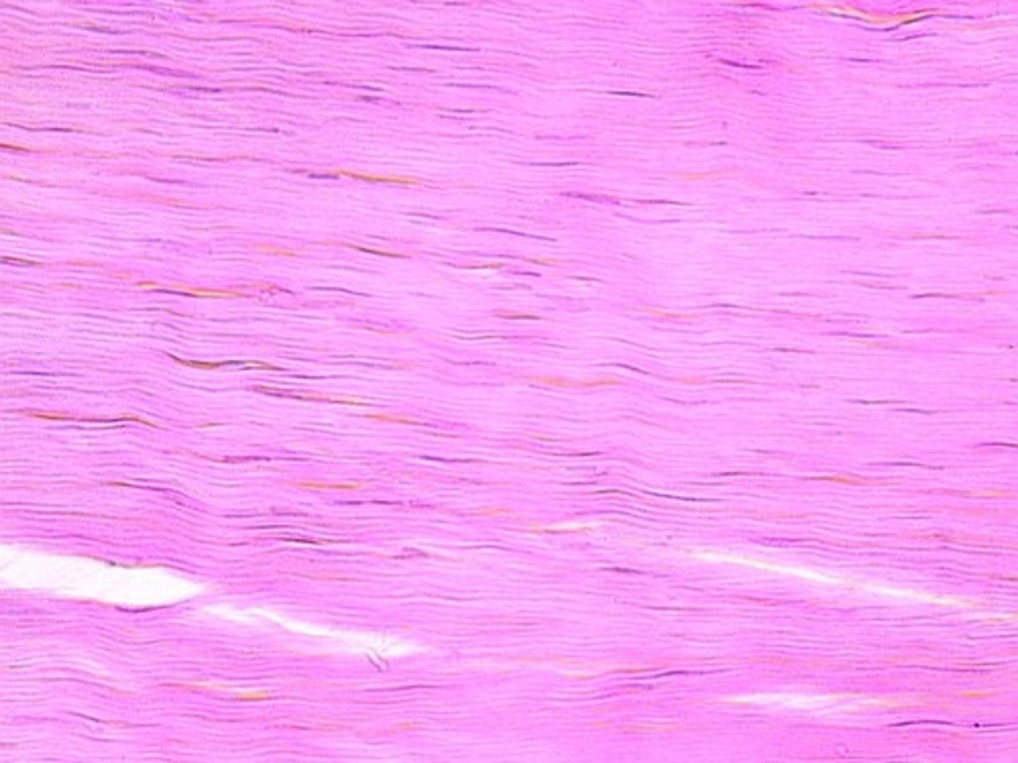
Dense Regular Tissue
- collagen fibers running parallel to one another
- resist stretching
- ligaments/muscle tendons
- Ligaments between vertebrae in vertebral column
- INCLUDES: collagen fibers, fibroblast nuclei
Dense Irregular Tissue
- random direction of fibers
- arrangement = more strength in all directions, but less in one
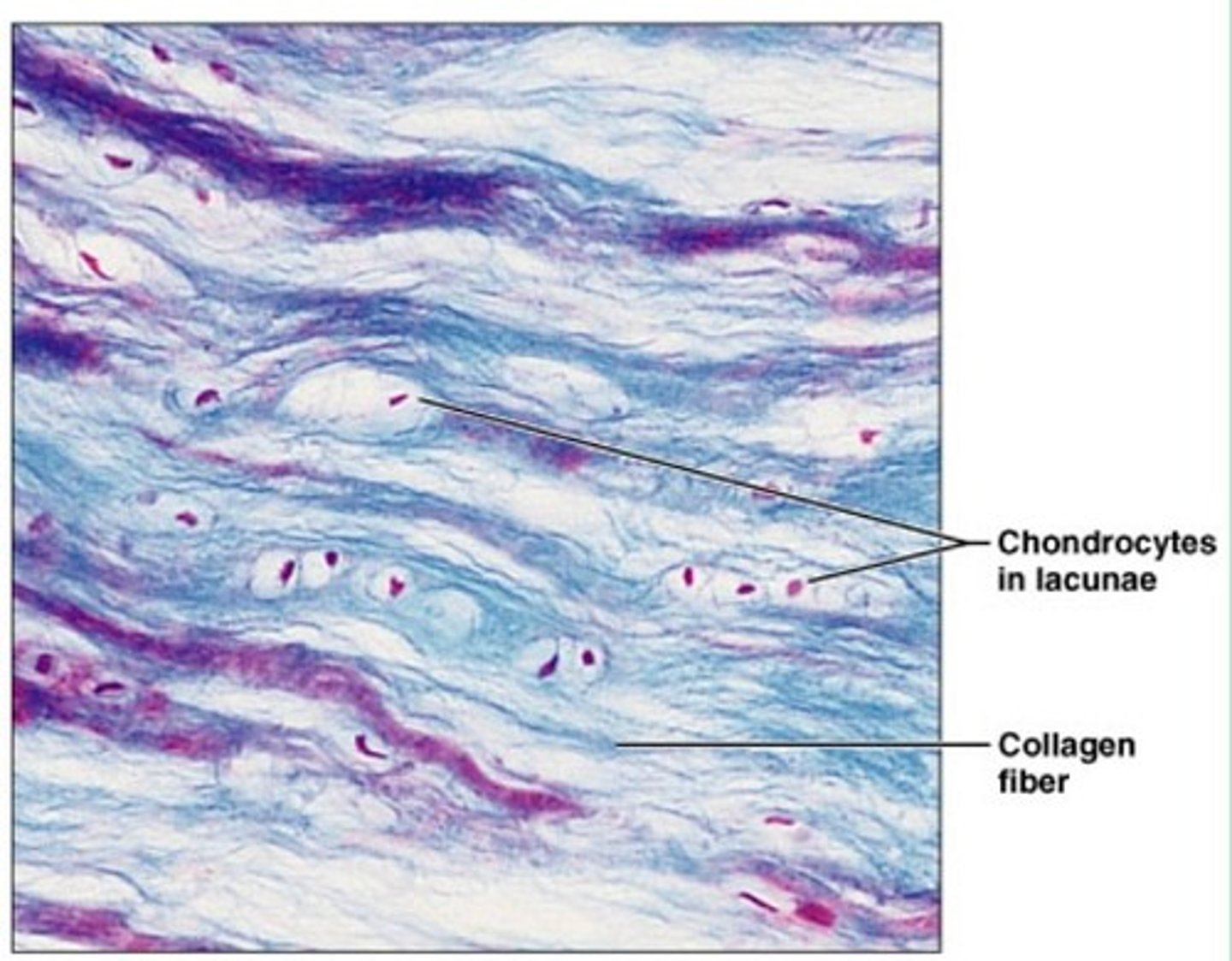
Hyaline Cartilage
- most common
- support AND flexibility
- matrix: short, dispersed collagen fibers; proteoglycans
- LOCATION: rib cage, nose, covers bones at joints

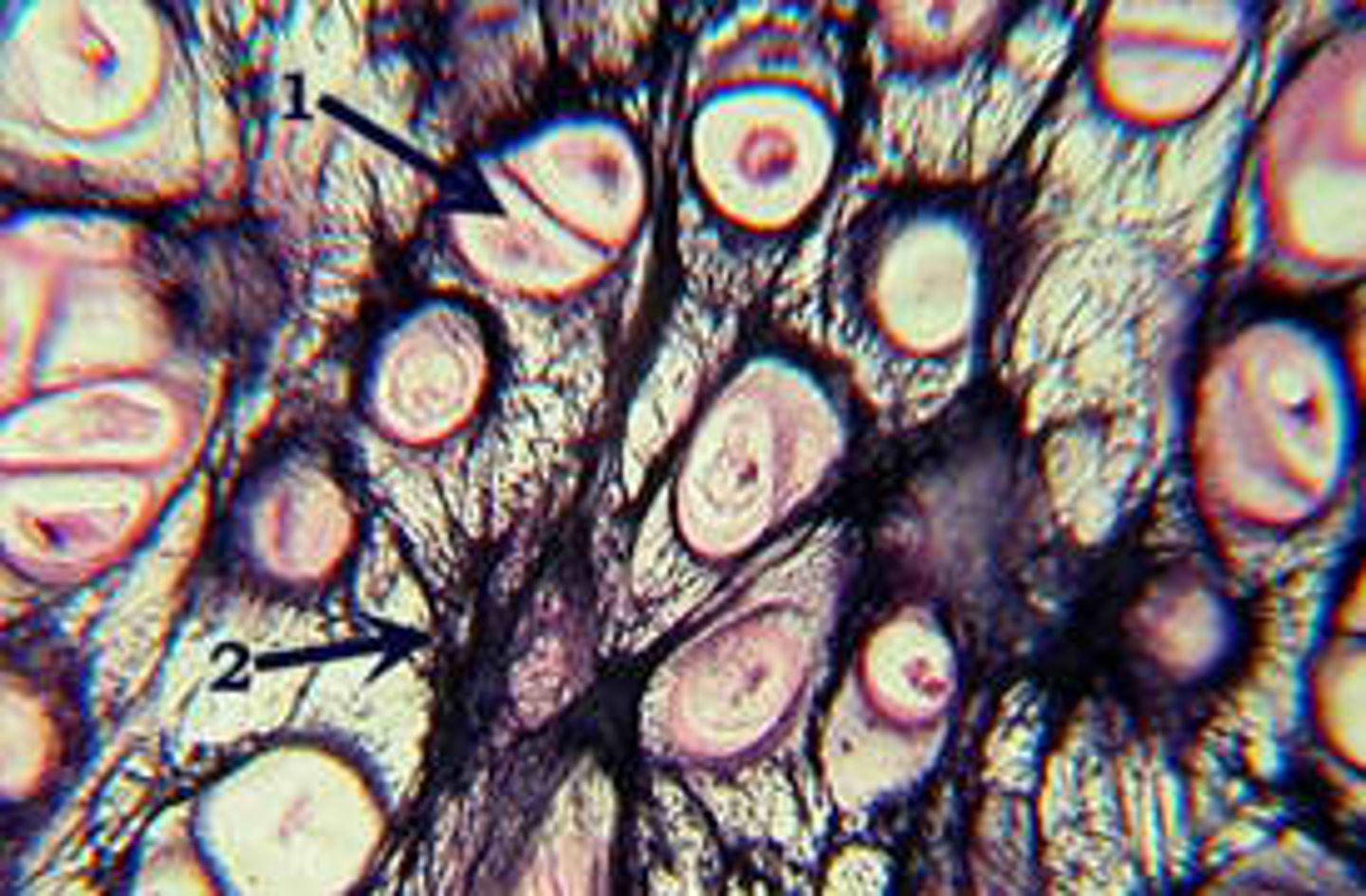
Fibrocartilage
- compressibility
MATRIX
- thick bundles of collagen fibers
FUNCTION
- absorbs pressure
- tough
LOCATION
- menisci in knee joint
- intervertebral discs
Elastic Cartilage
FUNCTION
- firm, elastic support
MATRIX
- elastic fibers AND collagen
- proteoglycans
LOCATION
- ear lobes
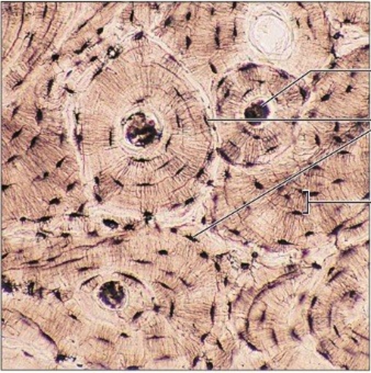
Bone CT
matrix = rigid (calcified)
- hardest CT
FUNCTION
- protects internal organs
- supports body
MATRIX
- collagen fibers covered in mineralized ground substance (HYDROXYAPATITE)
FUNCTION
- collagen helps to make bone more flexible and less brittle
- mineral crystals help increase bone rigidity and support
- osteocytes (bone cells) in the lacunae
- HEALS QUICKLY
Fluid Connective Tissue Types
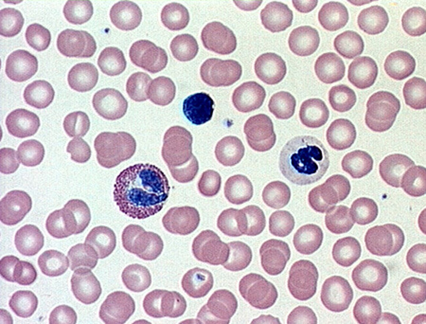
Blood - erythrocytes, leukocytes, platelets
- transport materials t/o body
- respond to illness/injury
Lymph - liquid matrix, leukocytes
- drains into blood vessels (delivering molecules to blood; i.e. absorbed fats from intestines?)
MATRIX
- watery
- salts, nutrients, dissolved proteins
Skeletal tissue
muscle tissue
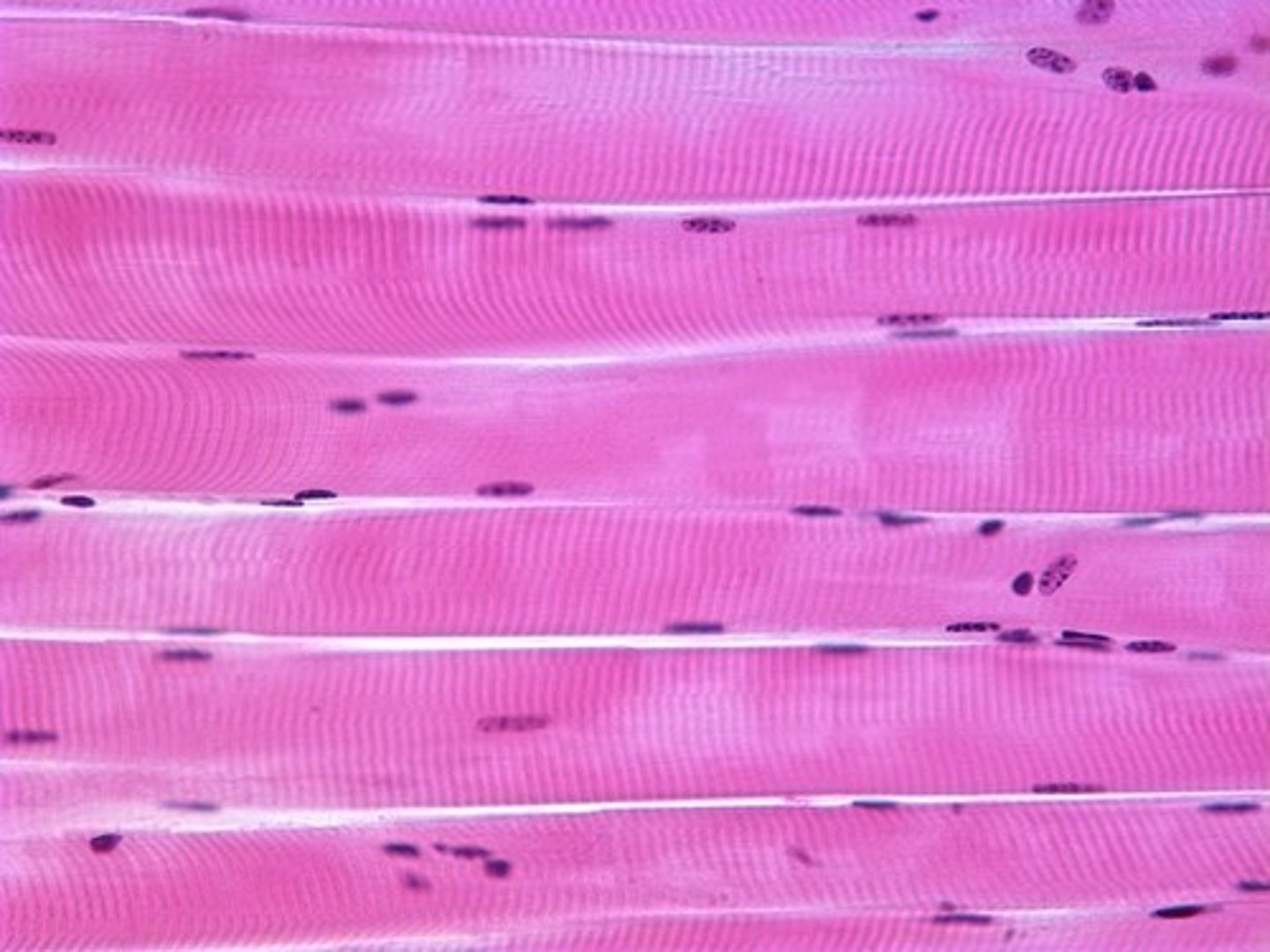
- long, cylindrical fiber, striated, peripheral nuclei
- 40% body mass
- VOLUNTARY MOVEMENT, produces HEAT, protects organs
- attached to bones and around entrance points in body
- multinucleated cells
FUNCTION
- locomotion, facial expressions, posture
- VOLUNTARY MOVEMENT, produces HEAT, protects organs
Epimysium (allows contraction of muscle while also maintaining structural integrity), Perimysium, Endomysium
Skeletal muscle structure
Layers of Connective Tissue:
Epimysium - wraps muscle; dense, irregular CT, contains many budles of muscle fascicles; contract, maintains structural integrity
Perimysium is the surrounding layer around muscle fascicles, includes endomysium and muscle fibers (small bundles inside endomysium); allows nervous system to trigger movement by activating a subset of muscle fibers within the fascicle)
Endomysium - inner-most layer of connective tissue; contains extracellular fluid, nutrients, blood vessels, and nerves needed to support muscle fiber
- Sarcolemma - surrounds muscle fiber?
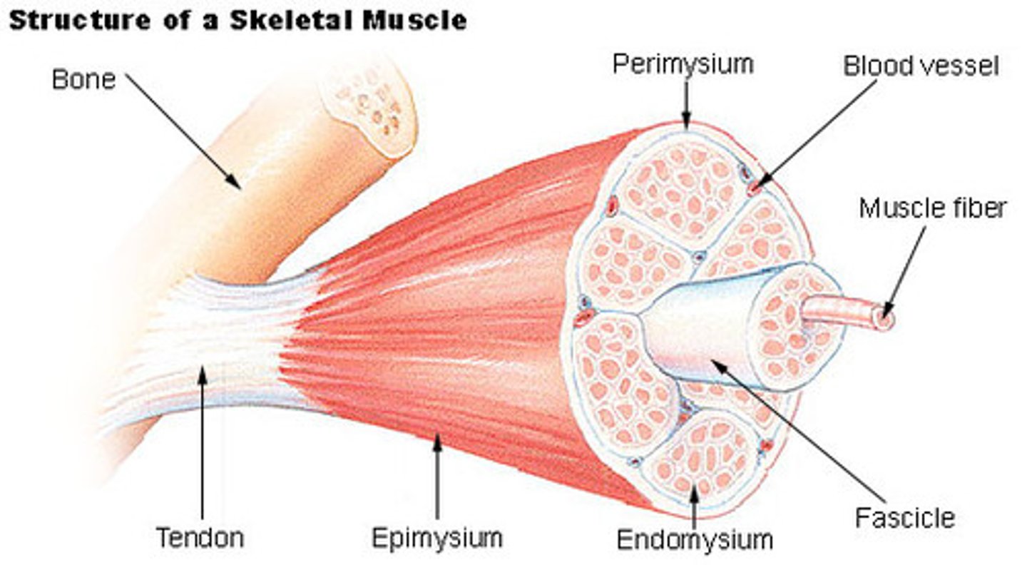
Aponeurosis (Fascia)
connective tissue between skin and bones
Parallel (non-fusiform)
skeletal muscle; fibers arranged in same direction along axis, no central belly
- i.e. Sartorius

Circular (Sphincters)
surrounds an opening to control the size of the opening
- i.e. Orbicularis oculi

Unipennate
tendons run through central region of muscle with fibers located on ONE side of the tendon
- i.e. Extensor digitorum
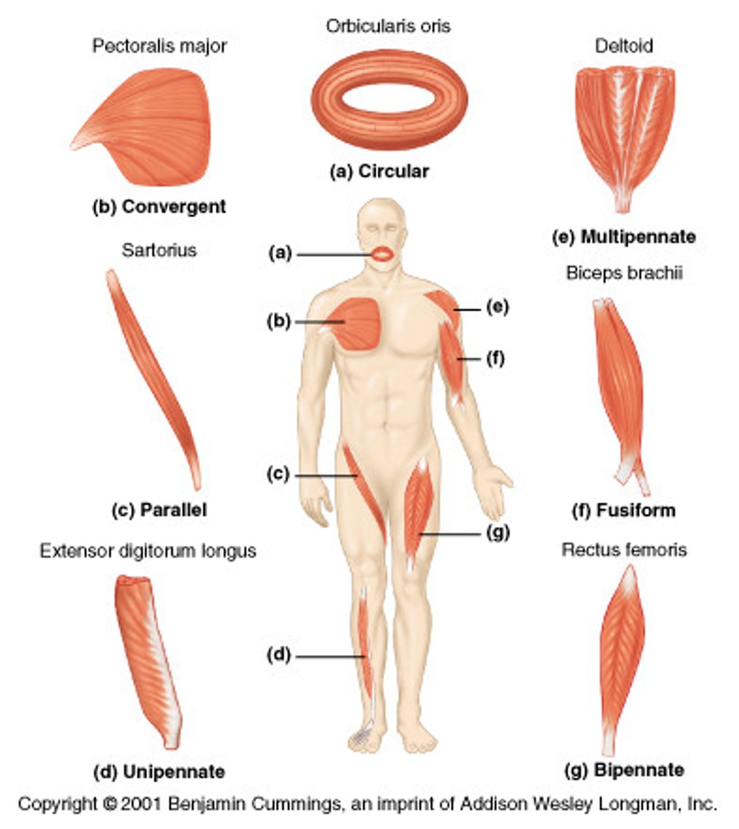
Bipennate
tendon runs down center of muscle with fibers located on both sides of the tendon (looks like a braid)
- i.e. Rectus femoris

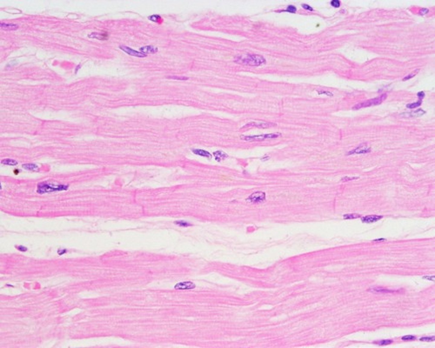
Cardiac tissue
short, branched, striated, single central nucleus
- contracts to move blood in the heart
- cardiomyocytes appear striated under microscope (attach to each other with intercalated discs (specialized cell junctions))
LOCATION
- heart (forms contractile walls of heart)
Multipennate
tendon runs through center of muscle with fibers wrapping tendon on all sides to form separate fascicles
- i.e. Deltoid

Convergent (Triangular)
widespread muscle fibers come to a single attachment point
- i.e. Pectoralis major

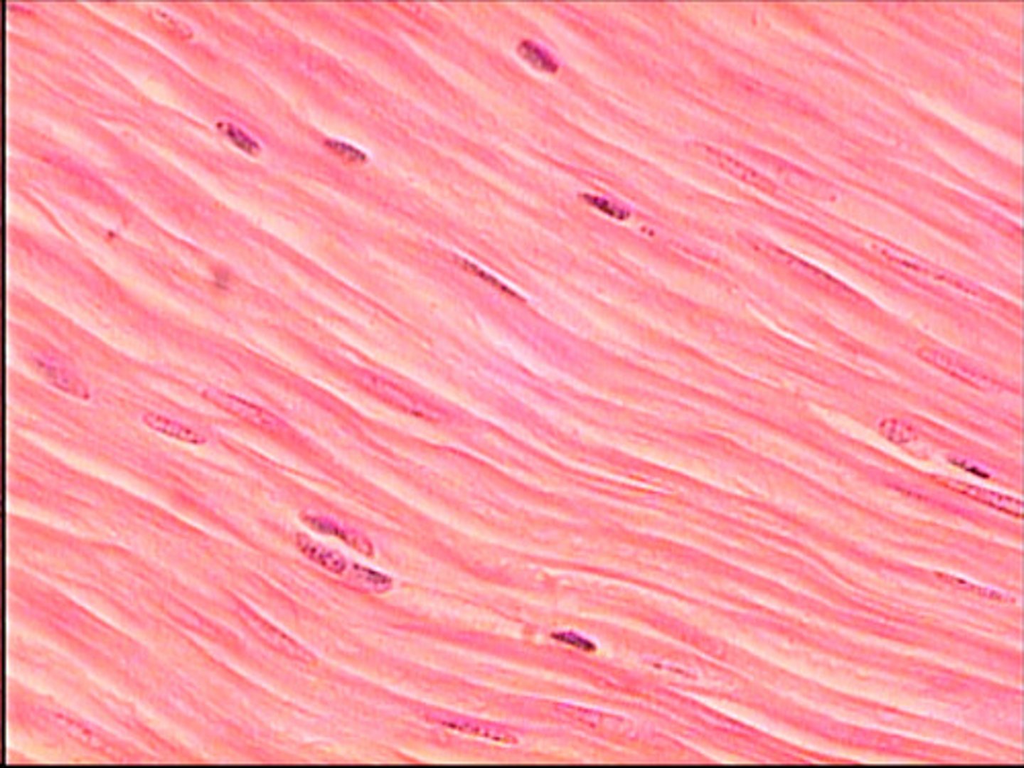
Smooth tissue
short spindle-shaped, single nucleus in each fiber
- INVOLUNTARY movement ( (food/air - respiration; flow of blood thru blood vessels)
LOCATION
- walls of major organs and passageways (digestive, urinary, repro; airways, arteries))
Nervous Tissue
excitable; able to propagate electrochemical signals in the form of nerve signals that allow for communication between different areas of the body
REGIONS
- Central (CNS)
- Peripheral (PNS)
Found in: brain, spinal cord, nerves
Axon
type of process; connects neuron to its target
- propagates nerve impulse
- often covered in myelin sheath (insulation; glial cells)
- axon terminal - connected to synapse, branches toward target cell
Nerves
bundles of axons in the periphery
- nervous AND connective tissue
- blood vessels (nourishment)
- associated with the region of the CNS they are connected to (CRANIAL vs. SPINAL nerves)
Epineurium
outer layer of nerves surrounding everything (fibrous connective tissue)
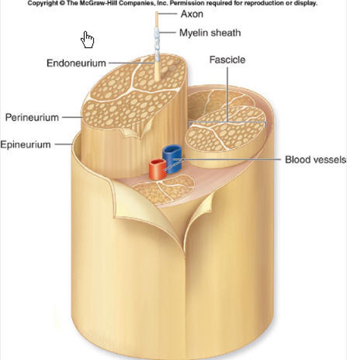
Perineurium
middle layer of fibrous connective tissue in nerves that encompasses the axon bundles (fascicles)
- Axion?
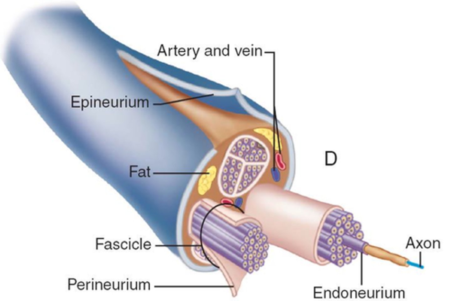
Endoneurium
inner-most layer of LOOSE connective tissue that encompasses individual axons

Epidermis
top layer of skin
- keratinized, stratified squamous epithelium
- NO BLOOD VESSELS (AVASCULAR)
- Keratinocytes = dominant cell type in all layers EXCEPT basale
- Thick skin: five layers
- Thin skin: four layers
Stratum corneum
first layer of epidermis
15-30 layers dead, keratinized keratinocytes
- bound together in sheets
Stratum lucidum
IN-BETWEEN layer of epidermis
- think layer of dead keratinocytes
- ONLY THICK SKIN
Stratum granulosum
SECOND layer of epidermis
3-5 layers keratinocytes
- thickened cell membranes (accumulate keratin, keratohylin granules, and glycolipids)
Stratum spinosum
THIRD layer of epidermis
8-10 layers of keratinocytes that begin to SYNTHESIZE KERATIN and GLYCOLIPIDS
(waterproofing)
Stratum basale
- inner-most layer; bottom (FOURTH/FIFTH layer)
PRODUCES
- Keratinocytes - TACTILE CELLS (sensory)
- MELANOCYTES (pigment)
- single layer cuboidal stem cells that produce KERATINOCYTES
Dermis
inner layer of skin
- contains BLOOD and LYMPH VESSELS, NERVES
- HAIR FOLLICLES, SWEAT GLANDS
PAPILLARY layer and RETICULAR
- ELASTIN and collagenous fibers (fibroblasts)
Papillary Layer
first layer of DERMIS
- loose, areolar connective tissue
- collagen/elastic fibers = loose mesh
- "finger" dermal papillae
- CONTAINS: fibroblasts, fat cells (adipocytes), and blood vessels; PHAGOCYTES (defense); Meissner corpuscles (touch receptors), nerve fibers, lymphatic capillaries
Reticular Layer
deeper layer of the DERMIS
- dense, irregular connective tissue
- VASCULARIZED (MANY NERVES)
- Net like - tight network of fibers
- Elastin fibers - ENABLES MOVEMENT of skin
- Collagen fibers - STRUCTURE (extend into papillary and hypodermis)
Hypodermis
subcutaneous layer/superficial fascia
- right below dermis
GOAL
- connects skin to underlying fascia of bones/muscles
- fat storage, insulation, cushioning
COMPOSITION
- well-vascularized, loose areolar connective tissue AND adipose tissue
Hair
accessory structure of integument
- keratinous filament
- epidermis (dead keratinized cells)
- Hair follicle - dermis
- Hair shaft
- Hair root
- Hair bulb
- Hair papilla (blood capillaries and nerve endings from dermis)
Arrector pili (piloerector muscle)
contracts in response to nerve signals from sympathetic nervous system
Eponychium
nail cuticle
Lunula
base of nail
- thick layer of epithelium over nail matrix
- crescent-shaped region
Sweat glands
Mecrocrine (Eccrine)
- hypotonic sweat, thermoregulation (homeostasis)
- palms of hands, soles of feet, forehead
Apocrine
- hairy (armpits, genital)
- LARGER, DEEPER
-thicker sweat
Diaphysis
tubular shaft of a long bone
- walls = COMPACT BONE (dense, hard)