Last Lab part 1
1/38
There's no tags or description
Looks like no tags are added yet.
Name | Mastery | Learn | Test | Matching | Spaced | Call with Kai |
|---|
No analytics yet
Send a link to your students to track their progress
39 Terms
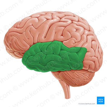
Which lobe is shown?
temporal
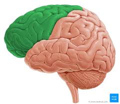
Which lobe is shown?
frontal
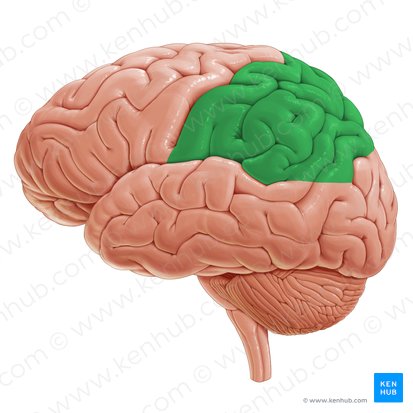
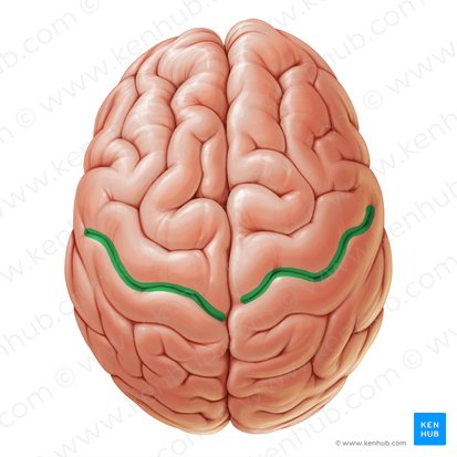
Which lobe is shown?
parietal
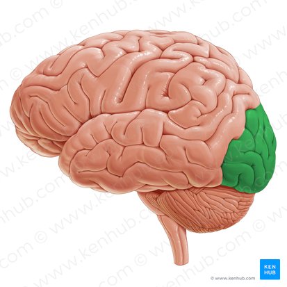
Which lobe is shown?
occipital
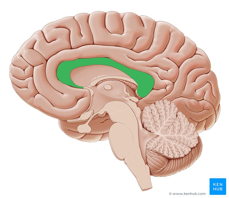
What structure is shown.
corpus callosum
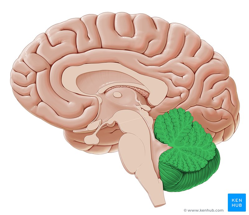
What structure is shown.
cerebellum
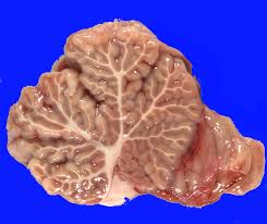
What structure is shown.
arbor vitae
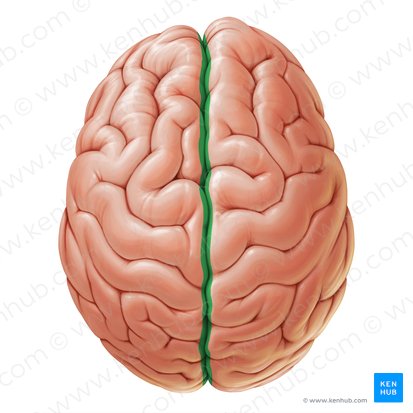
What structure is shown.
longitudinal fissue
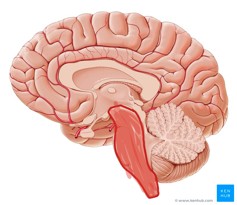
What structure is shown.
brain stem
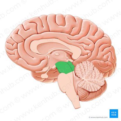
What structure is shown.
midbrain
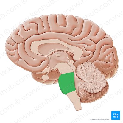
What structure is shown.
pons
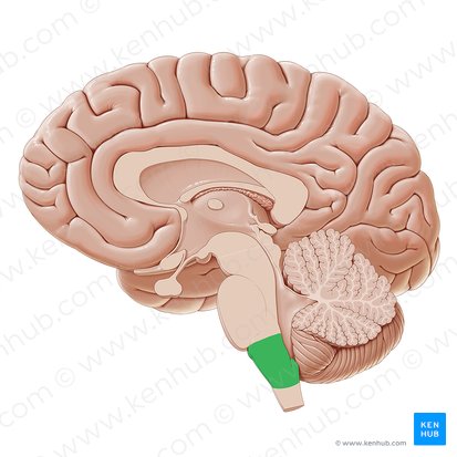
What structure is shown.
medulla oblongata
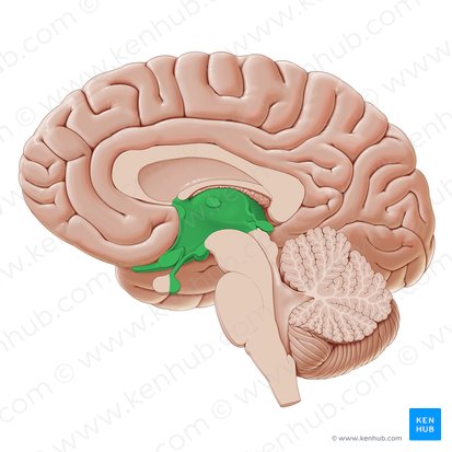
What structure is shown.
diencephalon
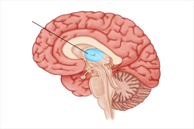
What structure is shown.
thalamus
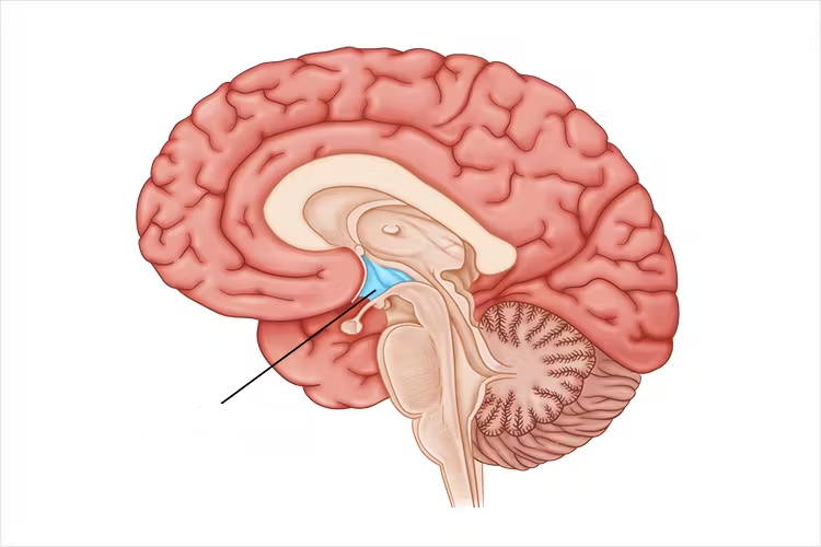
What structure is shown.
hypothalamus
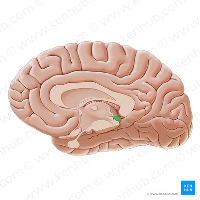
What structure is shown.
epithalamus/pineal

What is the green dot pointing to on the meninges.
dura mater
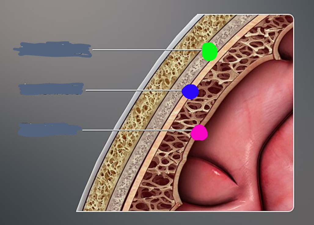
What is the blue dot pointing to on the meninges.
arachnoid

What is the pink dot pointing to on the meninges.
pia matter
What structure is shown.
sulcus/grooves
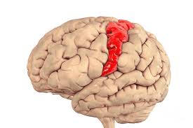
What structure is shown.
gyrus/bumps
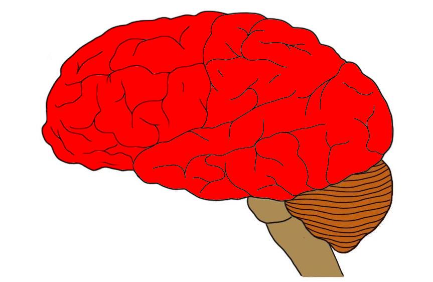
What structure is shown.
cerebrum
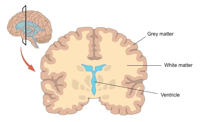
What are the names of the two colors shown in this image?
white and grey matter

Lable 1
grey matter

Lable 2
white matter

Lable 3
dorsal horns

Lable 4
ventral horns

Lable 5
central canal

Lable 6
dorsal root

Lable 7
ventral root

Lable 8
dorsal root ganglion

Lable 9
spinal nerve
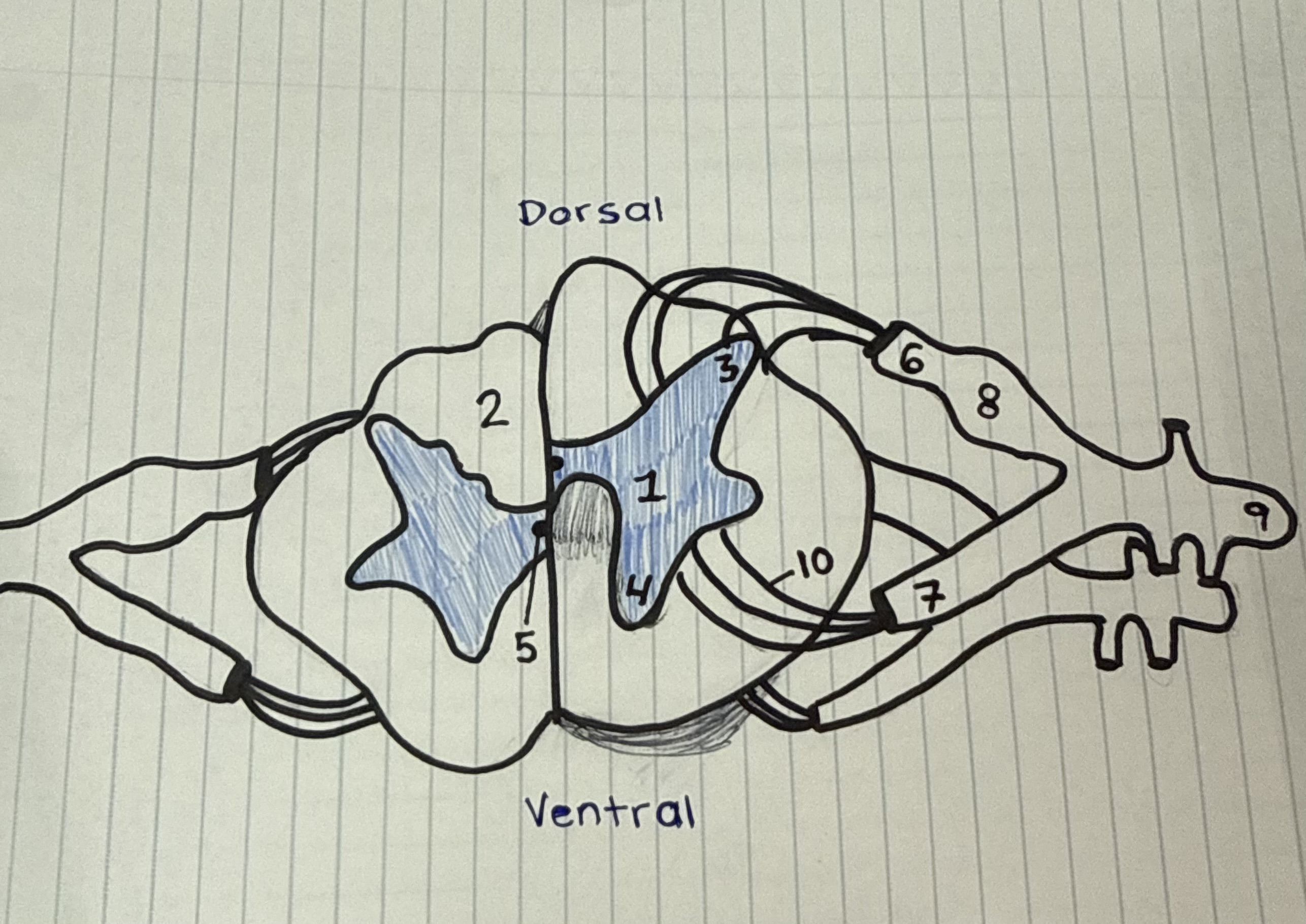
Lable 10
blood vessels

What is the green subdivision of the spine?
cervical c1-c8

What is the blue subdivision of the spine?
thoracic t1-t12
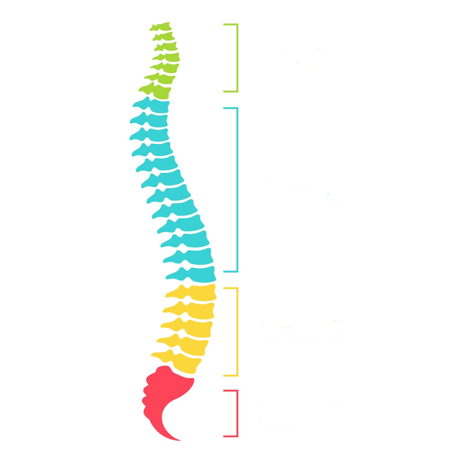
What is the yellow subdivision of the spine?
lumbar l1-l5

What is the red subdivision of the spine?
sacral s1-s5
Describe the relationship between the spinal cord, meninges, and vertabral column.
the vertabral column provides a boney canal that holds the 3 meningeal membranes which support and cushion the spinal cord with cerebralspinal fluid
What are the TWO structural divisions of the nervous system?
central nervous system and peripheal nervous system