Micro Exam 1
1/87
There's no tags or description
Looks like no tags are added yet.
Name | Mastery | Learn | Test | Matching | Spaced | Call with Kai |
|---|
No analytics yet
Send a link to your students to track their progress
88 Terms
Hippocrates
Is considered the “father of Western medicine”. He dismissed the idea that disease was caused by supernatural forces. He introduced the idea that diseases had natural causes from within patients or their environments.
Thucydides
Is considered the father of scientific history because he advocated for evidence-based analysis of cause-and-effect reasoning. Athenian plague, made the important observation that survivors did not get re-infected with the disease, this observation shows an early understanding of the concept of immunity.
Marcus Terentius Varro
One of the first people to propose the concept that things we cannot see (what we now call microorganisms) can cause disease. Because certain minute creatures [animalia minuta] grow there which cannot be seen by the eye, which float in the air and enter the body through the mouth and nose and there cause serious diseases.
Antonie van Leeuwenhoek
Was the first to develop a lens powerful enough to view microbes. Leeuwenhoek was able to observe single-celled organisms, which he described as “animalcules” or “wee little beasties,” swimming in a drop of rain water. From his drawings of these little organisms, we now know he was looking at bacteria and protists.
Louis Pasteur
Showed that individual microbial strains had unique properties and demonstrated that fermentation is caused by microorganisms. He also invented pasteurization, a process used to kill microorganisms responsible for spoilage, and developed vaccines for the treatment of diseases, including rabies, in animals and humans.
Robert Koch
Was the first to demonstrate the connection between a single, isolated microbe and a known human disease. For example, he discovered the bacteria that cause anthrax, cholera, and tuberculosis.
Taxonomy
The classification, description, identification, and naming of living organisms.
Carolus Linnaeus
Linnaeus published Systema Naturae, an 11-page booklet in which he proposed the Linnaean taxonomy, a system of categorizing and naming organisms using a standard format so scientists could discuss organisms using consistent terminology.
Linnaeus divided the natural world into three kingdoms:
animal, plant, and mineral
He grouped organisms using a hierarchy of increasingly specific levels and sublevels based on their similarities.
-Domain, kingdom, phylum class, order, family, genus, and species.
Ernst Haeckel
Proposed another kingdom to add to animalia and plantae, which was protista, for unicellular organisms. He later proposed a fourth kingdom, Monera, for unicellular organisms whose cells lack nuclei, like bacteria.
Prokaryotes
-Lack a membrane-bound nuclei in their cells, monera would be included in this kindgom
Eukaryotes
-Have membrane-bound nuclei in their cells, Fungi, Protista, Plantae, and Animalia would be included in this kingdom
Three domains above the level of Kingdom
Archaea, Bacteria, and Eukarya
Binomial nomenclature
A two-word naming system for identifying organisms by genus and specific epithet. For example, modern humans are in the genus Homo and have the specific epithet name sapiens
An object must measure about ____________ to be visible without a microscope
100 micrometer (µm)
Bacterial cells are typically
1 µm
Meter measurments
decimeter (dm) | 1/10 | 1 dm = 0.1 m = 10−1 m |
centimeter (cm) | 1/100 | 1 cm = 0.01 m = 10−2 m |
millimeter (mm) | 1/1000 | 1 mm = 0.001 m = 10−3 m |
micrometer (μm) | 1/1,000,000 | 1 μm = 0.000001 m = 10−6 m |
nanometer (nm) | 1/1,000,000,000 | 1 nm = 0.000000001 m = 10−9 m |
Acellular
Not composed of cells
The three domains of life
Archaea, Bacteria, and Eukarya
Microbes within the domains Bacteria and Archaea are all prokaryotes (their cells lack a nucleus), whereas microbes in the domain Eukarya are eukaryotes (their cells have a nucleus).
Bacteria
Most bacteria are harmless or helpful, but some are pathogens, causing disease in humans and other animals. Bacteria are prokaryotic because their genetic material (DNA) is not housed within a true nucleus. Most bacteria have cell walls that contain peptidoglycan.
Coccus
Spherical

Bacillus
Rod-shaped

Vibrio
Comma shaped rod
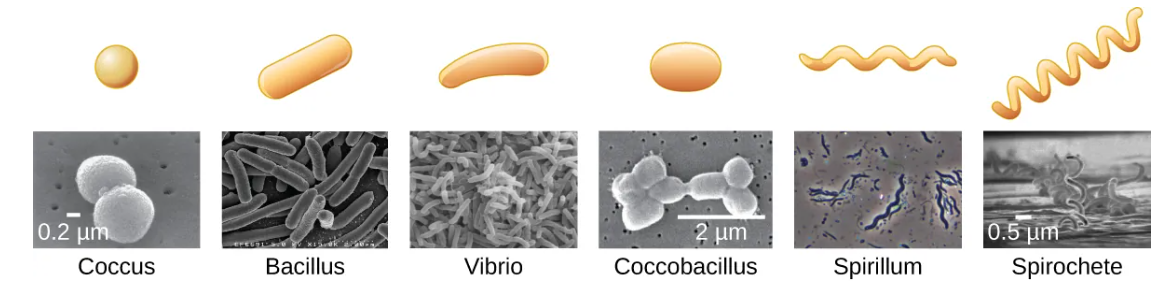
Spirillum
wavy, spirally

Spirochete
Corkscrew

Archeae
Unicellular prokaryotic organisms. Unlike most bacteria, archaeal cell walls do not contain peptidoglycan, but their cell walls are often composed of a similar substance called pseudopeptidoglycan. Like bacteria, archaea are found in nearly every habitat on earth, even extreme environments that are very cold, very hot, very basic, or very acidic.
Protists
An informal grouping of eukaryotes that are not plants, animals, or fungi. Some algae are protists and others are bacteria; all protozoa are examples of protists.
Algae
Mostly made up of protists that can be either unicellular or multicellular
Cyanobacteria
a type of bacteria, is also considered an algae, but these organisms are bacterial prokaryotes and therefore have a peptidoglycan-based cell wall, unlike the cellulose-based cell wall of the algal protists.
Protozoa
are protists that make up the backbone of many food webs by providing nutrients for other organisms. Protozoa are very diverse. Some protozoa move with help from hair-like structures called cilia or whip-like structures called flagella.
Fungi
are eukaryotes. Some multicellular fungi, such as mushrooms, resemble plants, but they are actually quite different. Fungi are not photosynthetic, and their cell walls are usually made out of chitin rather than cellulose.
Unicellular fungi
yeast
Molds
are made up of long filaments that form visible colonies
Helminths
Multicellular parasitic worms, not technically microorganisms, as most are large enough to see without a microscope. However, these worms fall within the field of microbiology because diseases caused by helminths involve microscopic eggs and larvae.
Viruses
acellular microorganisms, which means they are not composed of cells. Essentially, a virus consists of proteins and genetic material—either DNA or RNA, but never both—that
Bacteriology
the study of bacteria;
Mycology
the study of fungi;
protozoology
the study of protozoa
parasitology
the study of helminths and other parasites
Virology
The study of viruses
Immunology
the study of the immune system,
Wavelength
-The distance between one peak of a wave and the next peak.
Amplitude
-The height of each peak (or depth of each trough)
Frequency
-The rate of vibration of the wave, or the number of wavelengths within a specified time period
Reflection
Occurs when a wave bounces off of a material
Absorbance
-Occurs when a material captures the energy of a light wave.
Transmission
-Occurs when a wave travels through a material, like light through glass (the process of transmission is called transmittance).
Interference
-Creating complex patterns of motion.
Diffraction
Waves can also interact with small objects or openings by bending or scattering.
Refraction
Perhaps the most important behavior exhibited by light waves. Refraction is when light bends as it moves from one material to another. This happens because light travels at different speeds in different materials, causing it to change direction.
Refractive Index
The extent to which a material slows transmission speed relative to empty space
Image Point (focus)
We can think of a lens as an object with a curved boundary (or a collection of prisms) that collects all of the light that strikes it and refracts it so that it all meets at a single point called the image point (focus)
Focal point
The image point when light entering the lens is parallel
Focal length
The distance to the focal point
Electromagnetic radiation (EMR)
A type of energy that is all around us. Other forms of EMR include visible light, microwaves, X-rays, and radio waves
Dispersion
The seperation of colors and it occurs because, for a given material, the refractive index is different for different frequencies of light.
Magnification
The ability of a lens to enlarge the image of an object when compared to the real object.
Resolution
The ability to tell that two separate points or objects are separate. A low-resolution image appears fuzzy, whereas a high-resolution image appears sharp.
Numerical Aperture
-A measure of a lens’s ability to gather light. The higher the numerical aperture, the better the resolution.
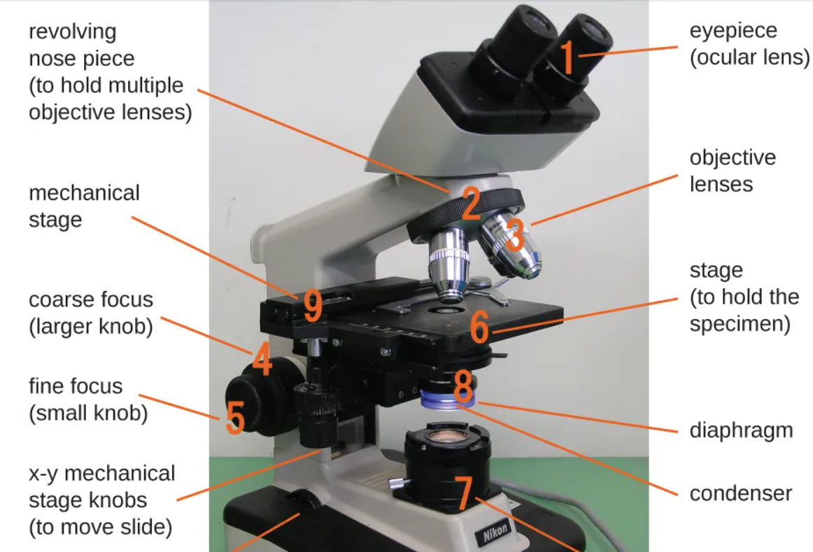
Galileo Galilei
Who used a compound microscope to examine insect parts. A compound microscope uses two sets of lenses to magnify objects, making it more powerful than a simple microscope, which uses only one lens.
Robert Hooke
Publishing in his book Micrographia (1665) many observations using compound microscopes. Viewing a thin sample of cork through his microscope, he was the first to observe the structures that we now know as cells
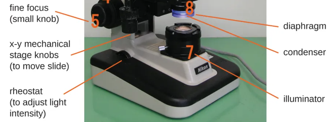
Brightfield microscope
A compound microscope with two or more lenses that produce a dark image on a bright background. Most commonly used microscope
Chromophores
Pigments that absorb and reflect particular wavelengths of light. Chromophores are artificially added to the specimen using stains, which serve to increase contrast and resolution.
Oil immersion lens
At very high magnifications, resolution may be compromised when light passes through the small amount of air between the specimen and the lens. This is due to the large difference between the refractive indices of air and glass; the air scatters the light rays before they can be focused by the lens. To solve this problem, a drop of oil can be used to fill the space between the specimen
Darkfield microscope
A darkfield microscope uses a special disk that blocks most of the light. Only light reflected or bent by the specimen reaches the lens, making the specimen appear bright against a dark background. (Used for things, like live things, that can’t be stained becaue they would die)
Phase-contrast microscopes
Uses refraction and interference caused by structures in a specimen to create high-contrast, high-resolution images without staining. It is the oldest and simplest type of microscope that creates an image by altering the wavelengths of light rays passing through the specimen. (often used to observe live specimens. Certain structures, such as organelles in eukaryotic cells and endospores in prokaryotic cells, are especially well visualized with phase-contrast microscopy)
Differential interference contrast (DIC) microscopes
A differential interference contrast (DIC) microscope uses two light beams to create high-contrast images. It makes live, unstained specimens appear three-dimensional, making it easier to see internal structures.
fluorescence microscope
Ues fluorescent chromophores called fluorochromes, which are capable of absorbing energy from a light source and then emitting this energy as visible light. (especially useful in clinical microbiology. They can be used to identify pathogens, to find particular species within an environment, or to find the locations of particular molecules and structures within a cell.)
immunofluorescence
which is used to identify certain disease-causing microbes by observing whether antibodies bind to them.
Direct immunofluorescence assay (DFA): Fluorescently labeled antibodies attach directly to the pathogen, allowing it to be seen under a fluorescent microscope.
Indirect immunofluorescence assay (IFA): Unlabeled primary antibodies bind to the pathogen, and fluorescent secondary antibodies bind to the primary antibodies. This produces a brighter image and makes the pathogen easier to detect.
Confocal microscope
uses a laser to scan multiple z-planes successively. This produces numerous two-dimensional, high-resolution images at various depths, which can be constructed into a three-dimensional image by a computer. (very useful for examining thick specimens such as biofilms, which can be examined alive and unfixed)
Two-Photon Microscopes
Uses a scanning technique, fluorochromes, and long-wavelength light (such as infrared) to visualize specimens. (useful for examining living cells within intact tissues—brain slices, embryos, whole organs, and even entire animals.)
Electron microscope (EM)
uses short-wavelength electron beams rather than light to increase magnification and resolution.
transmission electron microscope (TEM) and scanning electron microscope (SEM)
-Transmission Electron Microscope (TEM) Uses a beam of electrons that passes through a very thin specimen.Produces detailed images of internal structures. Requires the specimen to be thin, dehydrated, and placed in a vacuum. Can achieve very high magnification and resolution.
Scanning Electron Microscope (SEM)Uses electrons to scan the surface of a specimen. Produces detailed, three-dimensional-looking images of surfaces. Specimens are dried and coated with a thin layer of metal, such as gold. Used to view the outside of larger and smaller objects.
Key difference: TEM shows the inside of a specimen, while SEM shows the surface.
scanning probe microscope
does not use light or electrons, but rather very sharp probes that are passed over the surface of the specimen and interact with it directly. This produces information that can be assembled into images with magnifications up to 100,000,000⨯. Such large magnifications can be used to observe individual atoms on surfaces.
Scanning Tunneling Microscope (STM)
Uses a probe that moves just above a specimen's surface.
Measures tiny electrical currents between the probe and specimen.
Creates detailed images of surfaces, even showing individual atoms.
Atomic Force Microscope (AFM)
Uses a probe that moves over the specimen's surface.
Measures changes in the probe's height caused by forces between atoms.
Produces highly detailed images of surfaces at the atomic level.
Key difference: STM measures electrical current, while AFM measures changes in probe height caused by atomic forces.
Wet mount
A wet mount is made by placing a specimen in a drop of liquid on a microscope slide and covering it with a coverslip. The liquid may be water or a stain to improve visibility. This allows the specimen to be viewed under a microscope.
Fixation
Fixation is the process of attaching cells to a microscope slide. It kills microorganisms and preserves their structures for viewing. Fixation can be done by heating the specimen (heat-fixing) or by using chemicals such as formaldehyde, ethanol, or methanol.
Staining
is used to add color to specimens so they are easier to see under a microscope. Dyes contain colored ions called chromophores. If the colored ion is positive, it is a basic dye; if the colored ion is negative, it is an acidic dye.
Positive stain
a dye that will be absorbed by the cells or organisms being observed, adding color to objects of interest to make them stand out against the background.
Negative stain
Which is absorbed by the background but not by the cells or organisms in the specimen. Negative staining produces an outline or silhouette of the organisms against a colorful background
Simple staining
a single dye is used to emphasize particular structures in the specimen. A simple stain will generally make all of the organisms in a sample appear to be the same color, even if the sample contains more than one type of organism.
Differential staining
Differential staining uses multiple stains to distinguish between different types of organisms. As a result, different organisms may appear different colors. Common differential stains include Gram staining, acid-fast staining, endospore staining, flagella staining, and capsule staining.
Grma stain procedure
Gram Staining Steps:
Crystal violet is added, turning all cells purple.
Gram's iodine is added to help trap the purple stain inside the cells.
Alcohol or acetone is used to remove the stain from some cells.
Cells with thick cell walls stay purple.
Cells with thin cell walls lose the purple color.
Safranin is added, staining the decolorized cells pink.
Results:
Gram-positive bacteria = purple
Gram-negative bacteria = pink/red
Acid-fast stains
Acid-fast staining is used to identify bacteria with waxy cell walls containing mycolic acids. These bacteria retain the red carbolfuchsin stain even after being washed with acid-alcohol, while non–acid-fast cells are stained blue by methylene blue.
Acid-fast bacteria = red/pink
Non–acid-fast bacteria = blue
The Ziehl-Neelsen method uses heat during staining, while the Kinyoun method does not.
Capsule staining
Capsule staining is used to detect capsules, which are protective outer layers that help microbes cause disease. Because capsules do not absorb most dyes, a negative stain colors the background instead, making the capsule appear as a clear halo around the cell. Common stains used include India ink and nigrosin.
Endospore staining
ndospore staining is used to detect endospores, which help certain bacteria survive harsh conditions. The Schaeffer-Fulton method uses malachite green and heat to stain endospores green, then safranin to stain the rest of the cell pink.
Endospores = green
Vegetative cells = pink
This stain helps show the presence, shape, and location of endospores.
Flagella staining
Flagella staining is used to make thin flagella visible under a light microscope. A mordant is first applied to thicken the flagella, then a stain such as pararosaniline or basic fuchsin is added. This helps microbiologists see the number and location of flagella, which can be useful for identifying bacteria.