repro microanatomy
1/260
There's no tags or description
Looks like no tags are added yet.
Name | Mastery | Learn | Test | Matching | Spaced | Call with Kai |
|---|
No analytics yet
Send a link to your students to track their progress
261 Terms
surface epithelium
green 1
tunica albuginea
green 2

sub-surface epithelial structures
blue

ovary
red

uterine horn
orange

oviduct
yellow

uterine body
green

cervix
blue
mesometrium
purple

broad ligament
mesometrium is also called the _______
surface epithelium
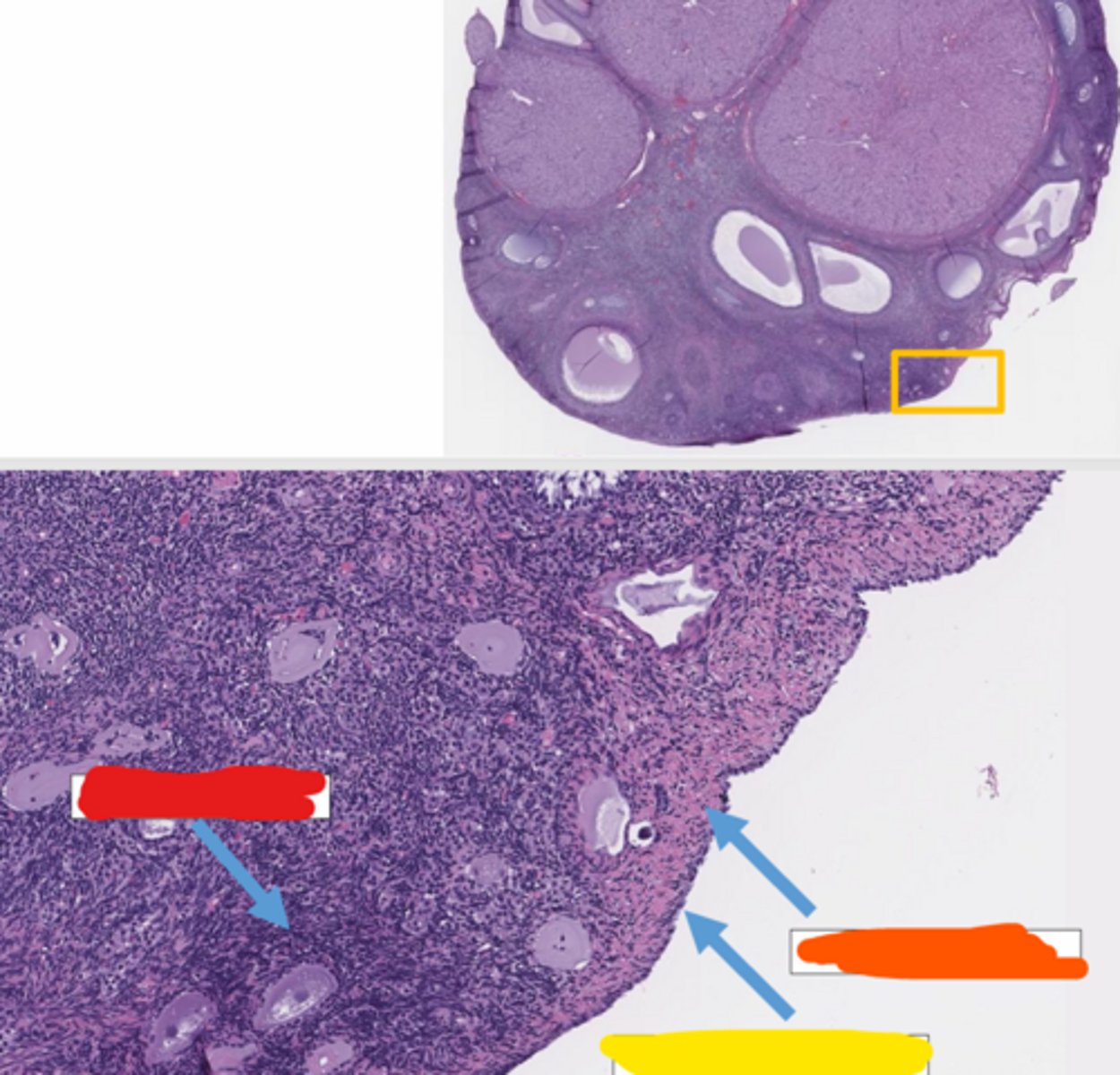
what is the outside layer of the overy called?
sub-surface epithelial structures (SES)
in dogs, the ovary has invaginations for ____
horses
in what species are the cortex and medulla inverted in the ovary?
cortex
in what part of the ovary does oogenesis occur?
follicles
oogenesis is the development of ______
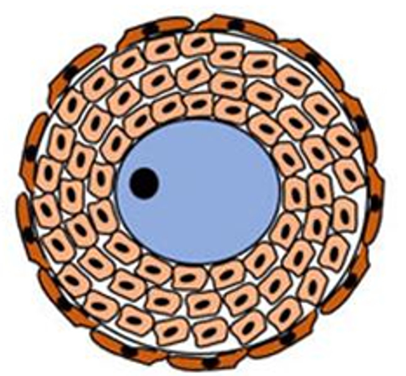
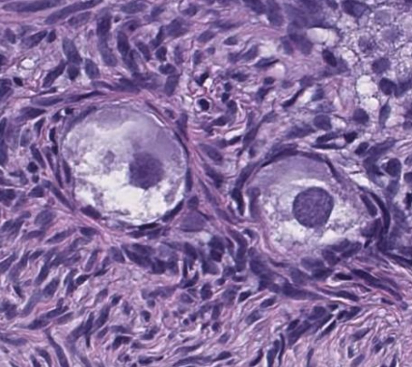
primordial, epithelial
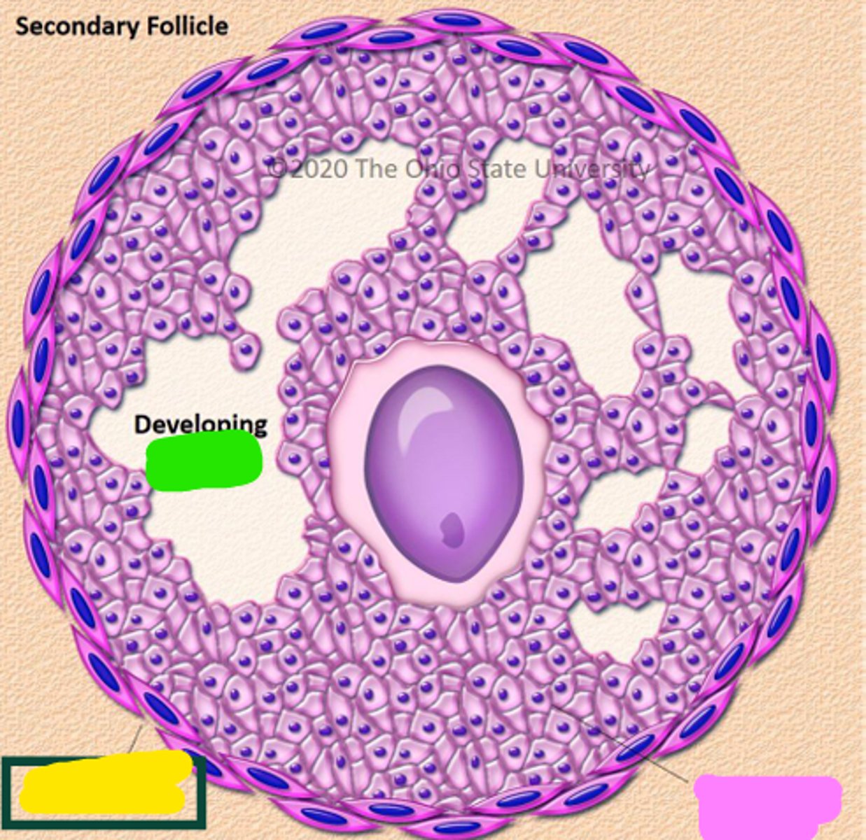
in the _____ follicle, _____ cells are surrounding the oocyte

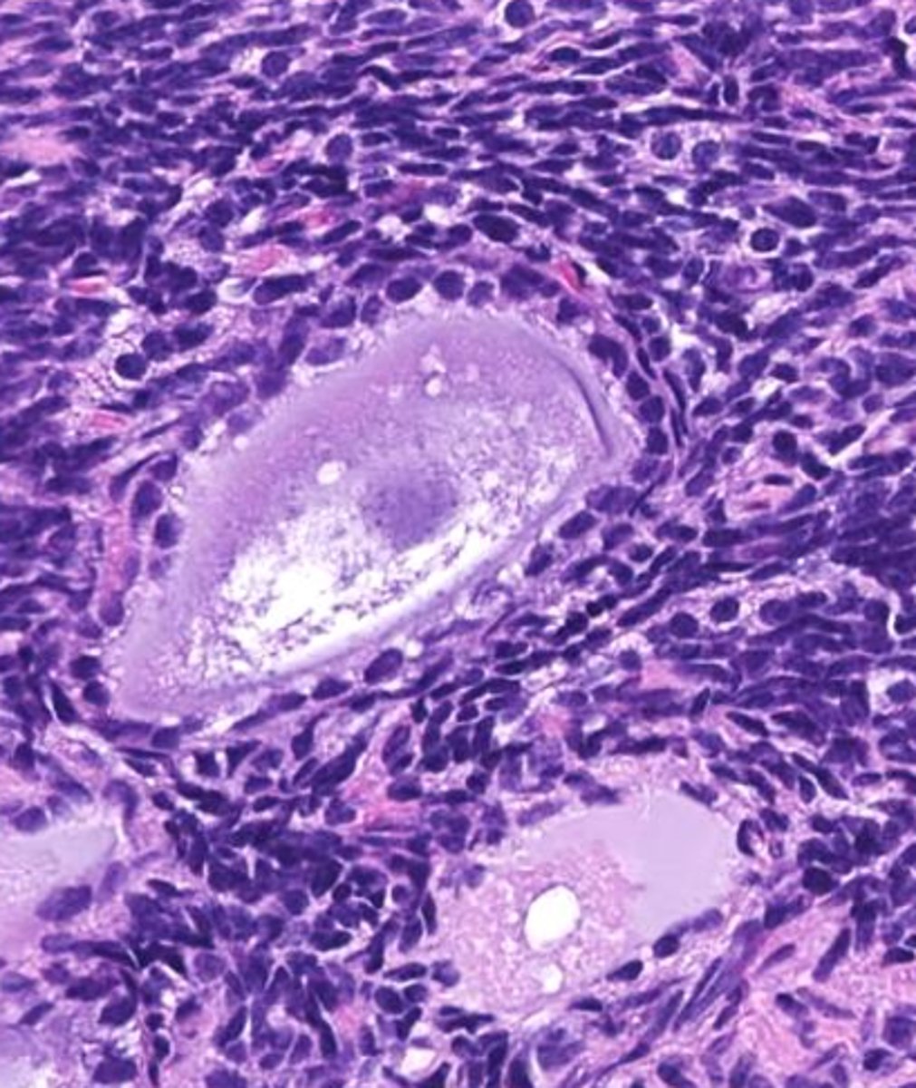
primary, granulosa
in the _____ follicle, _____ cells are surrounding the oocyte
secondary, granulosa, thecal
this is the _____ follicle: As the oocyte develops, the _____ cells proliferate and ______ cells are recruited from the stroma
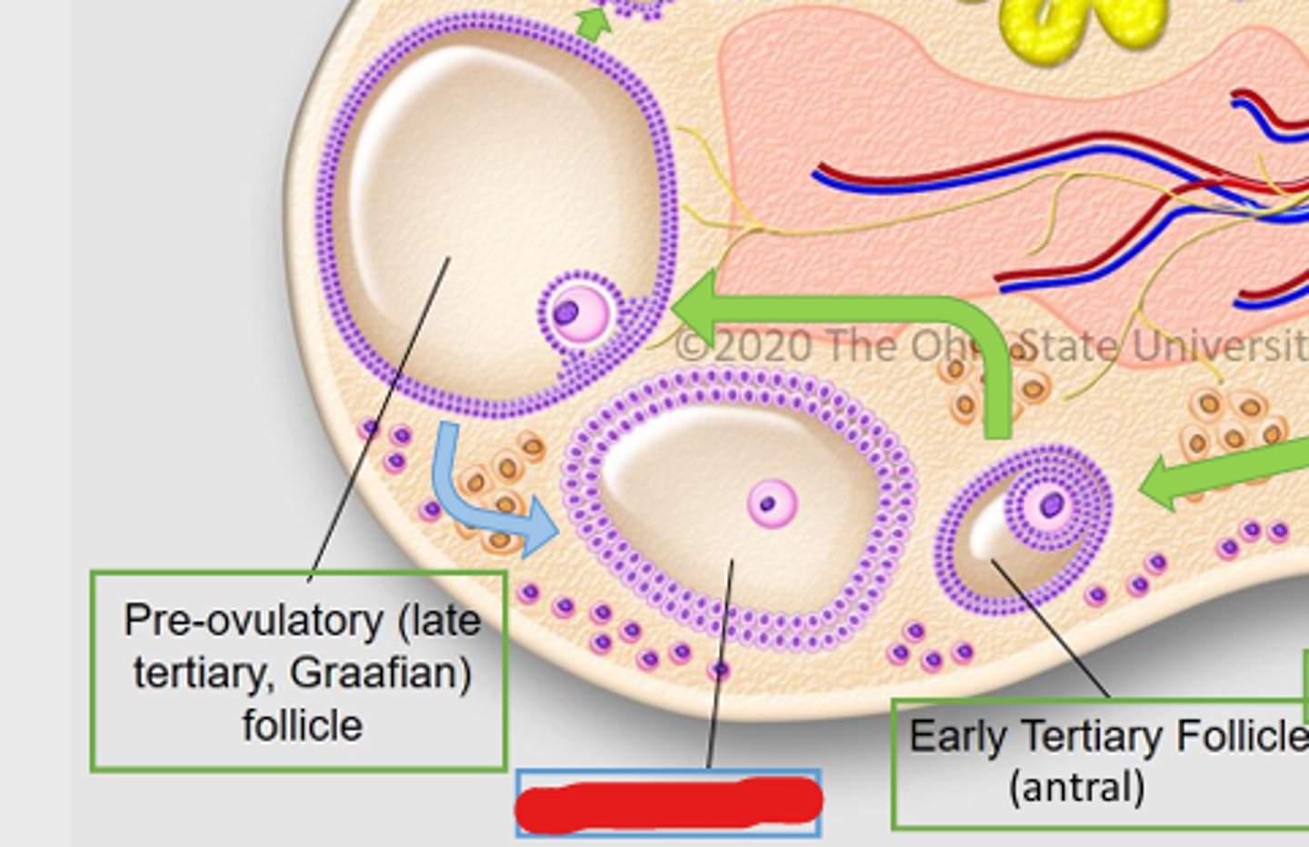
tertiary
this is a _____ follicle
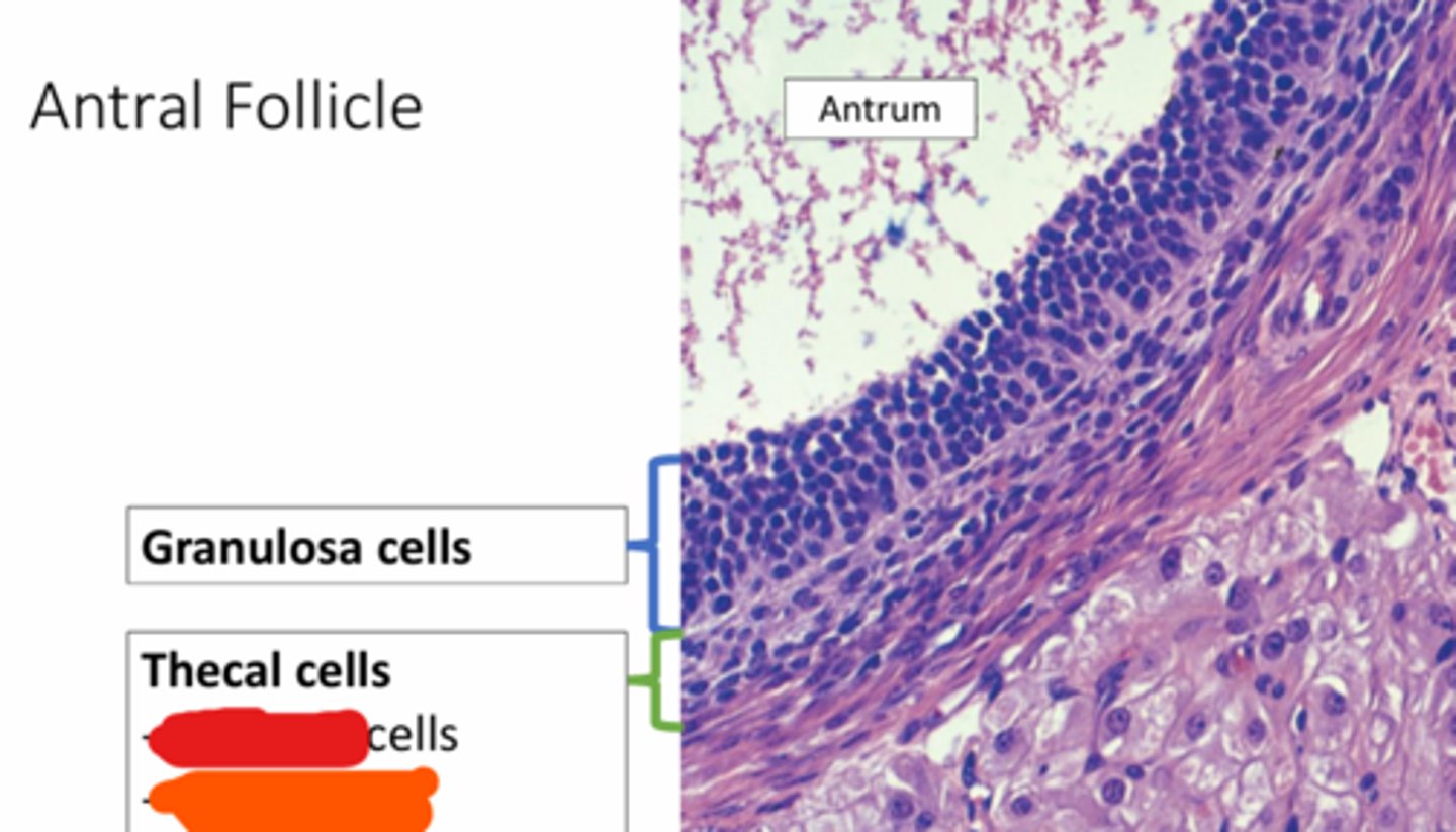
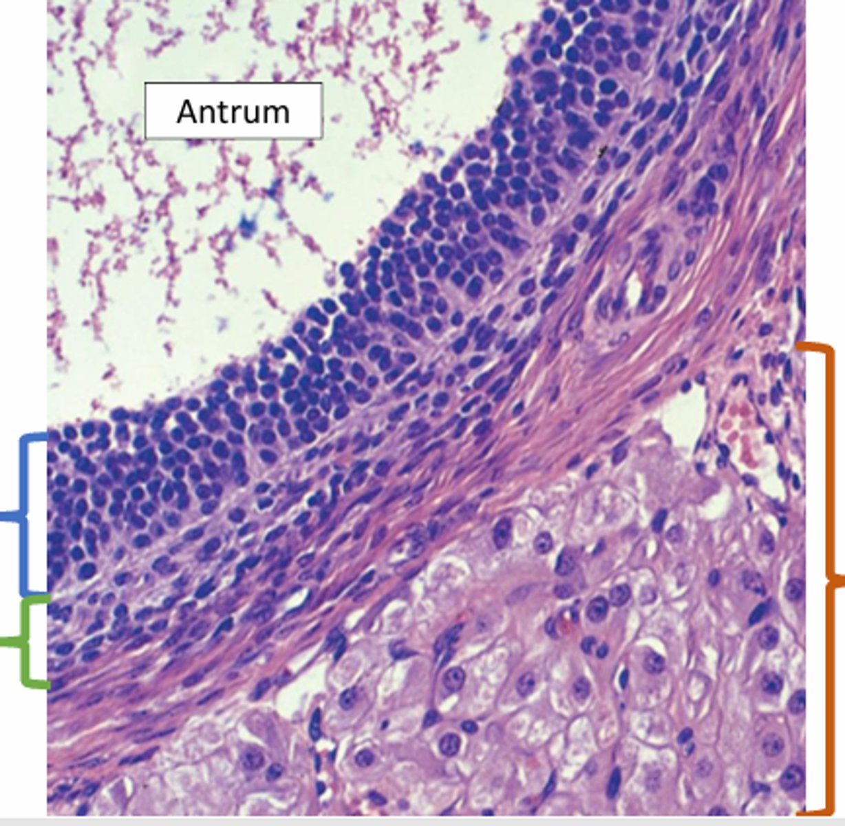
antrum
the fluid filled space, marked by the X, is called the _____

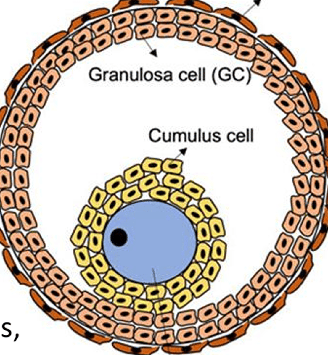
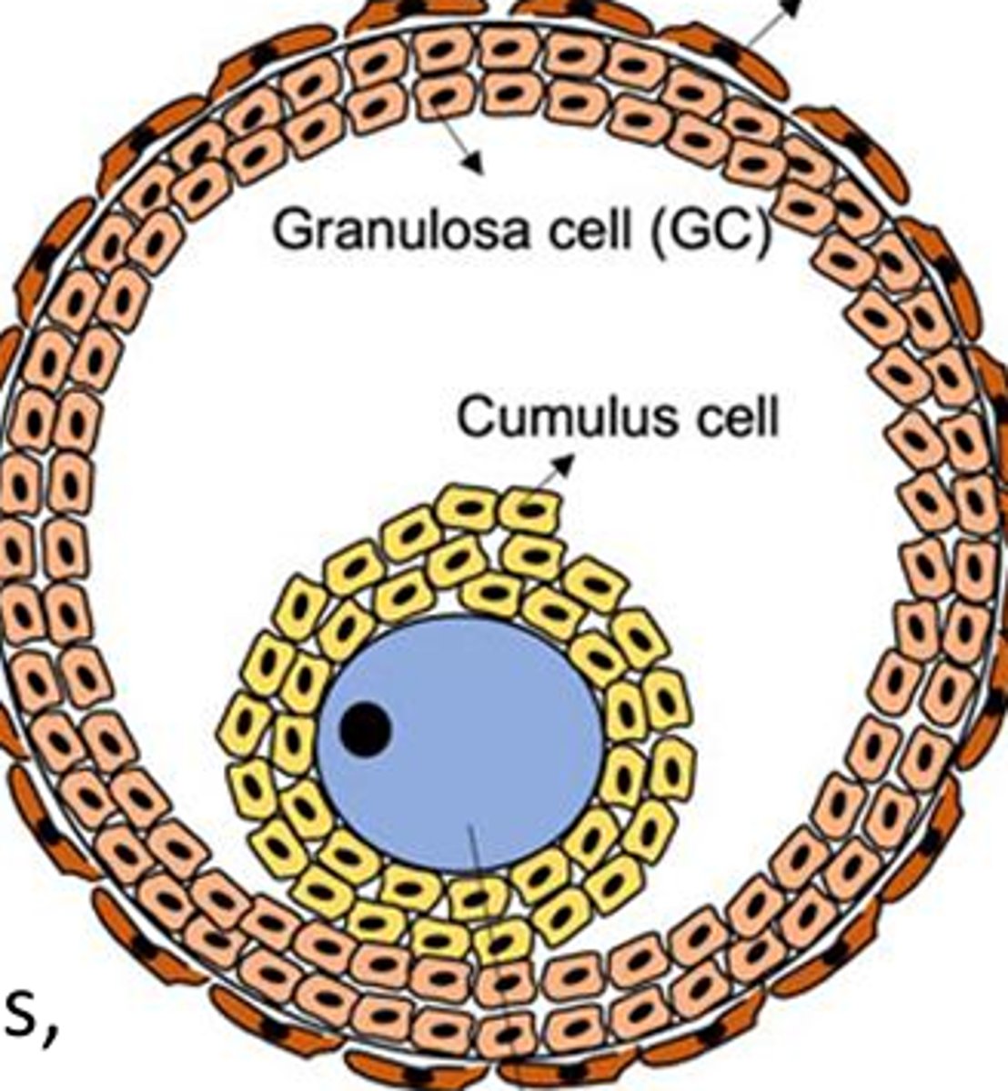
cumulus
the yellow cells surrounding the oocyte in the antral follicle are called ____ cells

Graafian
antral follicles, or tertiary follicles, are also called ______ follicles
atretic follicle
degenerating follicle
cortical stroma
red

tunica albuginea
orange
germinal epithelium
yellow

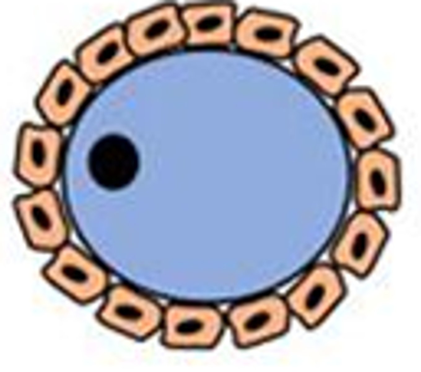
primordial
_____ follicle

follicular/epithelial
the flattened cells surrounding the primordial follicle are ______ cells
primary
_____ follicle

granulosa
As the oocyte develops in the primary follicle, the follicular cells become ______ cells
secondary
_____ follicle
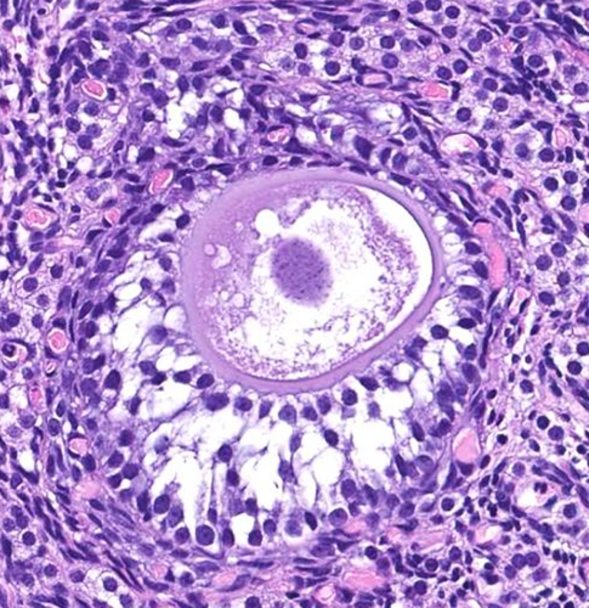
theca cells
yellow

antrum
green
granulosa cells
pink

theca cells
pink
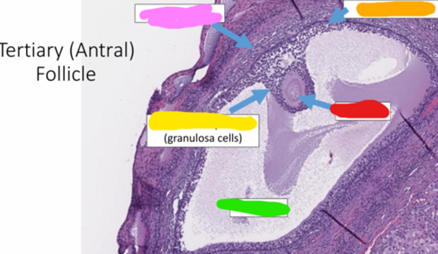
granulosa cells
orange

oocyte
red

antrum
green

cumulus oophorus
a mound of granulosa cells that covers the oocyte and secures it to the follicle wall (yellow)

stromal
red

spindleoid
orange
follicles
whats circled
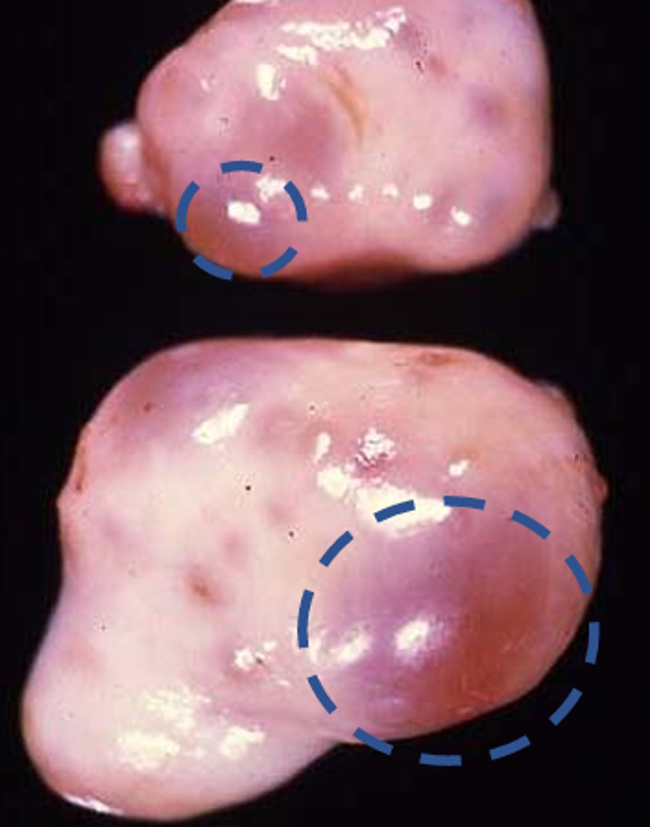
random
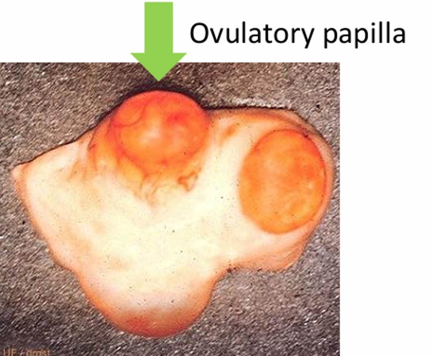
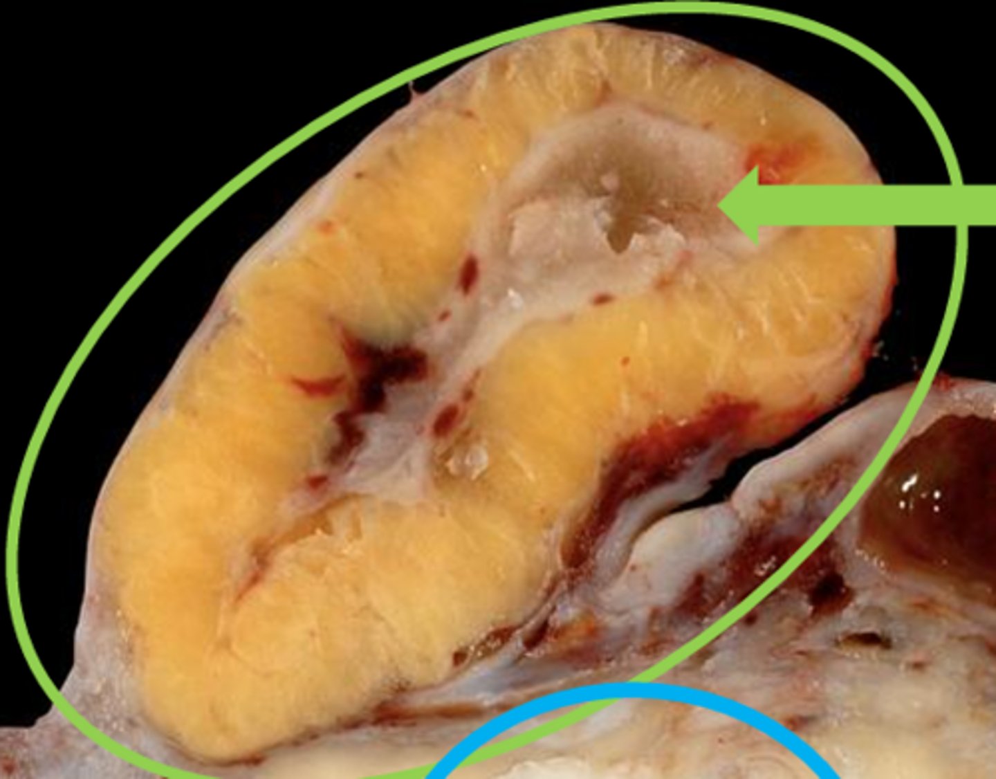
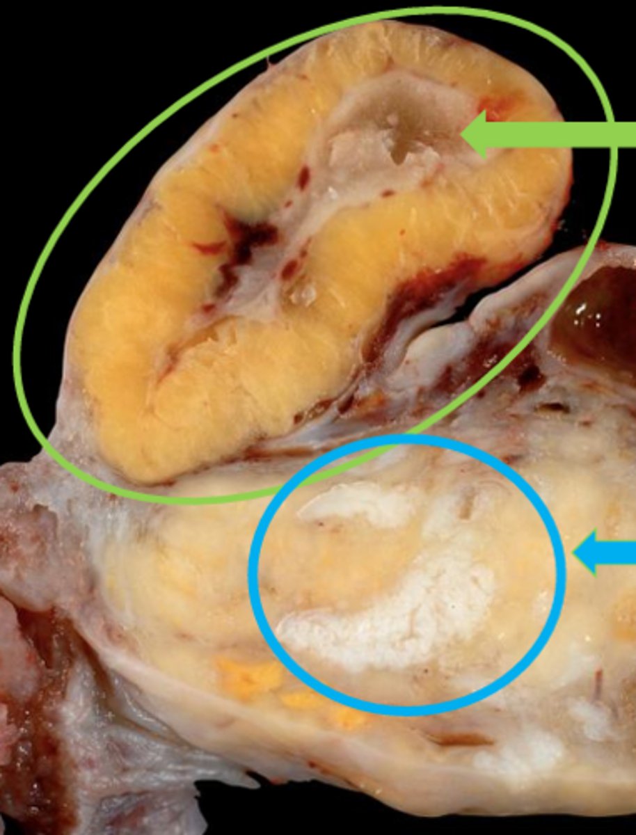
ovulation site is _____
graafian
mature (_____) follicles are the ones rupturing at the onset of ovulation
ovulation fossa, horses
orange, what species?
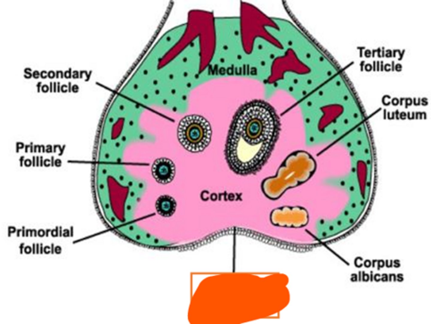
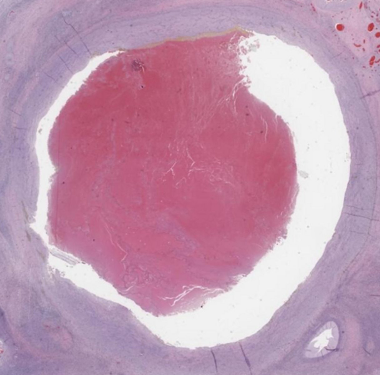
corpus hemorrhagicum
ruptured follicle filled with hemorrhage
ovulatory papilla
The _____ is the small, nipple-like protrusion on the ovarian surface marking the site where the follicle ruptured, which subsequently fills with blood to form the corpus hemorrhagicum.
ovulatory papilla
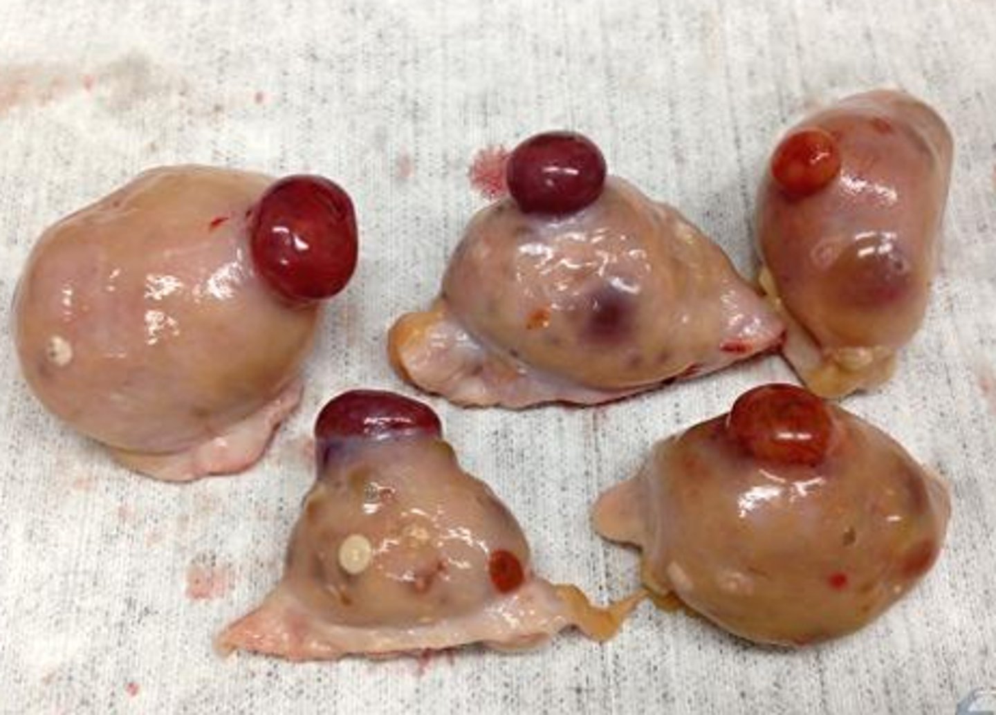
corpus hemorrhagicum
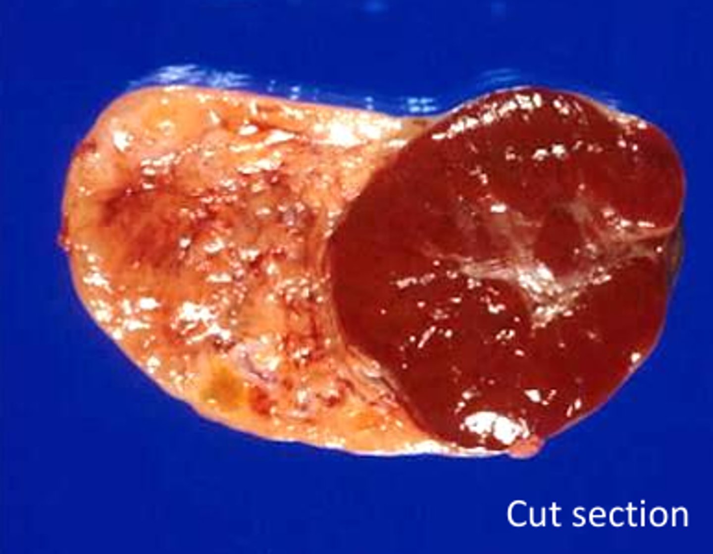
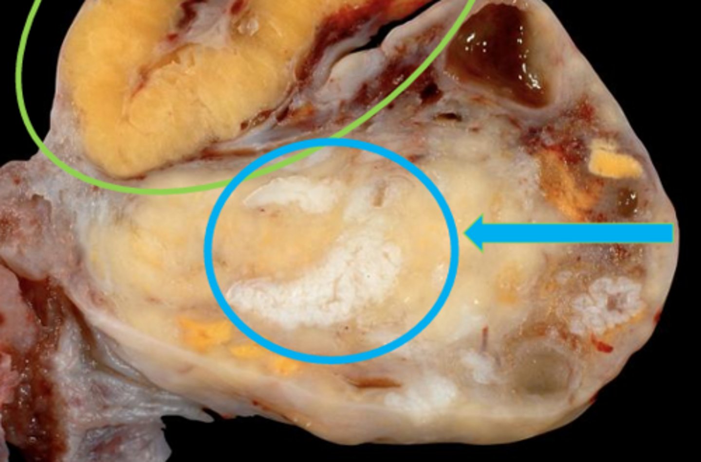
luteum
the corpus hemorrhagicum becomes the corpus _____
yellow
what color are luteal cells?
luteinization
the process of granulosa and thecal cells collapsing into the ruptured follicle and undergoing hypertrophy and hyperplasia
granulosa and thecal cells
what collapses into the ruptured follicle (corpus hemorrhagicum) and gets hypertrophied and has hyperplasia?
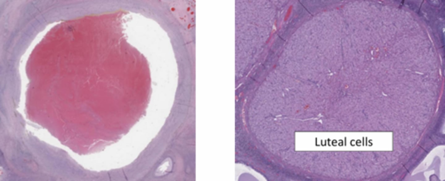
luteal cells
the red brackets indicate _____
progesterone
the corpus luteum, within this ovulatory papilla, produces ______
fertilization and implantation
the corpus luteum continues to develop if _______ occurs
luteal
______ cells, which produce progesterone to support pregnancy
luteal cells
these cells with vacuolated, eosinophilic cytoplasm
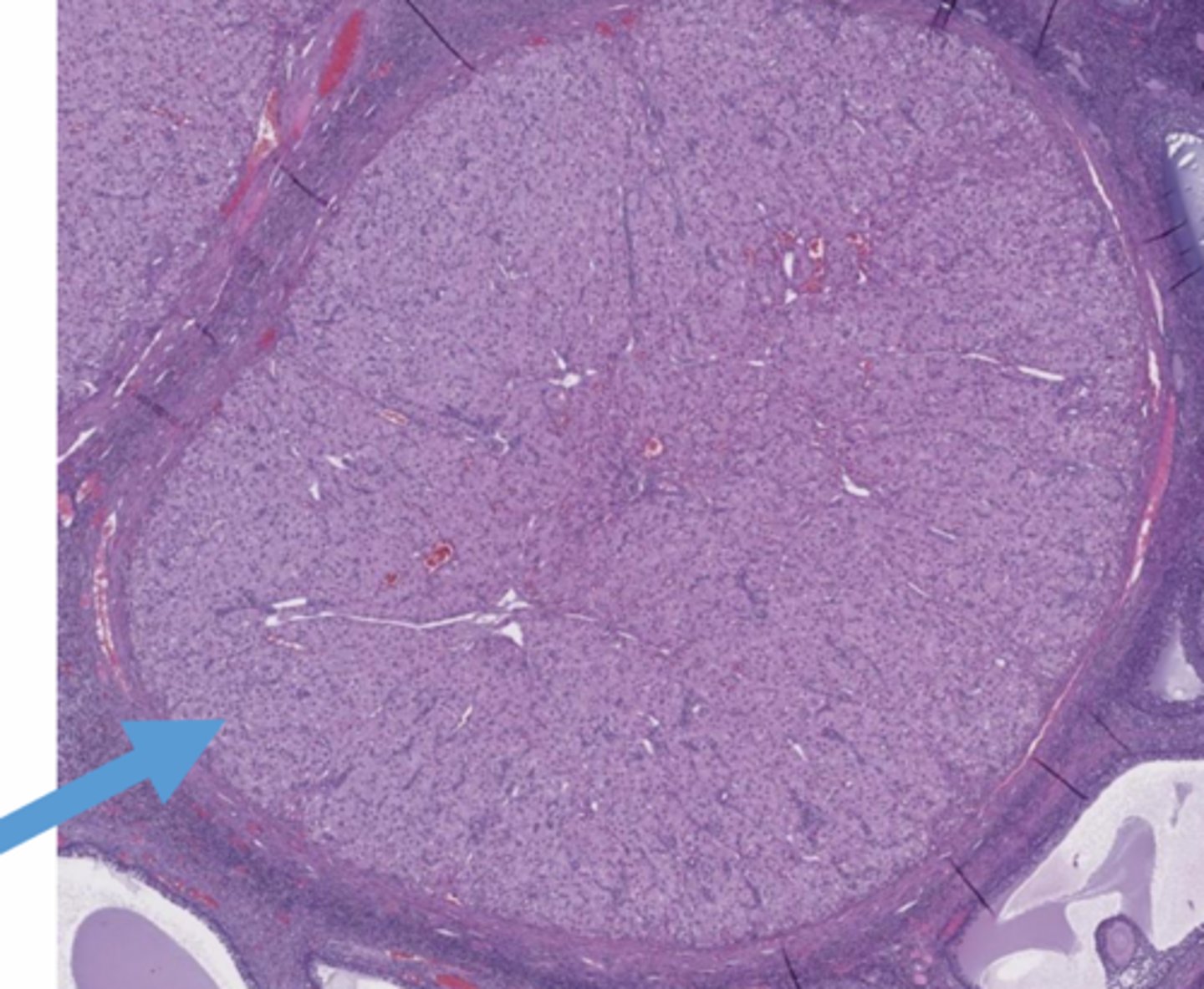
corpus luteum
the green arrow is pointing to ______ (central cavity (lacunae) is normal)
corpus albicans
the blue arrow is pointing to ____, which forms if fertilization DOES NOT occur (scar tissue!)
pregnancy
the corpus luteum (top) regresses and becomes corpus albicans at the conclusion of _____
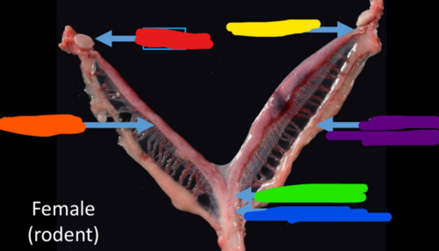
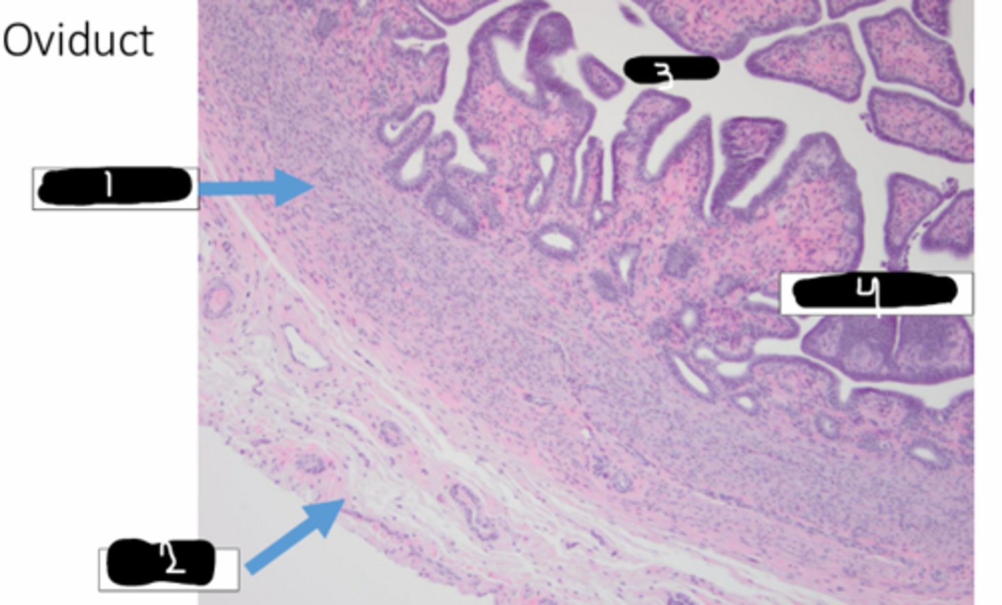
infundibulum
1

ampulla
2

isthmus
3
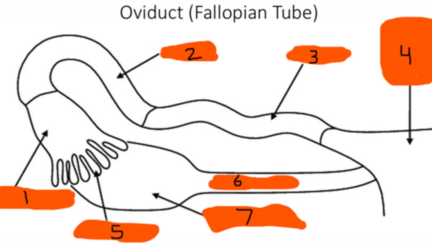
uterine horns and body
4

fimbria
5

ovarian pedicle
6

ovary
7

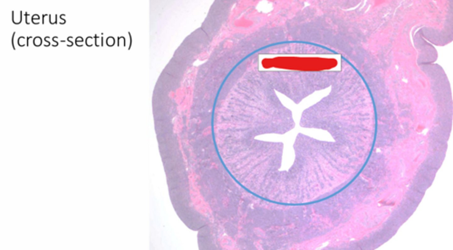
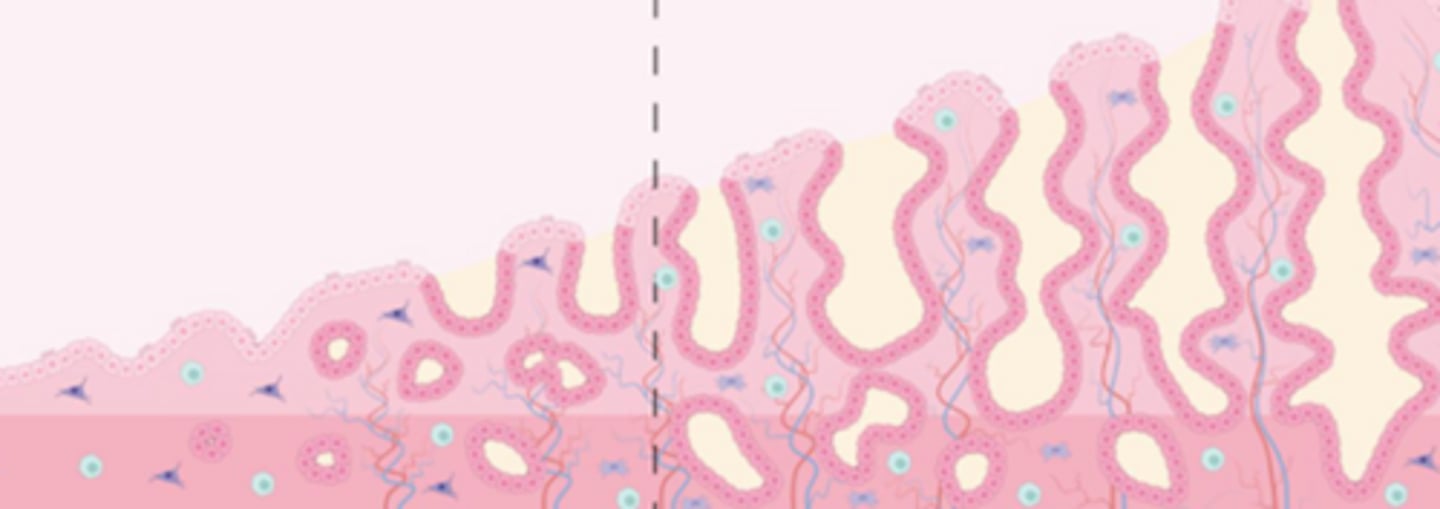
endometrium
3 Layers of Female Tubular Genitalia: the muscosal lining of epithelium (hint... its on the inside) RED
perimetrium
3 Layers of Female Tubular Genitalia: connective tissue (PURPLE)
infundibulum, ampulla, isthmus
components of the oviduct
infundibulum
this cross section of the oviduct would be found in the ______
isthmus
this cross section of the oviduct would be found in the ______
uterine horn/body junction
this cross section of the oviduct would be found in the ______
muscularis
1

serosa
2
lumen
3

endometrium
4

lamina propria
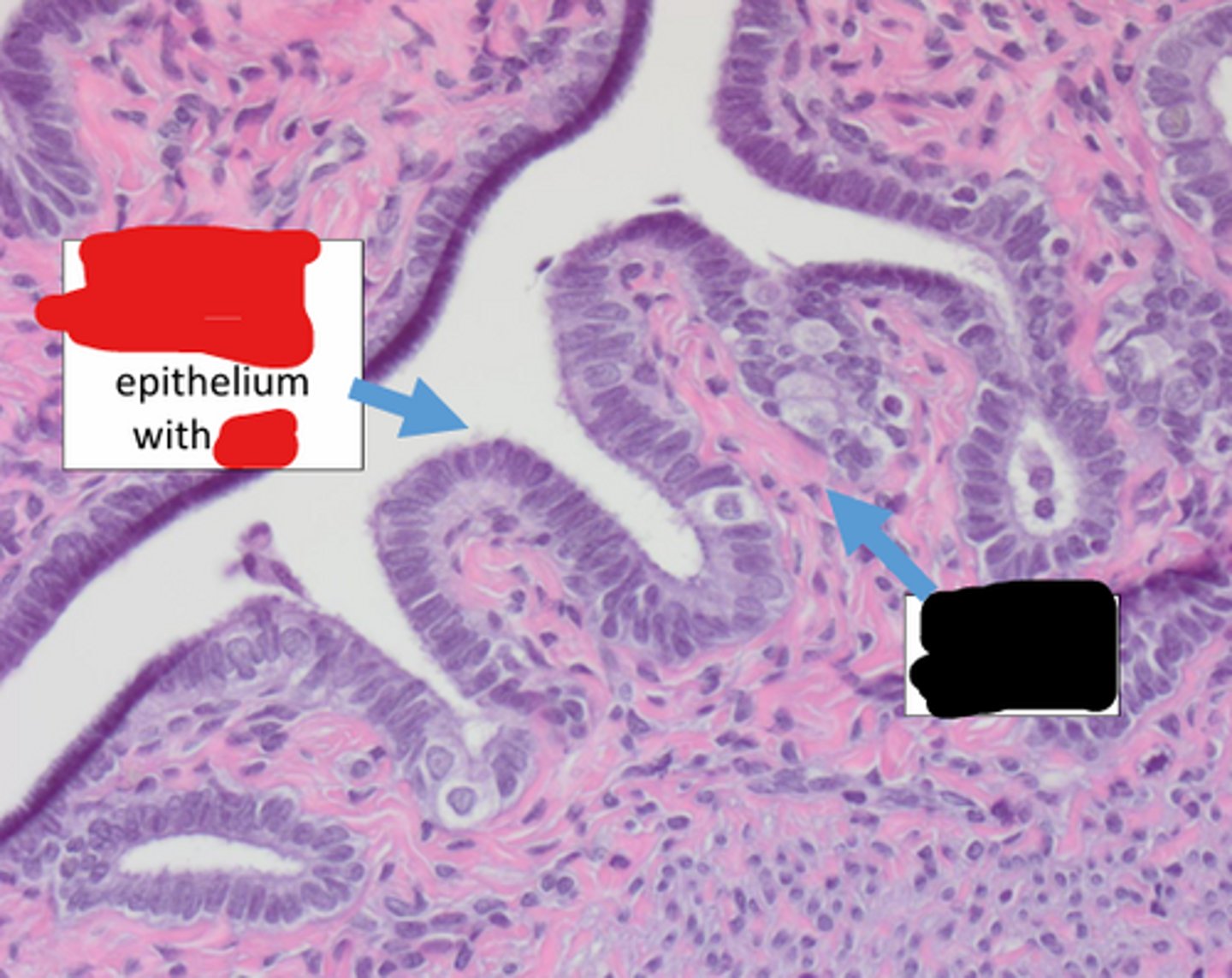
this is the oviduct. identify black

simple columnar, cilia
this is the oviduct. identify red
fallopian tube
the oviduct is also known as the _____
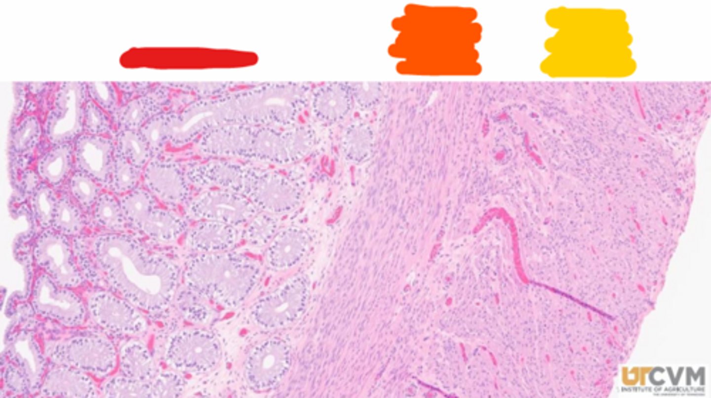
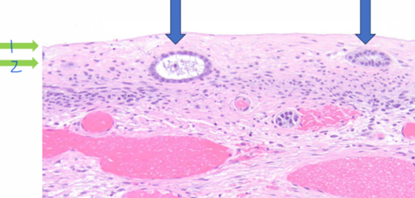
endometrium
the layer composing the uterus (horns and body)
columnar, tubular glands
the endometrium is simple _____ epithelium, supported by the _____ of the lamina propria that contains simple _____ that may branch or coil
endometrium
simple columnar
red

lamina propria
orange
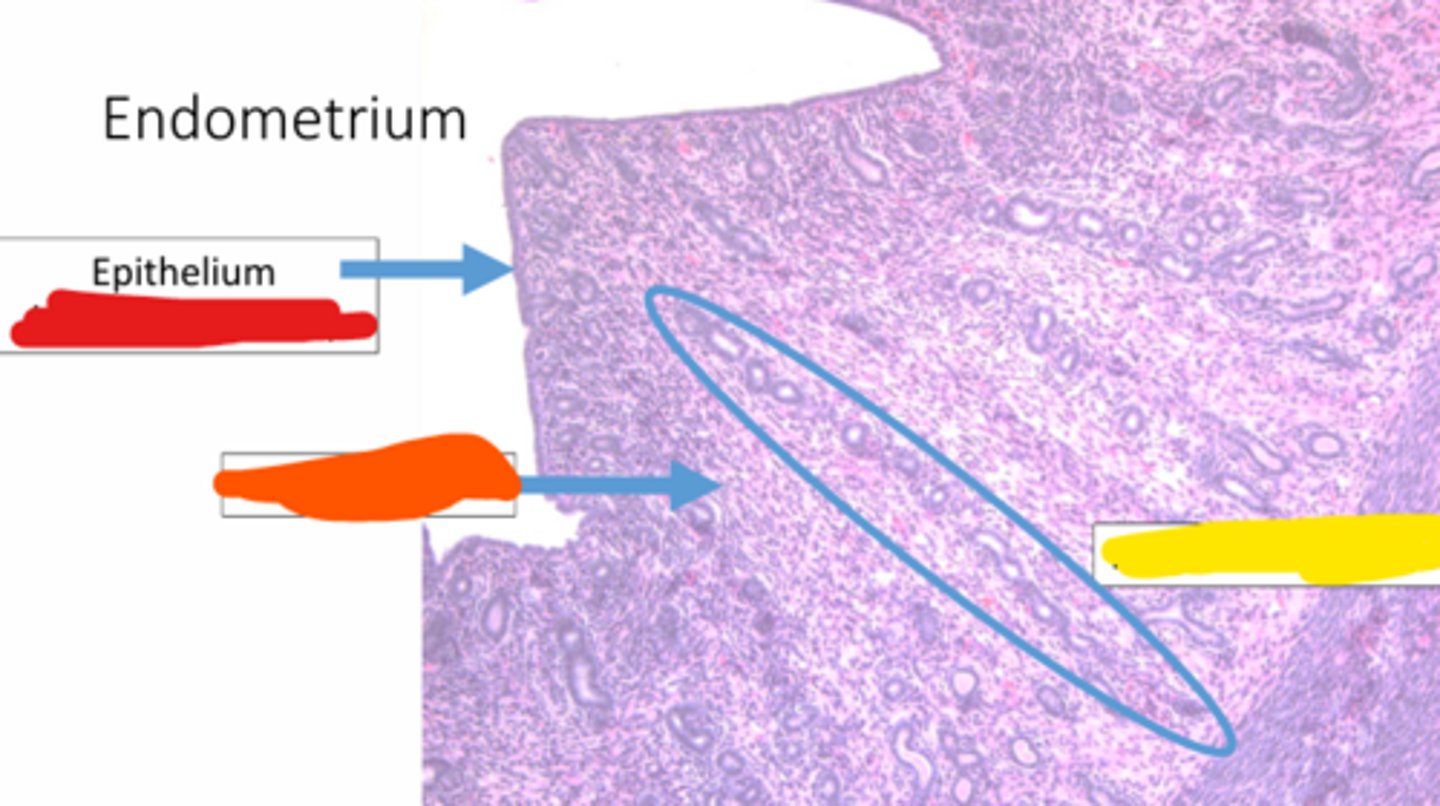
endometrial glands
yellow

endometrial glands
which component of the endometrium gets much larger as the estrus cycle progresses
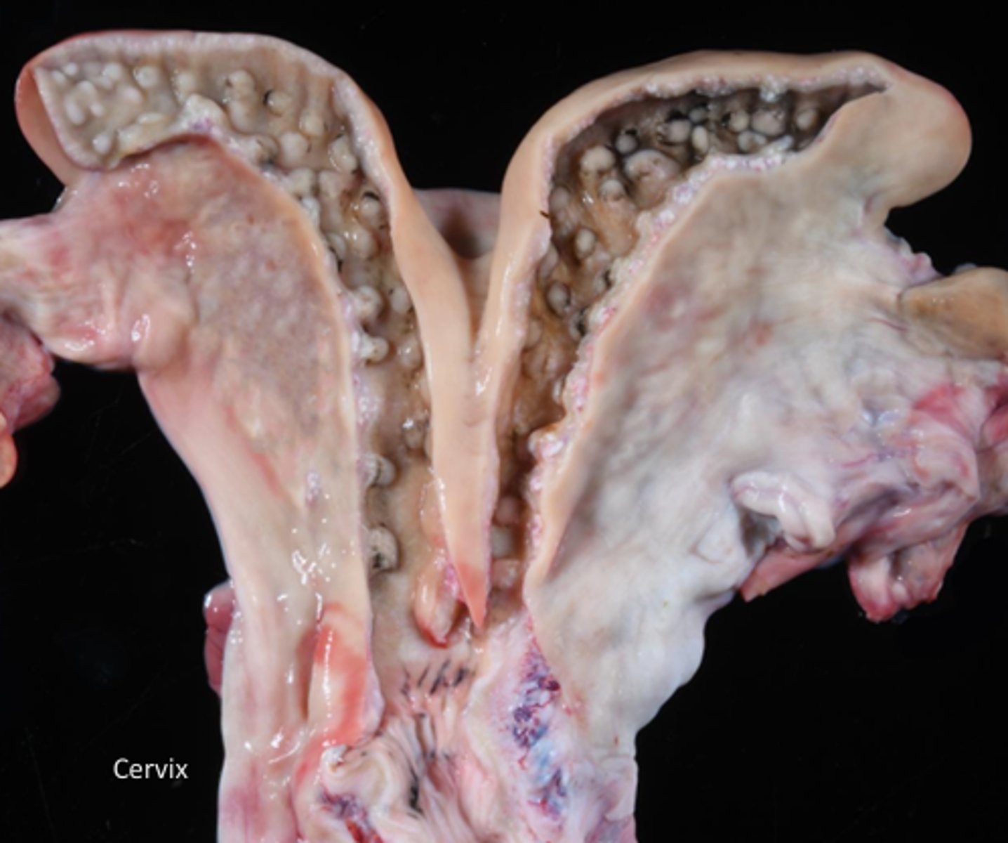
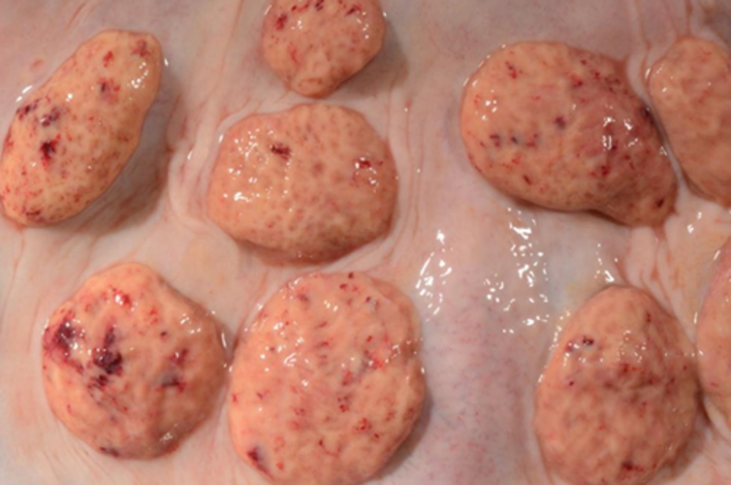
caruncles
sites of placental attachement, whose primary function is to fuse with fetal cotyledons to form placentones
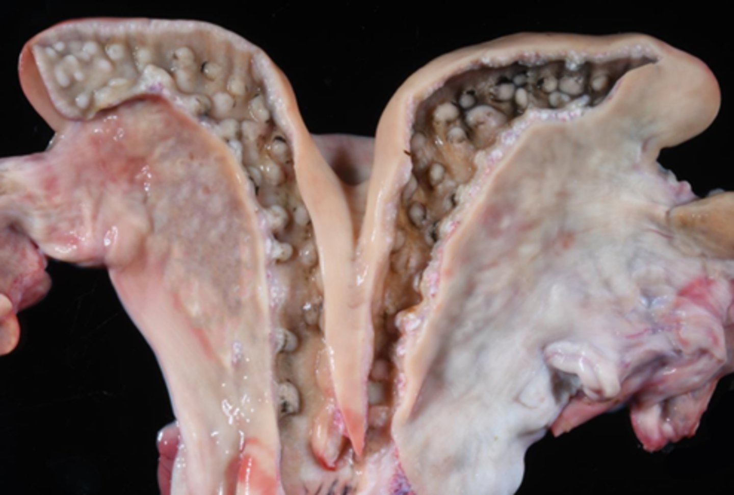
ruminants
these caruncles of the uterine horns (uterine body) are found in what species?
oviduct
fertilization occurs within the (oviduct/uterine horn)
uterine horn
the site of implantation and gestation (oviduct/uterine horn)
glands
what do caruncles lack?
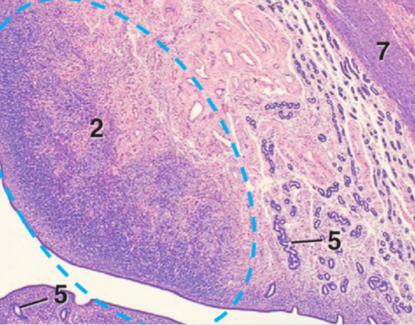
caruncles
______-pregnancy
vasculare
the inner and outer smooth muscle layers of the uterus are separated by the stratum ______
red

outer longitudinal myometrium
orange

stratum vasculare
yellow

inner circular myometrium
green
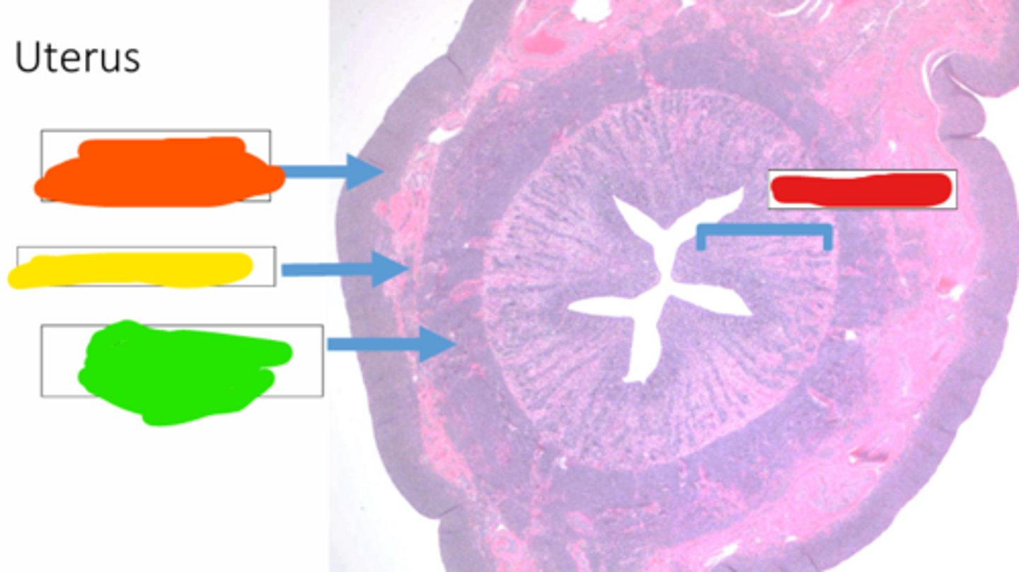
endometrium
red