Procedures
1/130
There's no tags or description
Looks like no tags are added yet.
Name | Mastery | Learn | Test | Matching | Spaced | Call with Kai |
|---|
No analytics yet
Send a link to your students to track their progress
131 Terms
opening between esophagus + stomach, controlled by cardiac sphincter
cardiac orifice
opening between stomach + small intestine, controlled by pyloric sphincter
pyloric orifice
muscosal folds of stomach, allow stomach to expand for nutrient absorption
rugae
sits posterior to body of stomach
fundus
sits anterior and inferior to fundus
body
sits posterior + distal to body of stomach
pylorus
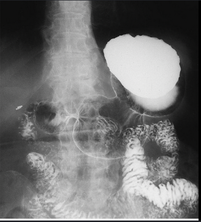
What does an UGI evaluate
esophagus, stomach, duodenum
Clinical indications for an UGI
peptic ulcer, hiatal hernia, bezoar, gastritis
mass formed from ingestion of hair, fingernails
bezoar
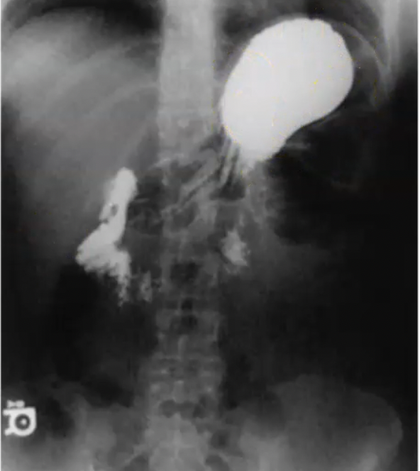
What projection is this? (barium in fundus)
AP
CR for AP stomach
midway between xiphoid tip and lower costal margin (L1)
What projection is this? (air in fundus)
PA
CR for PA stomach
L1 + 1” left of spine
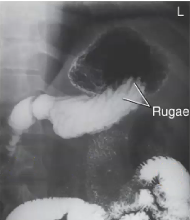
What projection is this? (air in fundus)
RAO
CR for RAO stomach
L1 midway between spine and lateral border of abdomen
Pt position for RAO stomach
40-70 oblique (45-55)
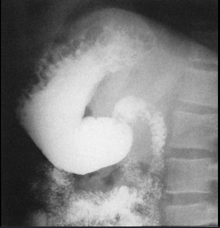
What projection is this?
Right lateral
What projection of an UGI shows the retrogastric space?
Right lateral
CR for Right lateral stomach
L1, 1.5” anterior to MCP
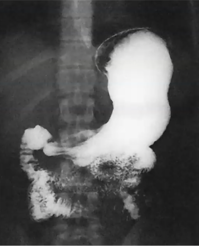
What projection is this? (barium in fundus)
LPO
CR for LPO stomach
L1, midway between midline of body + lateral border of abdomen
Pt position for LPO stomach
30-60 oblique (45)
What projection demonstrates an unobstructed view of the duodenal bulb?
LPO
extends from pyloric sphincter to ileocecal valve
small intestine
how long is the small intestine?
22 feet (7 meters)
shortest part of small intestine
Duodenum
How long is the duodenum?
8-10 inches (20-25 cm)
What portion of the small intestine contains major and minor duodenal papillae opening to ducts for liver or pancreas?
Duodenum
What is duojujenal flexure supported by?
Ligament of Treitz
How long is the Jejunum?
8 feet (2.5 meters)
how long is alimentary canal?
30 feet (9 meters)
What is the longest portion of the small intestine?
ileum
how long is the ileum?
11.5 feet (3.5 meters)
connect to large intestine by terminal ileum at ileocecal valve
ileum
What does a SBS evaluate?
stomach, small intestine + terminal ileum (TI)
Clinical indication for a SBS
crohns disease, SBO, abdominal pain, diarrhea
CR for scout KUB
2” above crest to include top of stomach
Why are images done prone for a SBS
compresses abdominal cavity to spread out intestines
uses fluoro and fspoon or compression paddle to visualize:
terminal ileum
What vertebral level is the xiphoid process located at?
T9-10
What vertebral level is the inferior costal margin located?
L2-3
What vertebral level is the iliac crest located?
L4-L5
done after gallbladder surgery to check for duct patency, look for retained stones, confirm no leaks or strictures
post op t tube cholangiography
tube left in common hepatic and bile ducts for post op drainiage
T-tube
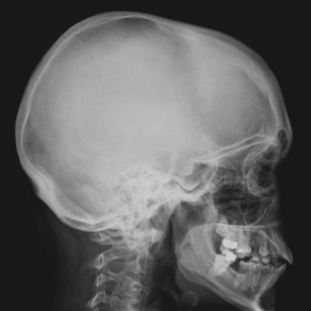
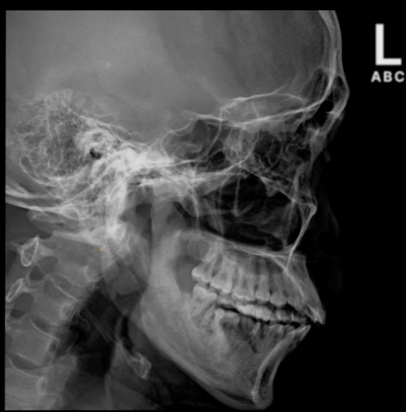
What does the lateral skull demonstrate?
superimposed orbital roofs, greater wings of sphenoid, sella turcica
What skull positioning line is perpendicular to front edge of IR to put patient in true lateral
IOML
CR for lateral skull
+, 2” ^ EAM
What projection demonstrates sphenoid sinus?
lateral skull
What projection demonstrates all 4 sinuses
lateral skull
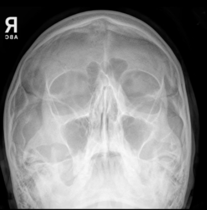
CR angle for PA Skull
0°
CR for PA skull
exits nasion
for the PA skull the OML is _ to the IR
+
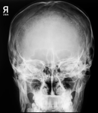
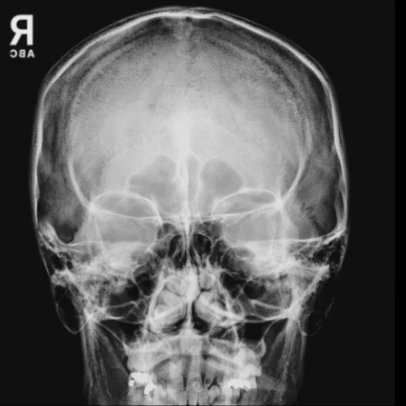
PA skull demonstrates:
symmetric petrous ridges, orbits filled by petrous ridges
CR angle for AP Axial Towne’s when using OML
30° caudad
CR angle for AP Axial Towne’s if pt unable to lower chin (using IOML)
15° caudad
CR for AP Axial Towne’s
2.5” ^ glabella
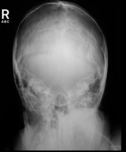
AP Axial Towne’s demonstrates:
symmetric petrous pyramids, dorsum sellae, posterior clinoid processes
CR angle for PA Axial Caldwell
15° caudad
CR for PA Axial Caldwell’s
exits nasion
for the PA Axial Caldwell’s the OML is _ to the IR
+
PA Axial Caldwell’s view demonstrates:
symmetric petrous ridges, petrous pyramids in lower 1/3 of orbit
CR angle for SMV full basal view
0°
for the SMV, the IOML is _ to the IR
parallel
CR for SMV
¾ anterior to EAM, through sella turcica, + to IOML
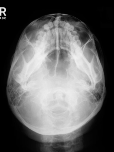
SMV demonstrates:
symmetrical mandibular condyles, foramen magnum
CR for lateral facial bones
b/w outer canthus and EAM
lateral facial bones demonstrates:
superimposed mandibular rami, sella trucica, superimposed orbital roofs,
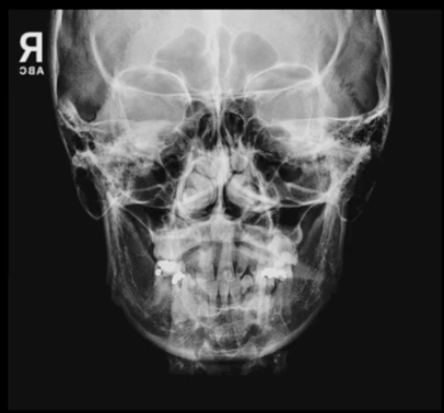
CR angle for water’s
0°
for the water’s view the OML forms a _ angle to the IR (chin on bucky)
37°
CR for water’s
exits acanthion
the MML is _ to the IR for the water’s view
+
the water’s for facial bones demonstrates:
petrous ridges projected below maxillary sinuses
What view demonstates the maxiallary sinuses?
water’s
Caldwell’s facial bones demonstrates:
symmetric petrous ridges lying in lower 1/3 of orbit
CR angle for modified water’s
0°
for the modified water’s view the OML forms a _ angle to the IR
55°
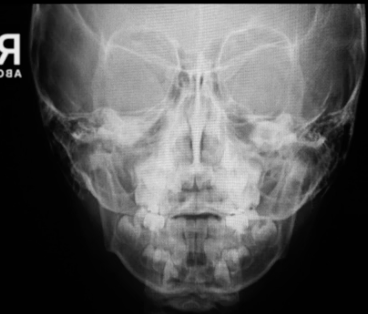
modified water’s facial bones view demonstrates:
petrous ridges projected below maxillary sinuses
aka parietoacanthial
water’s
CR angle for AP axial, reverse caldwell
15° cephalic
The lateral aspect of the obturator foramen is formed by:
ischium
CR for AP lumbar spine
1.5” ^ crest
Oblique C spine demonstrates:
intervertebral foramina + pedicles
CR for oblique lumbar spine
1.5” above crest + 2” medial to upside ASIS
CR for L5-S1 spot
1.5” distal to crest + 2” posterior to ASIS
for odontoid image, align:
incisor tooth to mastoid process
What level do the carotid arteries bifurcate?
C4
most stressed joint in the human body
knee
CR angle to free bladder neck of superimposition during a female cystourethrogram
5° caudad
Where do the ureters enter the bladder?
posterolateral
The wall of the esohpagus is composed of how many layers?
4
the concave medial border of the kidney where the blood vessels + ureter exit is called the:
hilum
How much lower should the IR be positioned when the upright position is used for projection of the stomach
3-6 in
Which projection of the stomach demonstrates its anterior and posterior surfaces?
lateral
Where is the compression applied during an IVU?
distal ends of ureters
the site where the ureters enter the bladder is termed the:
ureterovesical junction
for all projections of the esophagus, the top of the IR is positioned at:
the level of the mouth
How much is the body rotated for the PA oblique projections of the stomach and duodenum?
40-70°
CR angle for AP Axial bladder
10-15° caudad
how much is the knee flexed for a lateral projection?
20-30°
Where will the fibula be located on a properly positioned lateral radiograph of the ankle?
over posterior half of the tibia