Unit D3: Circulation and Immunity
1/50
Earn XP
Description and Tags
Circulation Topic 1
Name | Mastery | Learn | Test | Matching | Spaced | Call with Kai |
|---|
No analytics yet
Send a link to your students to track their progress
51 Terms
Roles of the circulatory system
-Transport gasses, nutrients, and waste throughout the body
-Regulate internal temp, distribution of hormones
-protect the body vs disease
Components of the circulatory system
Heart, Blood Vessels, Blood
Pulmonary Pathway
Circulates blood from the heart to the lungs and back
Systemic Pathway
Circulates blood from the heart to the body and back
Coronary Pathway
Circulates blood from the inside of the heart to the structure of the heart
What kind of organ is the heart?
Muscular
The heart’s pumps:
consists of 2 that are separated by the septum, they are parallel, and when the top contracts, the bottom will contract immidiately after
The action of the pulmonary pathway:
The pump on the right side of the heart receives deoxygenated blood returning from the body, and then pumps it into the lungs (alveoli). The alveoli then exchanges oxygen for carbon dioxide (to be breathed out), and then the newly oxygenated blood is pumped back to the heart
The action of the systemic pathway:
The left side of the heart receives the newly oxygenated blood and pumps it toward the brain and body. The oxygen in the blood is used for cellular respiration.
Action of the coronary pathway:
Oxygen and nutrients are delivered to the heart through capillaries embedded in the heart wall. The capillaries are supplied by two small arteries that branch off the aorta. Deoxygenated blood is carried away by the coronary veins.
The heart’s chambers
4 chambers; (TOP) right & left atrium, (BOTTOM) right and left ventricle.
Superior Vena Cava
Carries deoxygenated blood from the head & upper body to the right atrium
Inferior Vena Cava
Carries deoxygenated blood from all veins below the diaphragm to the right atrium
Where does oxygenated blood (from the lungs) go?
It enters the left atrium by way of the pulmonary veins (think pulmonary pathway)
Pulmonary Arteries
Carry deoxygenated blood from the right ventricle to the lungs
Pulmonary Veins
Carry oxygenated blood from the lungs to the left atrium
Artery
Any blood vessel that carries blood away from the heart
Vein
Any blood vessel that carries blood towards the heart
Coronary Arteries
Form an important branch of the aorta. The aorta (artery) carries oxygenated blood away from the heart. They also supply the muscle cells of the heart with oxygen and nutrients
Atrioventricular (AV) Valves
-Separate the atria from the ventricles
-prevent the flow of blood from the ventricles back into the atria
-Supported by bands of connective tissue called chordae tendinae
Semilunar Valves
-Separates the ventricles from the arteries
-Half moon shaped
-Prevent blood that has entered the arteries from flowing back into the ventricles
Septum
-A wall of tissue that separates the left & right sides of the heart
-Prevents oxygenated blood from mixing with deoxygenated blood
Why is the muscle surrounding the left ventricle thicker than the muscle surrounding the right ventricle?
Because the left ventricle is responsible for pumping blood out to the whole body, while the right only needs to pump out blood to the lungs
Angina
“Chest Pain”, occurs when too little oxygen reaches the heart (ex: blocked artery)
What kind of pattern does cardiac muscle display?
A striated pattern (like skeletal muscle) and a branching pattern
Myogenic Muscle
Muscle contractions or tissues that originate from within the muscle itself, rather than being triggered by external nerve impulses
The heart has the ability to contract without being stimulated by external nerves because of?
Myogenic muscle, which explains why the heart can beat for a short while when removed from the body
The heart’s tempo or beat rate is set by the:
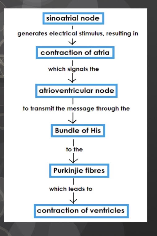
Sinoatrial Node
Sinoatrial Node definition
Bundle of specialised nerves and muscles located in the upper right atrium. Acts as a pacemaker, setting rhythm (@ about 70 bpm). Nerve impulses are carried from here to other muscle cells by modified muscle tissue
Atrioventricular (AV) Node
Serves as a conductor, passing nerve impulses via two large nerve fibres called purkinje fibers through the septum toward the ventricles.
Purkinje Fibres
Run along the septum, carrying impulses from the AV node to the bottom tip of the heart. These Fibres then carry impulses up along the outer walls of the ventricles back toward the atria.
Heart rate is influenced by what kind of nerves?
Autonomic Nerves (we do not have control over it)
Two regulatory nervous systems are? What do they do?
Sympathetic and Parasympathetic nervous systems. They conduct impulses from the brain to the SA node.
Sympathetic Nerves do what?
Increase heart rate, increasing blood flow to tissues. They are stimulated by times of stress.
Tachycardia
When the heart rate exceeds 100 bpm. Comes from exercise, caffeine/drug intake
What do the parasympathetic nerves do?
They are stimulated (during times of relaxation) to slow the heart rate
What causes the “lubb-dubb” sounds of the heart?
The closing of the heart valves
Diastole
Period of relaxation of the heart where the ventricles and atria are relaxed
Systole
Period of Contraction
Electrical Conduction System of the Heart
Coordinates the heartbeat
Step 1 of the Heart sounds
Relaxed Atria are full of blood. The atria contract, increasing the fluid pressure which forces the AV valves open
Step 2 of the heart sounds
Blood flows from the atria into the ventricles, where the now filled ventricles contract, forcing the AV valves shut.
Step 3 of the heart sounds
a heavy “lubb” sound is produced and pushes blood through the semilunar valves, into the arteries
Step 4 of the heart sounds
Closing of semilunar valves create the lighter “dubb” sound
Heart Murmur
-Occurs when blood leaks past the closed heart valve because of an improper seal.
-The AV valves (especially the left one; bicuspid) are susceptible to defects
What is the gurgling sound that can be heard with a stethoscope?
The rush of blood from the ventricle back into the atrium.
The more cardiac muscle is stretched…
The stronger is the force of contraction
Electrocardiogram (ECG)
Used to record the electrical activity of the heart
P on an ECG =
Contraction of Atria
QRS on an ECG =
Contraction of ventricles
T on an ECG =
Recovery of ventricles