Chapter 33 - Muscles of the Abdominal and Pelvic Walls
1/16
There's no tags or description
Looks like no tags are added yet.
Name | Mastery | Learn | Test | Matching | Spaced | Call with Kai |
|---|
No analytics yet
Send a link to your students to track their progress
17 Terms
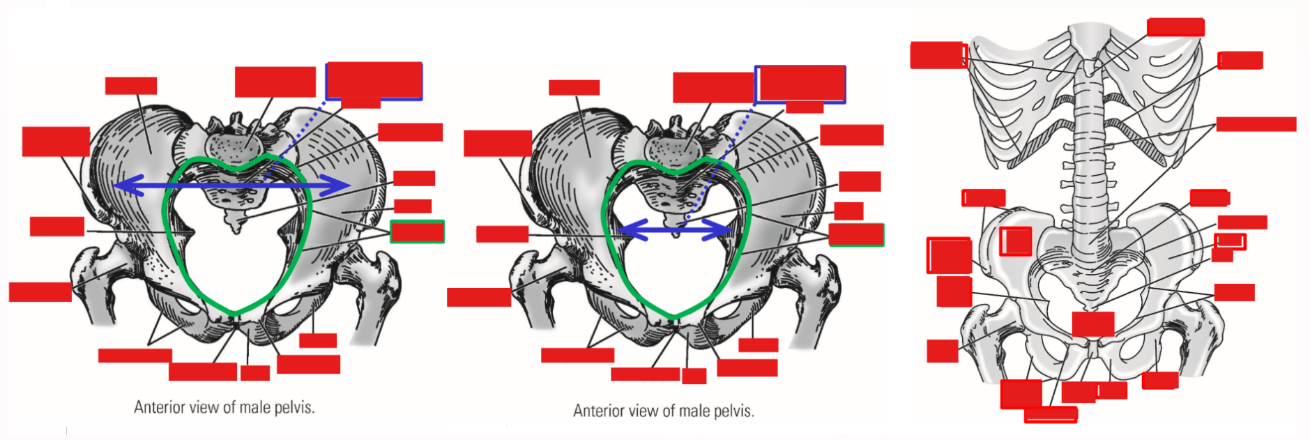
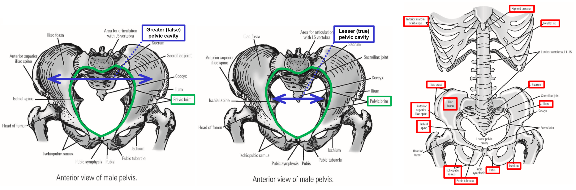
Bony Landmarks
–Rib cage
Sternum - inferior tip is xiphoid process
12 pairs of ribs, with costal cartilages
12 thoracic vertebrae
–Pelvis - two hipbones and the sacrum
–Hipbone
Ilium - iliac crest, iliac fossa, anterior superior iliac spine
Ischium - ischial spine, ischial tuberosity
Pubis - pubic symphysis, pubic tubercle
Ischiopubic ramus
→ Subpubic angle
–Pelvic brim
Borders - superior margin of pubis, inferior margin of iliac fossa, and superior margin of sacrum
Separates greater and lesser pelvic regions
–Greater (false) pelvis (greater pelvic cavity)
Area above pelvic brim
Contents: small intestine, sigmoid colon
–Lesser (true) pelvis (lesser pelvic cavity)
Narrow area below pelvic brim
Contents: pelvic organs (bladder, rectum, uterus, vagina, prostate gland)
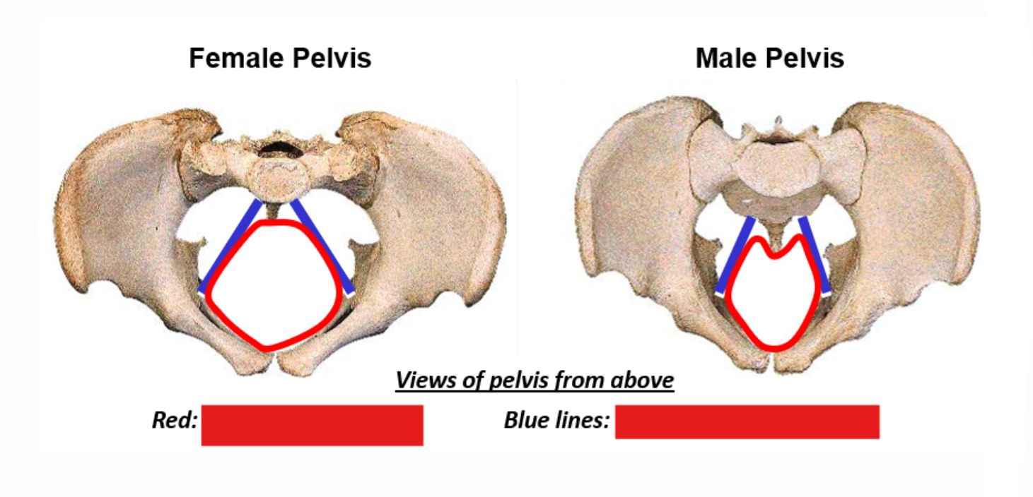
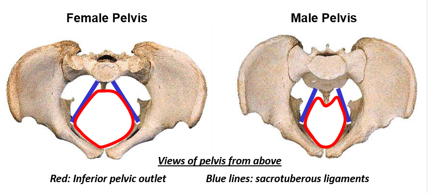
–Inferior pelvic outlet
Borders - pubic symphysis, ischiopubic rami, ischial tuberosities, sacrotuberous ligaments, coccyx
Pelvic outlet of female is wider and rounder compared to male outlet
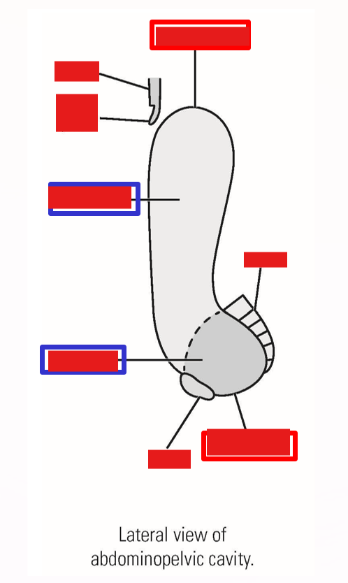
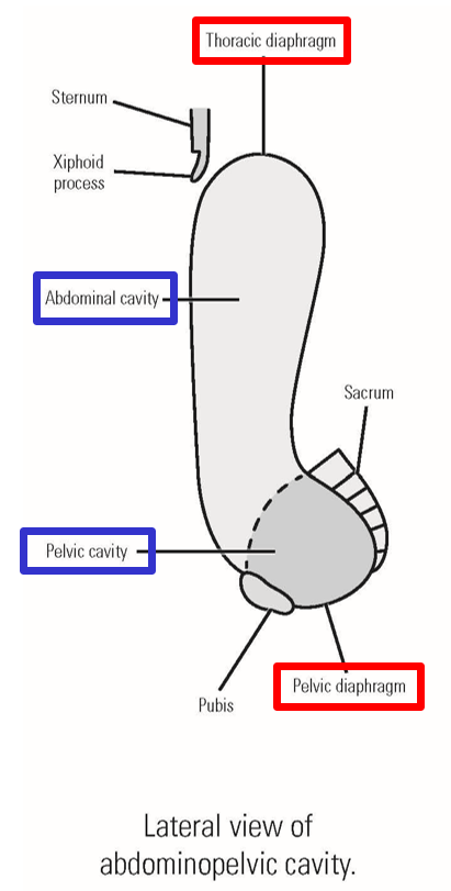
Abdominopelvic Cavity
–The abdominal cavity and pelvic cavity form a continuous body cavity
Pelvis is tilted anteriorly at 45° angle
–Borders:
Superior: rib cage & thoracic diaphragm
Inferior: pelvis & pelvic diaphragm
Anterior/Lateral: abdominal wall muscles
Posterior: vertebral column, m. psoas major, m. quadratus lumborum
Muscles of Abdominal Wall
Abdominal wall - consists of 4 muscles:
–Three layers of muscles that begin laterally and extend to anterior mid-line
M. External abdominal oblique
M. Internal abdominal oblique
M. Transverse abdominis
–One muscle of anterior abdominal wall
M. Rectus abdominis
–Three lateral muscles extend anteriorly - become aponeurotic (transition to broad, flat tendon)
Each lateral muscle inserts into its paired muscle from opposite side (form raphe)
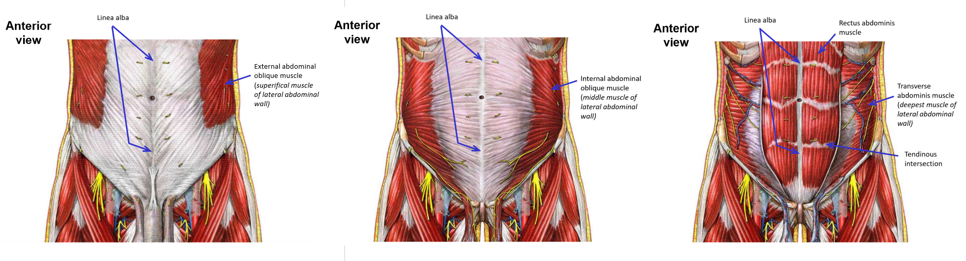
–Linea alba - anterior, mid-line raphe formed by all three lateral muscles
Serves as insertion for all lateral muscles
Extends down anterior mid-line of abdominal wall, from xiphoid process of sternum to pubic symphysis of pelvis
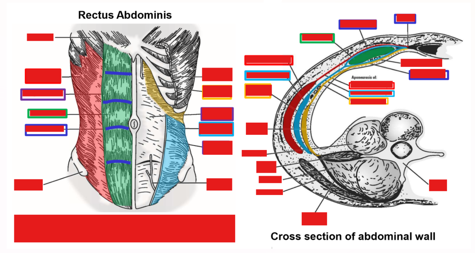
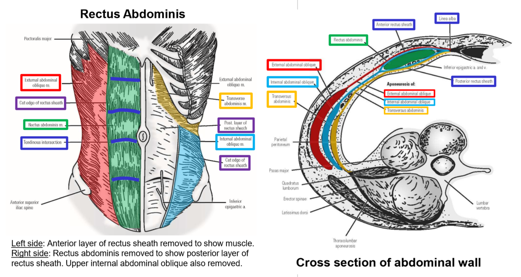
M. Rectus abdominis
Elongated, anterior abdominal wall muscle that runs vertically (from pelvis to rib cage), parallel to anterior mid-line
Surrounded by rectus sheath - dense connective tissue covering formed by aponeurosis from all lateral abdominal wall muscles
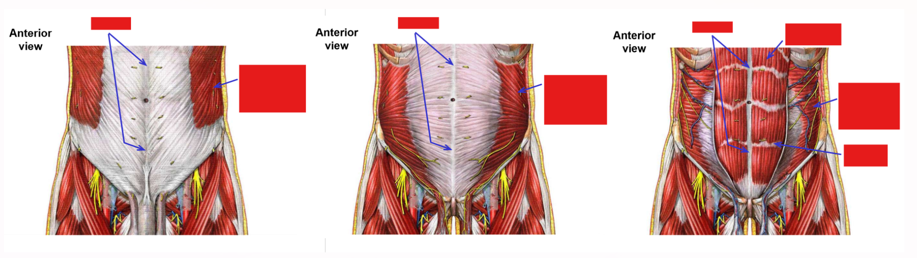
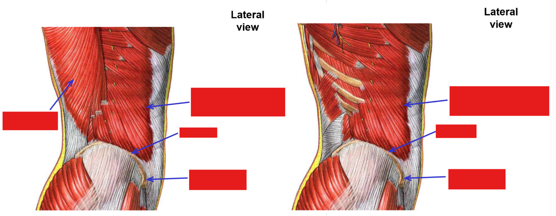
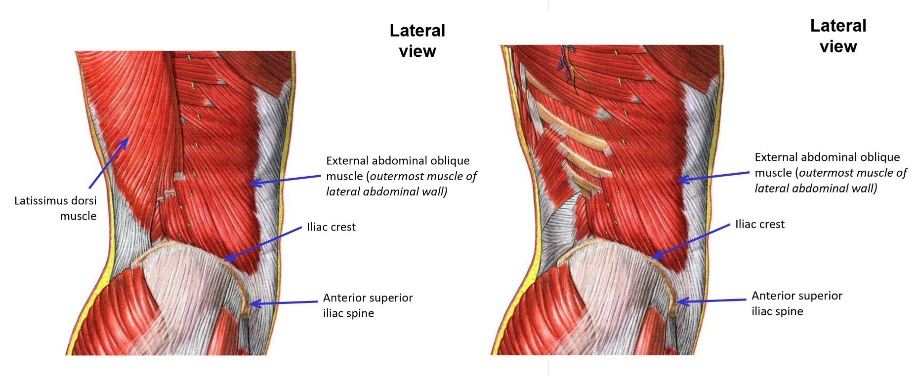
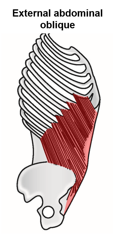
M. External abdominal oblique
–Origin: lower margin of rib cage
–Insertion: linea alba, iliac crest, anterior superior iliac spine, pubic tubercle
Most superficial of lateral abdominal wall muscles
Muscle fibers run in down and medial direction
Inferior free margin (spanning between anterior superior iliac spine and pubic tubercle of pelvis) forms the inguinal ligament
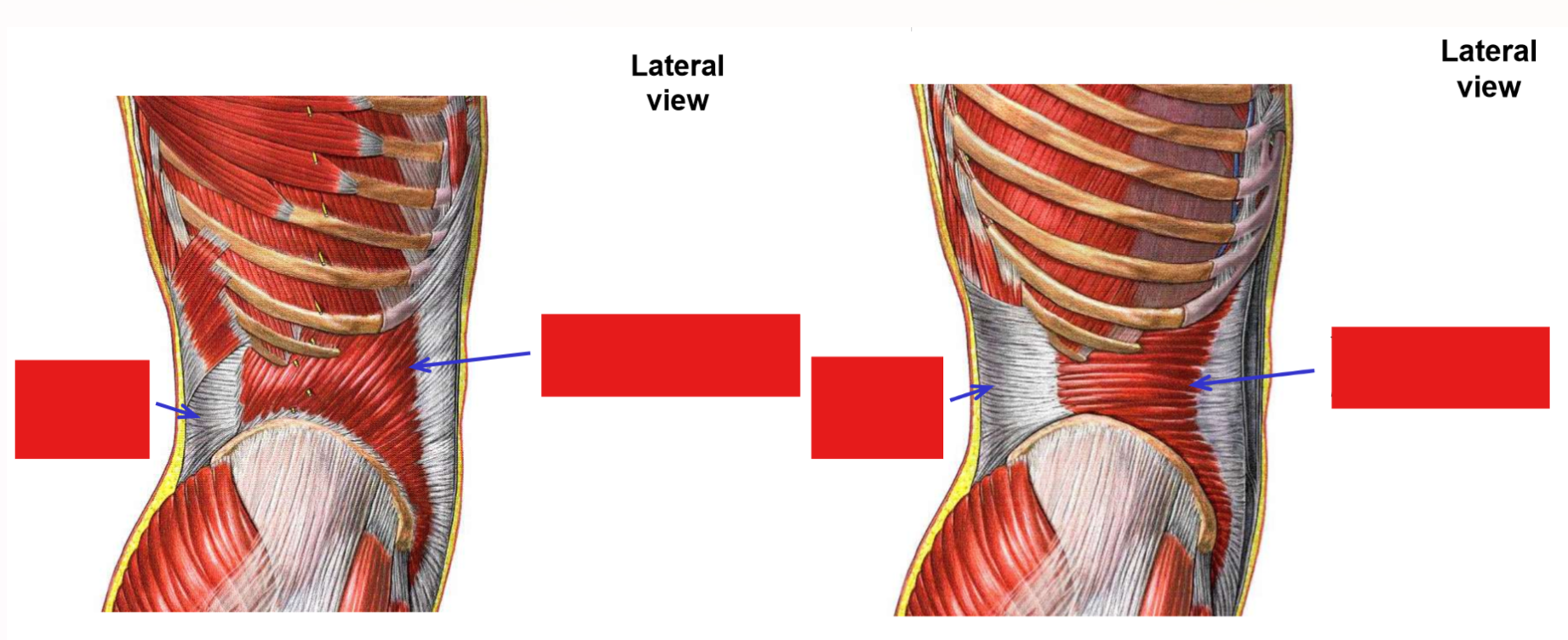
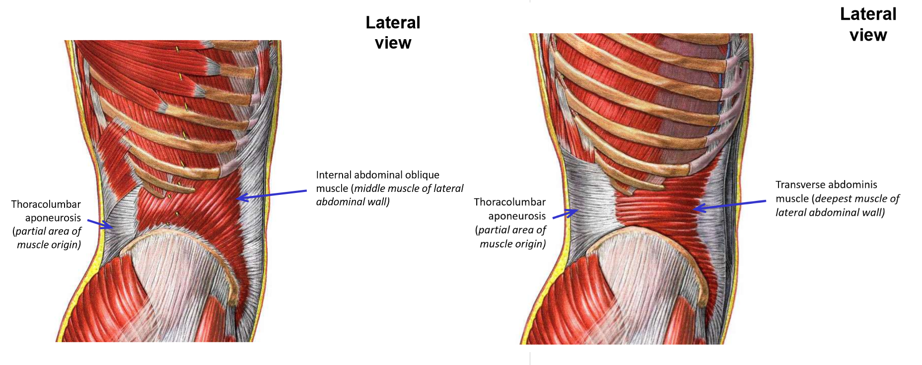
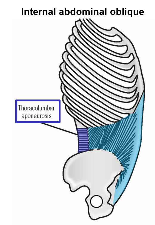
M. Internal abdominal oblique
–Origin: iliac crest, thoracolumbar aponeurosis
Thoracolumbar aponeurosis - broad tendinous structure arising from spinous processes of lower thoracic and all lumbar vertebrae
–Insertion: lower rib cage and linea alba
Middle layer of lateral abdominal wall muscles
Muscle fibers run in up and medial direction
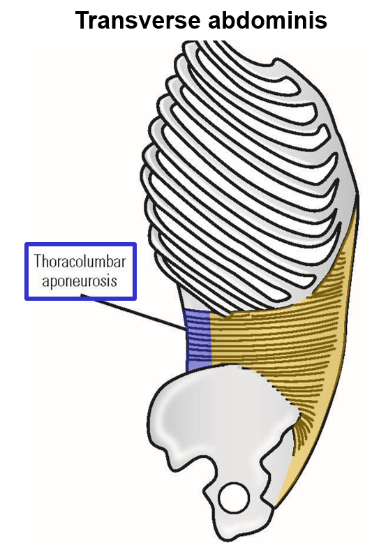
M. Transverse abdominis
–Origin: lower rib cage, thoracolumbar aponeurosis, iliac crest
–Insertion: linea alba
Deepest layer of lateral abdominal wall muscles
Muscle fibers run in transverse (horizontal) direction
M. Rectus abdominis
–Origin: pubic portion of pelvis
–Insertion: xiphoid process of sternum, lower rib cage
Muscle runs vertically, parallel to anterior mid-line of abdomen
Muscle divided into segments by tendinous intersections (prevent “bowstringing” of muscle)
–Muscle is covered on both anterior (superficial) and posterior (deep) sides by rectus sheath
Sheath formed by fused aponeurosis from all three lateral muscles of abdominal wall
Fused aponeurosis then passes both superficial and deep to rectus abdominis muscle
Both layers of rectus sheath come together at anterior mid-line (medial to rectus abdominis) to insert into linea alba
M. Rectus abdominis
Anteriorly flexes trunk of body
Actions of abdominal wall muscles
All four muscles serve to contain the abdominal organs and compress abdominal wall during respiration, defecation, urination, vomiting, or childbirth
Actions of External and internal abdominal oblique muscles
Contraction of both muscles on one side only - lateral flexion of body trunk toward that side
Contraction of both muscles on both right & left sides simultaneously - anterior flexion of body trunk
Primary muscles for rotation of body trunk
Contraction of external abdominal oblique on one side plus contraction of internal abdominal oblique on opposite side produces rotation of body trunk toward internal abdominal oblique side
Example: contraction of right external abdominal oblique plus contraction of left internal abdominal oblique rotates body trunk toward left side