PSYC 301 MT2 - Perception
1/24
There's no tags or description
Looks like no tags are added yet.
Name | Mastery | Learn | Test | Matching | Spaced | Call with Kai |
|---|
No analytics yet
Send a link to your students to track their progress
25 Terms
sensation
detection of internal/external stimuli
raw info about environment is made available to brain through senses
perception
awareness and interpetation of sensory info by brain
sensation vs. perception
perceptual deficits can exist without sensory loss
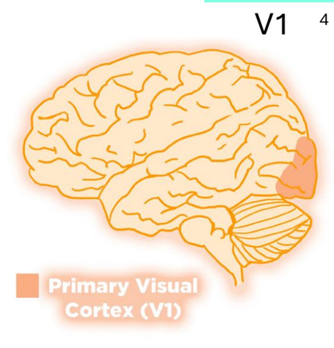
primary visual cortex (V1)
visual info travels from retina → subcortical relays → V1, first cortical stage
V1 damage
some patients with V1 lesions show blindsight:
e.g. can guess visual stimuli (forced-choice discrimination) or navigate obstacles above chance, despite reporting blindness
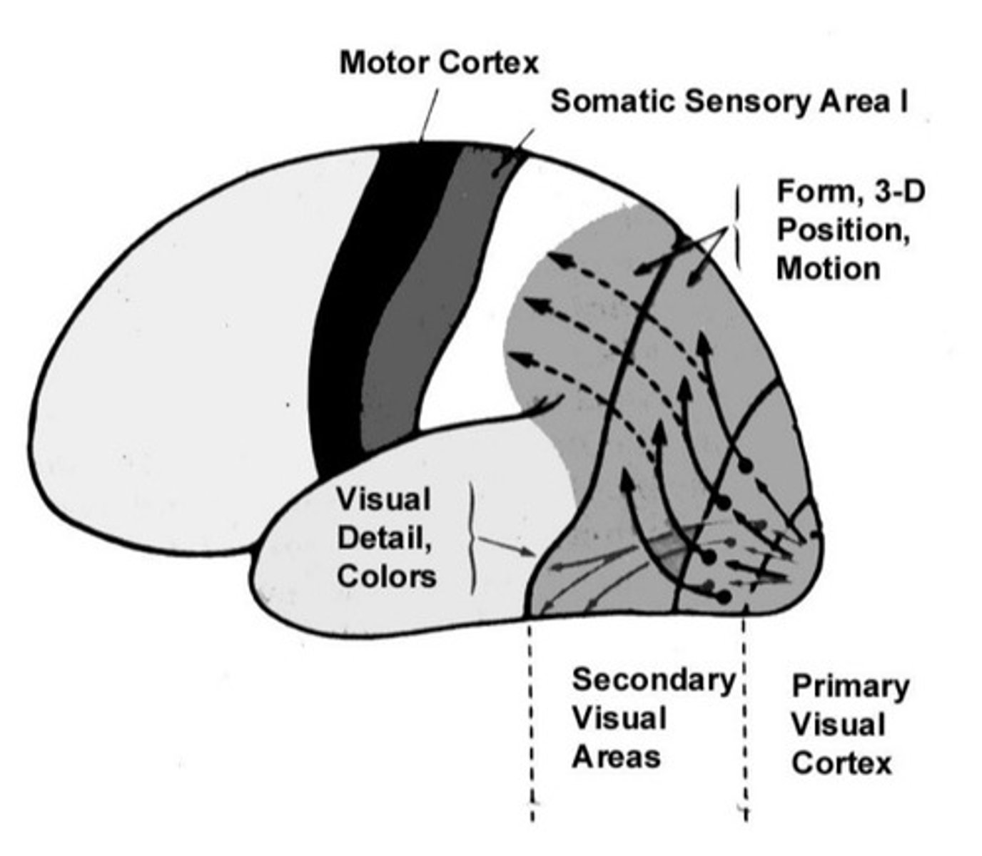
higher visual cortices I: secondary visual cortices (~24)
get visual input from V1 → analyze form, motion, shape, colour
secondary visual cortices V5 damage
akinetopsia: bilateral damage can cause selective loss of visual motion perception (i.e motion blindness)
higher visual cortices II
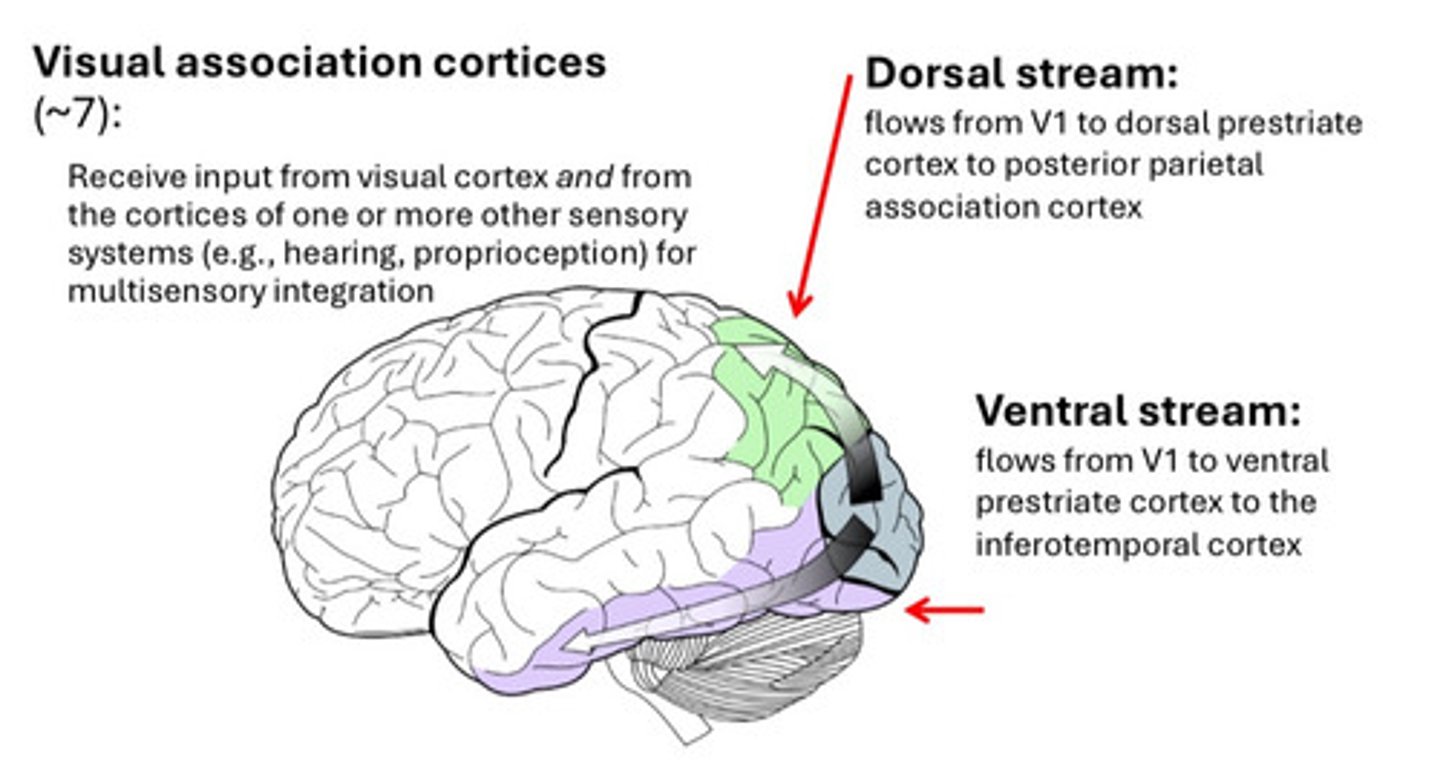
1. visual association cortices (~7): integrate input from visual cortex with other sensory systems (auditory, proprioceptive) for multisensory perception
2. dorsal stream: V1 → dorsal prestriate cortex → posterior parietal association cortex
3. ventral stream: V1 → ventral prestriate cortex → inferotemporal
functions of two streams - theory 1: what vs. where
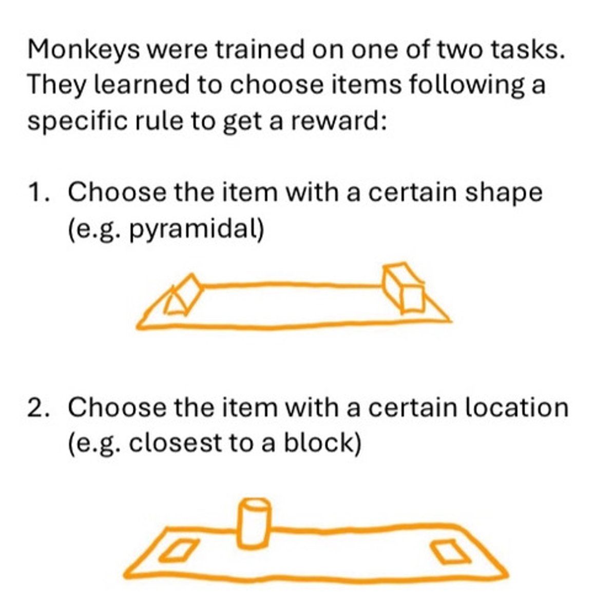
monkey studies:
posterior parietal lesion (dorsal) → poor spatial/location discrimination → "where" pathway
inferotemporal cortex lesion (ventral) → poor object discrimination → "what" pathway
human support:
dorsal damage → can describe objects but can't reach accurately
ventral damage → can reach for objects but can't identify them
functions of two streams - theory 2: action vs. perception
patient DF: carbon-monoxide poisoning → bilateral ventral-stream lesions
ventral lesion → visual form agnosia (cannot identify shapes but dorsal, visuomotor abilities spared (can grasp objects correctly)
-
streams differ not from the type of information carried but the use of that information
dorsal: direct behavioural interaction with objects
ventral: conscious perception
functions of two streams - theory 2: action vs. perception - opposite pattern
dorsal damage → action → optic ataxia (poor visually guided reaching)
agnosia
inability to recognize objects/shapes (with no evidence of significant memory loss) → perceptual loss (not sensory)
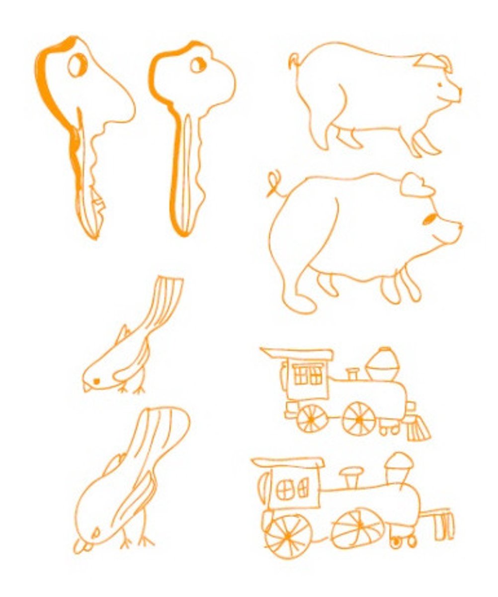
agnosia - types - apperceptive agnosia
apperceptive agnosia: recognition failure due to impaired perceptual processing
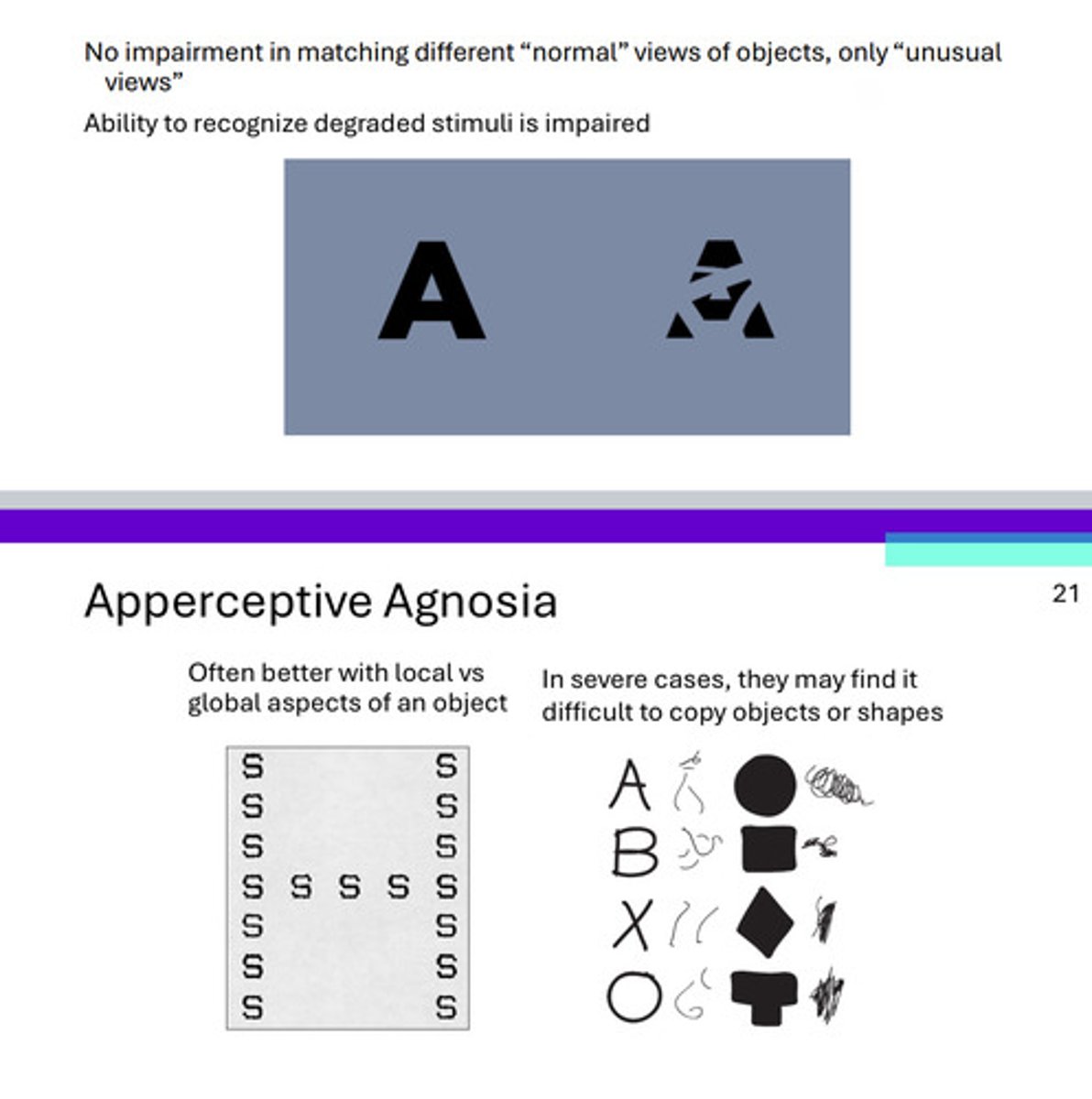
agnosia - types - apperceptive agnosia - features
can't recognize objects from unusual views or degraded images
often rely on local features instead of holistic form
severe cases: difficulty copying shapes or matching objects

agnosia - types - associative agnosia
normal perception but can’t link perception to meaning
ventral not dorsal stream (e.g. hands know what to do with the object but can’t only use the visual information from photo of lock to determine what it is)
agnosia - types - associative agnosia - features
can copy drawings but can't name or recognize them visually
when told the name of objects, can describe it accurately → stored knowledge intact
agnosia - types - prosopagnosia
failure to recognize faces, with intact object recognition
agnosia - types - prosopagnosia - features
can describe features (eyes, hair, etc.) but can't identify the person
affects both familiar (retrograde) and new (anterograde) faces
can still recognize people by voice, clothing, or gait
agnosia - types - prosopagnosia - treatment
training programs and coping strategies (e.g. using other features)
agnosia - types - prosopagnosia - structures affected
bilateral occipito-temporal damage
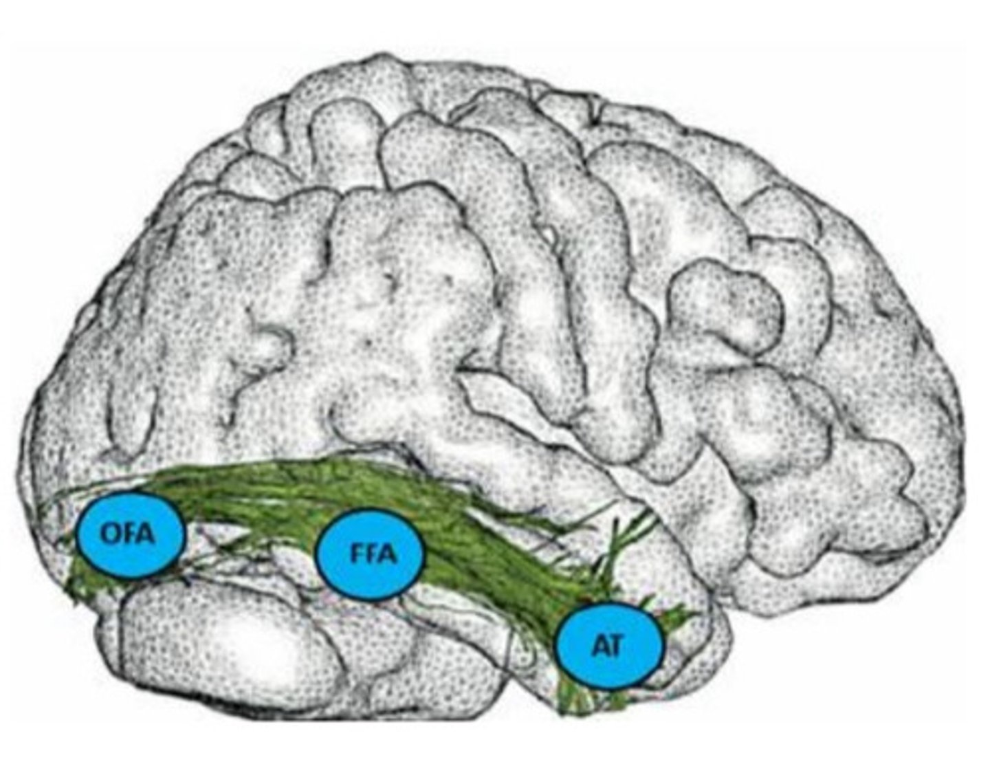
face processing - structures
network of occipito-temporal areas:
1. occipital face area (OFA)
2. fusiform face area (FFA)
3. anterior temporal cortex (AT)

is FFA purely for faces?
FFA also active for visual expertise (any category you’ve become an expert at recognizing visually)
e.g. bird or car experts
prosopometamorphopsia
disorder where faces appear distorted (e.g. warped, drooping, stretched)
prosopometamorphopsia - features
only affects faces, not other objects
can involve one or both sides of the face
prosopometamorphopsia - structures affected
linked to abnormal activation in face-processing network (not absence of it)
can be experimentally induced via stimulation through intracranial electrodes in right inferior temporal lobe