BIO271 - Lecture 5 - Sensory Physiology: Photoreception
1/161
There's no tags or description
Looks like no tags are added yet.
Name | Mastery | Learn | Test | Matching | Spaced | Call with Kai |
|---|
No analytics yet
Send a link to your students to track their progress
162 Terms
what is photoreception?
-ability to detect a small proportion of the electromagnetic spectrum from ultraviolet to near infrared
what do most electromagnetic waves not have?
-most of these do not travel through water
what are the only 2 things that can penetrate deeper water?
only:
visible light
very long wavelength of electromagnetic (EM) radiation
what do animals detect?
-they detect a narrow band of the EM spectrum in the visible range, which suggests the possibility that photoreceptors evolved in aquatic organisms
what can other vertebrates perceive?
-they can perceive different ranges of photoreception
what is the range of visible light that humans can detect?
-the range is from 350 nm - 750 nm
photoreceptors
-organs that range from single light-sensitive cells to complex, image forming eyes
what are the 2 major types of photoreceptors?
rhabdomeric photoreceptors
ciliary photoreceptors
rhabdomeric photoreceptors
-apical surface is covered with multiple out-foldings called microvillar projections
-found exclusively in invertebrates
ciliary photoreceptors
-have single, highly folded cilium; folds form disks that contain photopigments
-found in exclusively in vertebrates/mammals
eyespots
-are single cells or regions of a cell that contain photosensitive pigment, (ex. protist Euglena)
eyes
-are complex organs consisting of group of cells specialized for different functions
-include both multiple photoreceptor cells and separate pigment cells
-provide information such as light direction and contrast between light and dark
-some of these can form focused images
what are protist euglena?
-a unicellular, eukaryotic microorganism belonging to the kingdom Protista that inhabit freshwater and brackish environments
-have a unique mixotropic nature, possessing chlorophyll for photosynthesis (plant-like), while also being able to ingest nutrients (animal-like)
what are the 4 major types of animal eyes?
flat-sheet eyes
cup-shaped eyes
vesicular eyes
convex eyes
flat sheet eyes
-primitive retina
-provide some sense of light direction and intensity
-most often seen in larval forms or as accessory eyes in adults (some snails)
layer organization:
photoreceptor cells → pigment layer → primary afferent neurons
photoreceptor cells (flat sheet eyes)
-perceive light
pigment layer (flat sheet eyes)
-absorb light
primary afferent neurons (flat sheet eyes)
-sending info about the light source to the integrating center
cup-shaped eyes
-retinal sheet is folded to form a narrow aperture
-better discrimination of light direction and intensity
-seen in the Nautilus (mollusk)
-has a whole where light coming from different directions can be detected
layer organization: (cup-shaped)
hole → pigment layer → photoreceptor cells → afferent neurons
what animal has the most complex kind of cup-shaped eye?
-the Nautilus (mollusk)
vesicular eyes
-use a lens in the aperture to improve clarity and intensity
-lens refracts light and focuses it onto a single point on the retina
-found in some mollusks
-vertebrates have complex types of this eye with a lens that can generate a sharp, focused image
-provide the best resolution of images because it has a lens
layer organization:
lens → photoreceptor cells (retina) → afferent neurons
lens
-focuses the light on the retina
-provides directionality
-refracts light and focuses it onto a single point on the retina
-aka crystalline cone
convex eyes
-photoreceptors radiate outward forming a convex retina to perceive light
-present in annelids, mollusks, and arthropods
layer organization: (convex shape)
photoreceptor cells (retina) → afferent neurons
what are the most complex convex eyes?
-compound eyes are the most complex of this type
compound eyes
-most complex convex eyes found in insects and crustaceans
-consist of up to several thousand light detectors called ommatidia
-very effective at detecting movement
ommatidia
-light detectors
what does the ommatidium consist of?
in this orientation:
-a cornea
-a crystalline cone
-several rhabdomeric photoreceptors (retinular cells)
-retinular cells arrange radially with microvilli inward forming rhabdom
what do ommatidium give?
-they give a system where light is well funneled
what are the 2 types of compound eyes?
apposition compound eyes
super-position compound eyes
**two ways to form images
apposition compound eyes
-ommatidium operate independently; afferent neurons make interconnection to generate an integrated image from all ommatidia
-found in diurnal insects (→ lot of light present, has a 1:1 ratio)
(ex. dragonfly)
super-position compound eyes
-ommatidia work together to form a bright image on the retina
-function well in dim light
-found in nocturnal insects and crustaceans (→not a lot of light present, can signal to 1 sensory neuron, where there aren't a lot of photoreceptors available)
-being mapped onto 1 sensory neuron
(ex. crustaceans)
how do you improve an image?
-to improve an image, you want more ommatidium
what is insect vision like (compound eyes)?
-very blurry
-can see edges of objects but not a clear picture
vertebrate eye
-forms bright, focused images
what are the components of the anterior chamber in vertebrate eyes?
sclera
cornea
iris
pupil
lens
ciliary body
sclera
-the white of the eye
-protective part and where tears are formed
cornea
-transparent layer of sclera
iris
-two layers of pigmented smooth muscle
-regulates the size of the pupil
pupil
-opening in iris
-our apeture (we can change its size)
ciliary body
-muscles for changing lens shape (contains ciliary muscles)
what are the 2 cavities the eye is divided into?
aqueous humor
vitreous humor
what are the 2 cavities of the eye separated by?
-they are divided by the lens and ciliary body
aqueous humor
-watery fluid in the anterior chamber
vitreous humor
-gelatinous mass in the posterior chamber
what are the components of the posterior chamber in vertebrate eyes?
retina
choroid
tapetum
retina
-contains photoreceptors
choroid
-vascular layer of eye
tapetum
-layer in the choroid of nocturnal animals that reflects light
blind spot
-where the optic nerve (behind the optic disk) exits the eye→ there are no rods and cones
what is the flow of light in the vertebrate eye?
flow:
light → cornea → aqueous humor → pupil → lens → vitreous humor → retina
refraction
-bending of light rays
-occurs when light waves pass at an oblique angle into two mediums of different densities
-consider the optical density of water vs air
how do concave lens refract light rays?
-they scatter light rays
how do convex lens refract light rays?
-they cause light rays to converge (creating a focal point from a focal length)
focal length/distance
-is the distance from the center of the lens to the focal point
-distance from a lens to its focal point
what are the 2 lens that undergo refraction of light?
concave lens
convex lens
focal point
-point at which light waves converge
what happens when light enters the eye?
-it is refracted by both the convex surface of the cornea and the convex surface of the lens
how is an image focused on the retina?
-an image focused on retina is upside down and reversed from left to right (so opposite from how we perceive it, we perceive an object in the right orientation)
in terrestrial vertebrates, how des refraction occur?
-in these types of vertebrates, most of the refraction occurs between the air and the cornea
image accommodation
-the brain is sending signals to the ciliary body to adjust the light
-is the process by which the eye adjusts the shape of the lens to keep objects in focus
-this is lost over time with age
what is the orientation of light rays from a distant object?
-the light rays from this distance are parallel when they strike the eye, and focal length is short
what is the orientation of light rays from a nearby object?
-the light rays from this distant are not parallel. The focal length increases and the image is not focused on the retina (its behind the retina)
what does the lens changing shape alter and help fix?
-it alters the focal length and helps bring the image of a nearby object into focus on the retina in the process of accommodation
what must incoming light rays do to produce a clear image?
-they must converge on the retina to produce this
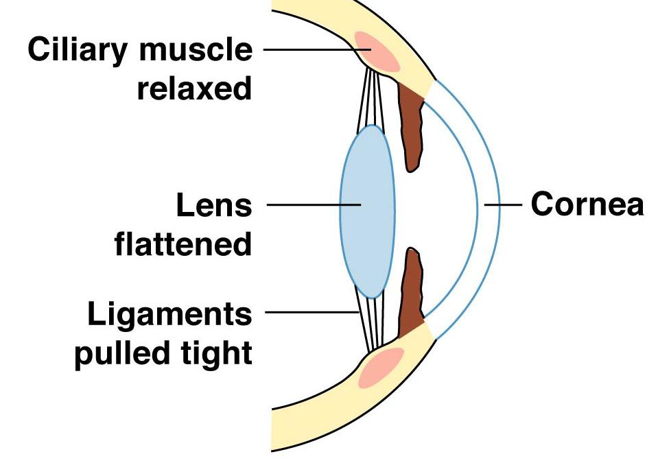
what happens when the ciliary muscle is relaxed?
-when it is relaxed, the ligaments pull on and flatten the lens
-occurs when focusing on distant objects
-causes muscle to widen
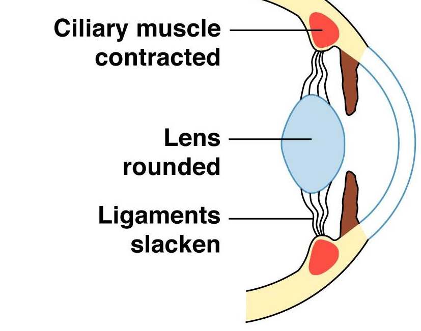
what happens with the ciliary muscle contracts?
-it releases tension on the ligaments and the lens becomes more rounded and thicker
-occurs when focusing on a nearby object
-causes muscle to constrict
hyperopia
-aka far-sightedness
-occurs when the focal point falls behind the retina
myopia
-aka near-sightedness
-occurs when the focal point falls in front of the retina
how is hyperopia corrected?
-it is corrected with convex lens
-because it pre-converges light rays before they enter the eye, shifting the focus forward onto the retina for clearer near vision
how is myopia corrected?
-it is corrected with a concave lens
-because it diverges (spreads out) light rays before they enter the eye, shifting the focal point backward so it lands directly on the retina, rather than in front of it, which causes distant objects to appear blurry
the retina
-complicated circuit of cells
-arranged into several layers
-rods and cones are at the back and their tips face backwards
optic nerve
-axons of ganglion cells join together to form this
-exits the retina at the optic disk (“blind spot”)
horizontal cell
-important for lateral inhibition
-GABA releasing inhibitory cells
what is the orientation of the retina (+eye) in vertebrates?
order:
light → ganglion cells + (amacrine cells)→ bipolar cells + (horizontal cells) → photoreceptors (rods and cones) → pigment epithelium
macula lutea (ML)
-is part of the retina responsible for producing sharp visual images
what is the middle of the macula lutea (ML) called?
-it is called the fovea
fovea
-a small depression in the center of the ML where overlying bipolar and ganglion cells are pushed to the side
-contains only cones
-provides the sharpest images
-highest density of cones, no rods in the very center
rods
-sensitive to black and white images
-ciliary photoreceptor
-outer segment rod-shaped
-sensitive to very dim light
-has one type of photopigment
signal processing:
-principle of convergence – as many as 100 rods synapse with a single bipolar cell → many bipolar cells synapse with a ganglion cell
-large visual field because of its high amount
-fuzzy image
cones
-are less sensitive to contrasts of light
-help us perceive colour
-ciliary photoreceptor
-outer segment cone shaped
-sensitive to brighter light
-up to 3 types of photopigments in mammals
signal processing:
-have a 1:1 ratio
-One cone synapses with one bipolar cell which connects to one ganglion cell
-small visual field
-high resolution image
-concentrated in the fovea
where does light not need to pass?
-light does not need to pass through bipolar and ganglion cells, or blood vessels
age-related macular degeneration (AMD)
-a very common age-related disease
-can be due to the loss of the pigment epithelium or the abnormal development of blood vessels under the macula
(blood vessels can become leaky or cause inflammation which can lead to blindness)
-causes the loss of photoreceptors in the macula lutea
what are all mammalian photoreceptors?
-they are all ciliary photoreceptors
what are the 2 types of ciliary photoreceptors?
rods
cones
**epithelial cell type
what are the 3 structural components of photoreceptors?
outer segment
inner segment
synaptic terminal
outer segment
-series of disks containing photopigments
inner segment
-the cell body, which contains the nucleus
synaptic terminal
-makes connections with neurons in the retina
epithelial cell type
-cell that releases a neurotransmitter to a neuron, which can fire an AP, since it can not fire can AP
photopigments
-molecules that absorb energy from photons
-have 2 covalently bonded parts
what are the 2 covalently bonded parts of a photopigment?
chromophore
opsin
chromophore
-pigment that is a derivative of vitamin A, (ex. retinal)
opsin
-G-protein-coupled receptors
-a rod
what is the general process of photoreception?
chromophore absorbs energy from photon
chromophore changes shape
photoreceptor protein changes shape
signal transduction cascade
change in membrane potential (leads to a graded potential)
bleaching
-when activated retinal no longer bonds to opsin, thereby activating opsin
-called this once opsin is activated
what are the visual pigments in rods called?
-they are called rhodopsin
what are the visual pigments on cones called?
-they are called
erythrolabe,
cholorolabe
cyanolabe
rhodopsin
-decomposes in presence of light
-triggers a complex series of reactions that initiate nerve impulses
-impulses travel along optic nerve
erythrolabe
-responds to red pigment
chlorolabe
-responds to green pigment
cyanolabe
-responds to blue pigment