Special Senses: the Ear
1/35
There's no tags or description
Looks like no tags are added yet.
Name | Mastery | Learn | Test | Matching | Spaced | Call with Kai |
|---|
No analytics yet
Send a link to your students to track their progress
36 Terms
function of the outer ear
funnels vibrations to the tympanic membrane (eardrum)

components of the outer ear
auricle (pinna)
external auditory canal

auricle (pinna)
shell-shaped structure formed by elastic cartilage that funnels vibrations to the external auditory canal

external auditory canal
passage leading from the auricle, through the temporal bone, to the tympanic membrane

location of the middle ear
tympanic cavity

components of the middle ear
tympanic membrane
pharyngotympanic (auditory) tube
auditory ossicles
muscles: tympanic reflex (stapedius and tensor tympani)

tympanic membrane
“ear drum” that separates the external auditory canal from the tympanic cavity; transfers vibrations to the auditory ossicle

pharyngotympanic (auditory) tube
passageway to the nasopharynx and functions to equalize pressure between the middle and outer ear
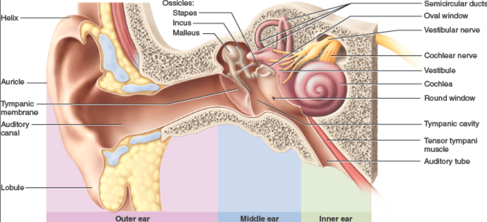
auditory ossicles
smallest bones in the human body
function: transfer vibrations from the tympanic membrane to the inner ear
components: malleus, incus, stapes

malleus
part of the auditory ossicles; “hammer” that is found between tympanic membrane and incus; connected to tensor tympani muscle

incus
part of the auditory ossicles; “anvil” found between malleus and stapes

stapes
part of the auditory ossicles; “stirrup” that is found between incus and oval window; connected to the stapedius muscle

tympanic reflex in the middle ear
muscle group (stapedius and tensor tympani) that muffles the transfer of vibrations during loud sounds by altering tension on malleus and stapes

stapedius
part of the tympanic reflex; reduces motion of stapes to lessen effect on the inner ear
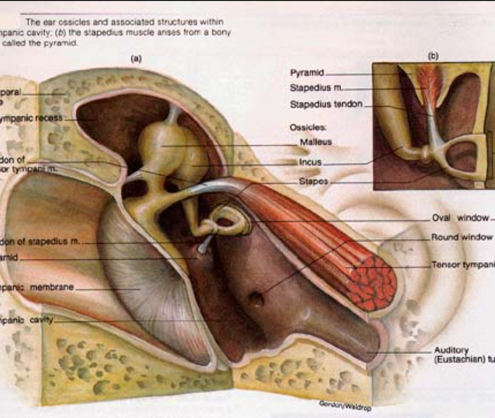
tensor tympani
part of the tympanic reflex muscle group; pulls malleus away from eardrum

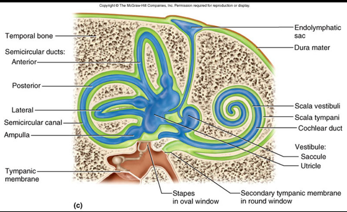
location of the inner ear
in the temporal bone passageways called the bony labyrinth (the bony labyrinth houses the membranous labyrinth)

perilymph
part of the inner ear; cushioning fluid found between the bony and membranous labyrinth

endolymph
part of the inner ear; fluid found within the membranous labyrinth
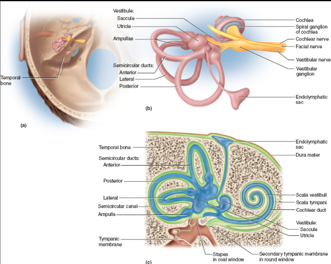
cochlea
part of the inner ear; coiled, snail-shell-shaped organ for hearing that arises from the vestibule
oval window: connected to the stapes; transfers vibrations to cochlea
round window: found at the end of the cochlea acting as a pressure relief

scala vestibuli
part of cochlea that is filled with perilymph; begins near the oval window

scala media (cochlear duct)
part of cochlea that is filled with endolymph; contains Spiral Organ of Corti
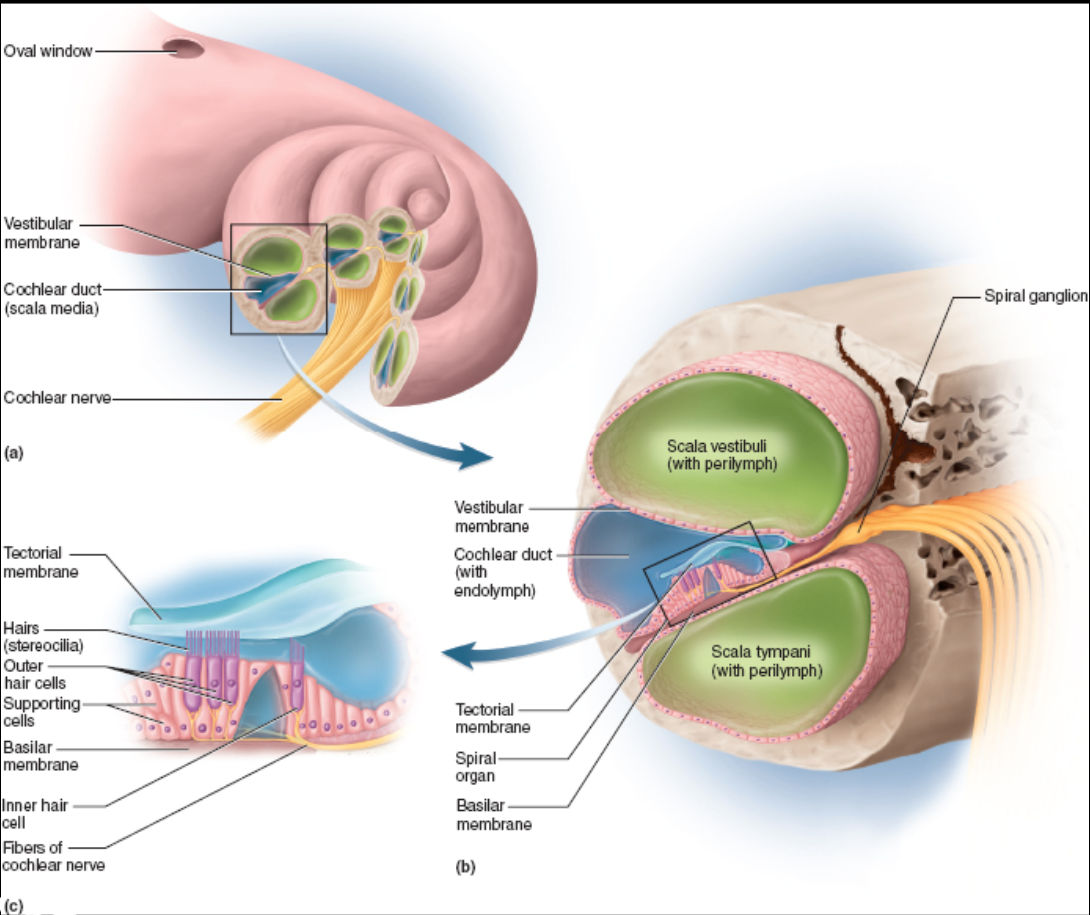
scala tympani
part of cochlea that is filled with perilymph; ends at the round window

vestibular membrane of the cochlea
separates the scala vestibuli from the cochlear duct

tectorial membrane of the cochlea
feather-shaped gelatinous substance that acts to open channels for depolarization; part of the Spiral Organ of Corti

basilar membrane of the cochlea
separates the cochlear duct from the scala tympani
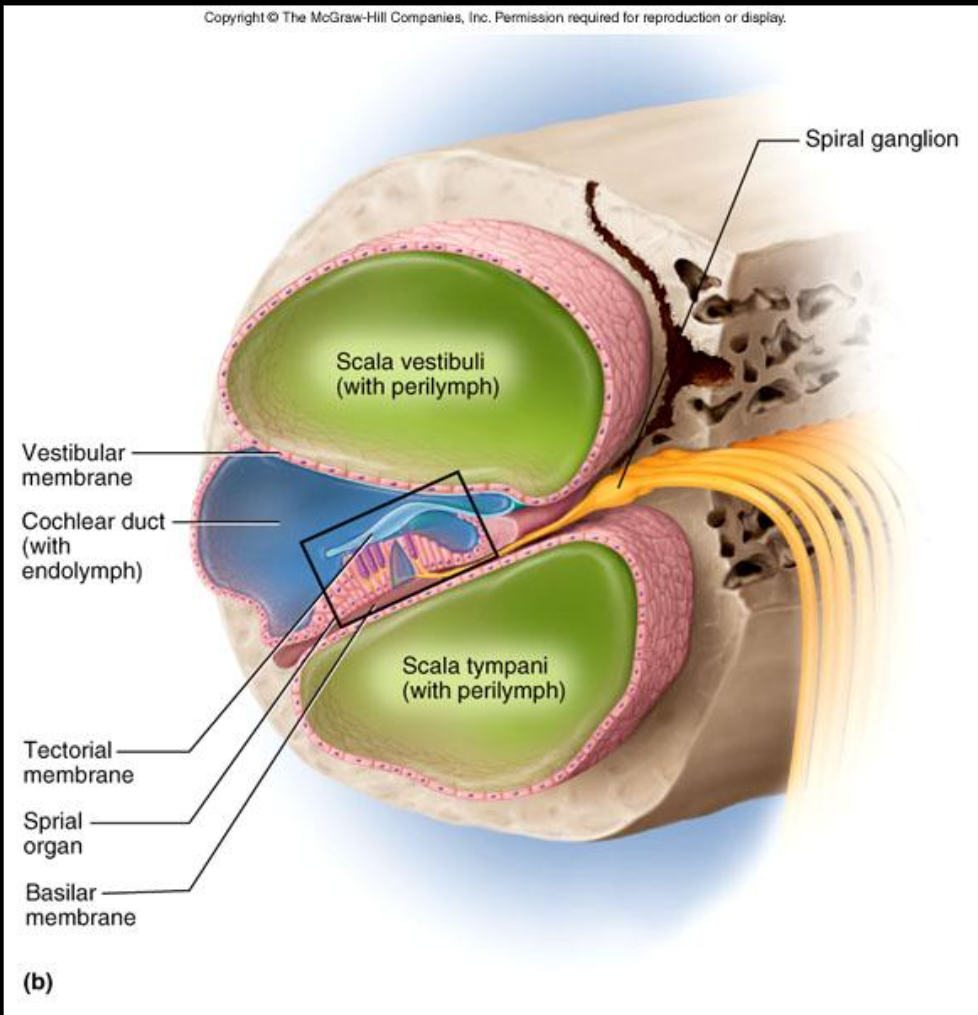
spiral organ of corti
structure of the cochlea that converts vibrations to neural impulses; stereocilia (hairs) vibrate, pressing up against the tectorial membrane in order to open channels allowing for depolarization

physiological workflow of hearing
vibrations are captured by the auricle and funneled by the external auditory canal to the tympanic membrane. the tympanic membrane transfers vibrations through the auditory ossicles to the oval window. vibrations travel through perilymph (fluid), ascending up the scala vestibuli (which vibrates the vestibular membrane). the vibration of the scala vestibuli vibrates the endolymph (fluid), causing movement of the stereocilia (hairs) against the tectorial membrane (depolarizing the neural cells). vibrations travel through the basilar membrane to the scala tympani, out through the round window.

Vestibule
chamber that contains the organs of equilibrium

oval window
the beginning of the inner ear that is attached to the stapes

saccule
portion of the Vestibule that leads to the cochlea; contains maculae and is responsible for linear acceleration

utricle
portion of the Vestibule that leads to the Semicircular Canals; contains Maculae and is responsible for linear acceleration
maculae
equilibrium receptors found in the Vestibule that respond to gravitational pull and changes in head position

static equilibrium
the body is stationary but the head is tilted; perceived by Macula found in the Saccule and Utricle
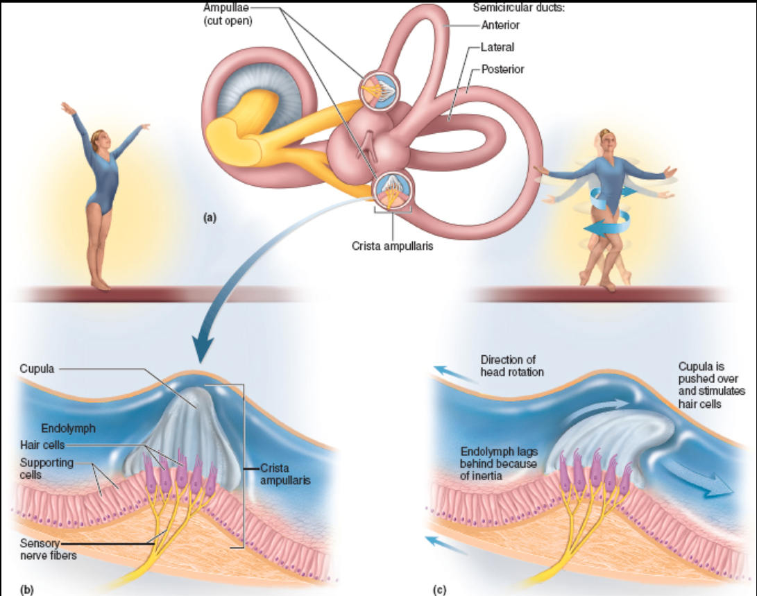
Semicircular Canals
responsible for sense of equilibrium
made up of anterior, posterior, and lateral ducts, which respond to angular movement of the head

dynamic equilibrium
perception of motion or angular acceleration; perceived by the semicircular canals

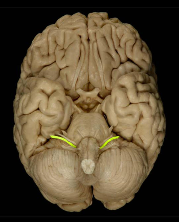
vestibulocochlear nerve (CN VIII)
formed from the Vestibular Nerve and Cochlear Nerve
composition: mixed
function: hearing and balance
damage: deafness and impaired balance