Mass transport
1/70
Earn XP
Description and Tags
Name | Mastery | Learn | Test | Matching | Spaced | Call with Kai |
|---|
No analytics yet
Send a link to your students to track their progress
71 Terms
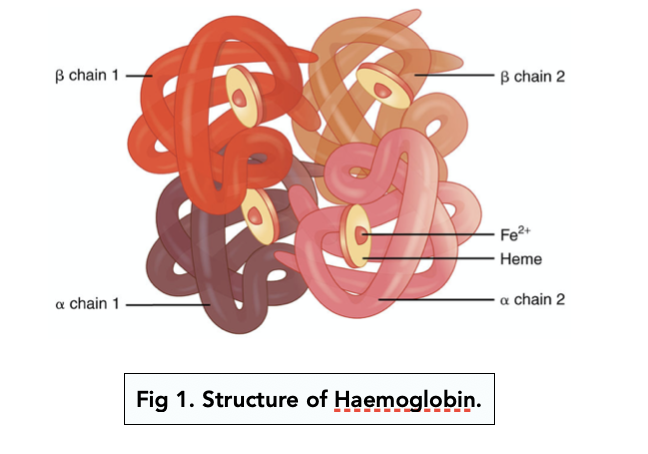
Describe the structure of haemoglobin.
Four polypeptide chains, each chain has a haem group attached where there is an iron ion.
Where exactly in a molecule of haemoglobin does oxygen bind?
Oxygen binds to the haem groups in haemoglobin.
True or false? Oxygen is loaded in regions of low oxygen partial pressure and is unloaded in regions of high oxygen partial pressure.
False. OXYGEN IS LOADED IN REGIONS OF HIGH OXYGEN PARTIAL PRESSURE AND UNLOADED IN REGIONS OF LOW OXYGEN PARTIAL PRESSURE.
Where in the body is there usually a high partial pressure of oxygen?
In the alveoli in the lungs.
Where in the body is there usually a low partial pressure of oxygen?
In respiring tissues.
What is this?
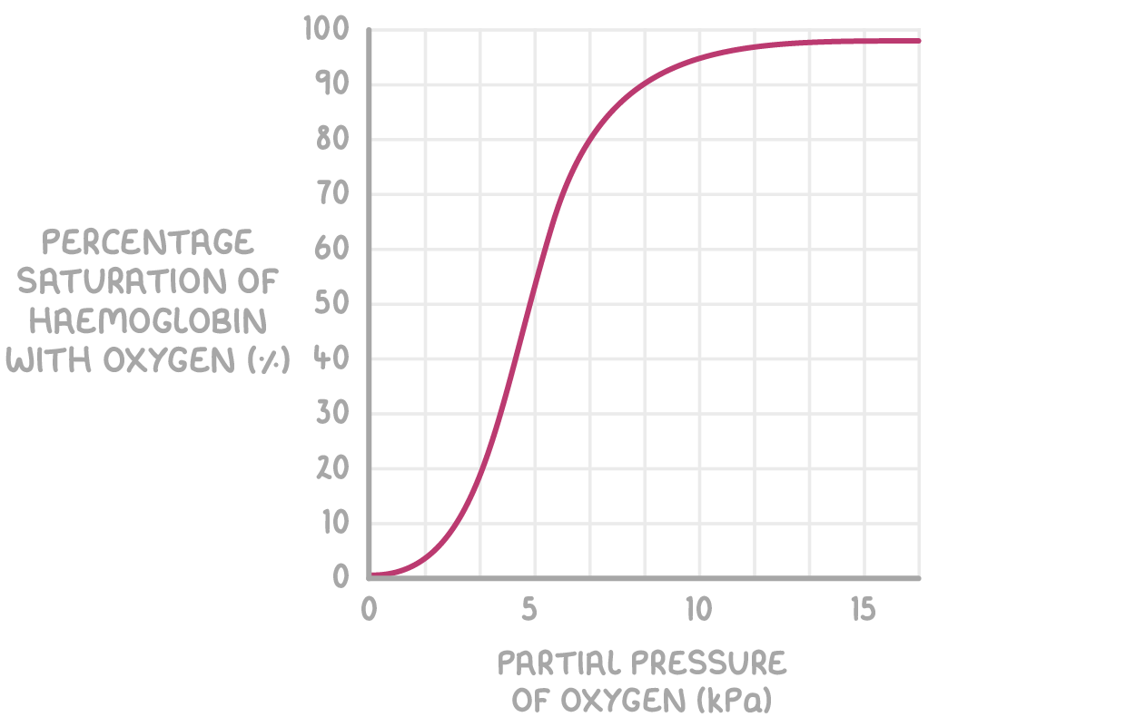
Oxyhaemoglobin dissociation curve.
What does affinity to oxygen mean?
The attraction and ability to bind to oxygen.
What is the maximum number of oxygen molecules that a haemoglobin molecule can load at a time and why?
4 oxygen molecules, haemoglobin has four haem groups for oxygen to bind to.
Describe the four different regions on an oxyhaemoglobin dissociation curve (each region for each oxygen molecule bound to the haemoglobin).
1) When one oxygen molecule has bound to the haemoglobin, haemoglobin changes shape in a way that makes it easier to more oxygen molecules to bind to it (by exposing the other haem groups).
2/3) The differently shaped haemoglobin makes it easier for the second and third oxygen molecule to bind to it so haemoglobin’s affinity for oxygen increases.
4) It is harder for the fourth oxygen to bind to haemoglobin because there is only a ¼ chance that the oxygen molecule with come into contact with a haem group.
Describe haemoglobin’s affinity for oxygen in areas with a high partial pressure of oxygen.
High affinity for oxygen.
Describe haemoglobin’s affinity for oxygen in areas with a low partial pressure of oxygen.
Low affinity for oxygen.
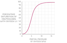
Explain the Bohr effect.
Carbon dioxide dissolves to form carbonic acid, this decreases the pH of the tissue. This decrease in pH slightly changes the shape of haemoglobin meaning haemoglobin’s affinity for oxygen decreases meaning oxygen is released more easily into the tissue where oxygen is needed so that aerobic respiration can continue.
How does a decrease in tissue pH (increase carbon dioxide partial pressure) affect the position of the oxyhaemoglobin dissociation curve and why?
A decrease in pH causes the oxyhaemoglobin curve to shift right, this indicates a decrease in haemoglobin’s affinity for oxygen meaning it more readily unloads oxygen where it is needed (the tissues that respire a lot and therefore produce lots of carbon dioxide which causes the decrease in pH).
True or false? Different animals have differently adapted haemoglobin depending on their environment.
True.
How is the haemoglobin of animals living in low oxygen environments adapted and why?
The haemoglobin of animals living in low oxygen environments has a higher affinity for oxygen, this means that there is an oxygen store and this oxygen only dissociates from haemoglobin when all the rest of the oxygen has been used up by cells.
True or false? Mammals have open circulatory systems.
False. MAMMALS HAVE CLOSED CIRCULATORY SYSTEMS.
True or false? Mammals have double circulation systems.
True.
What is a closed circulatory system?
A circulatory system in which blood remains within the blood vessels.
What is a double circulatory system?
A circulatory system in which the blood passes through the heart twice in each circuit (one circuit delivers blood to the lungs and the other circuit delivers blood to the rest of the body).
Why do mammals need a double circulatory system?
To manage the pressure of blood flow around the body.
Why does blood flow through the lungs at a lower pressure?
To avoid damage to the capillaries in the alveoli.
So that blood moves through the lungs at a slower pace giving more time for gas exchange.
Why is blood pumped through the rest of the body at a higher pressure than in the lungs?
Pumping blood through the body at a higher pressure ensures that blood reaches all of the respiring cells.
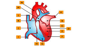
Label this diagram of the heart (ignore D).
A) Pulmonary vein.
B) Left atrium.
C) Left atrioventricular valve (biscupid valve).
E) Left ventricle.
F) Septum.
G) Right ventricle.
H) Right atrioventricular valve (triscupid valve).
I) Vena cava (superior).
J) Right atrium.
K) Semi-lunar valve.
L) Vena cava (inferior).
M) Pulmonary artery (inferior)
N) Aorta.
Where does blood go to and from in the pulmonary artery? Is this blood oxygenated or deoxygenated?
Blood goes from the heart into the lungs. This blood is deoxygenated.
Where does blood go to and from in the pulmonary vein? Is this blood oxygenated or deoxygenated?
Blood goes from the lungs to the heart. This blood is oxygenated.
Where does blood go to and from in the aorta? Is this blood oxygenated or deoxygenated?
Blood goes from the heart to the rest of the body in the aorta. This blood is oxygenated.
Where does blood go to and from in the vena cava? Is this blood oxygenated or deoxygenated?
Blood goes from the rest of the body into the heart in the vena cava. This blood is deoxygenated.
Where does blood go to and from in the renal vein? Is this blood oxygenated or deoxygenated?
Blood goes from the kidneys to the heart in the renal vein. This blood is deoxygenated.
Where does blood go to and from in the renal artery? Is this blood oxygenated or deoxygenated?
Blood goes from the heart into the kidneys through the renal artery. This blood is oxygenated.
What is this?
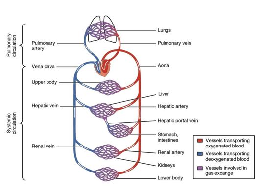
The circulatory system of a mammal.
How do arteries branch of into capillaries?
Arteries become arterioles when then become capillaries.
Give and explain three reasons why arteries are well adapted to their function.
They contain lots of elastic tissue (this helps maintain smooth blood flow and a consistent blood pressure, stretch and recoil in response to heart).
They have thick muscular walls (to maintain/withstand high blood pressure).
They have smooth endothelium (to reduce friction).
True or false? Arterioles have very thin muscular walls.
False. ARTERIOLES HAVE VERY THICK MUSCULAR WALLS.
What is the advantage of arterioles having thick muscular walls?
They are more easily able to contract and relax to change blood pressure of blood moving from the arteries into the capillaries, if blood moves at such a high pressure in the capillaries the capillaries will be damaged.
Give and explain three adaptations of veins.
Thinner muscular walls (so that blood travels at a lower pressure).
Less elastic tissue (blood travels at a lower pressure so there is less need for recoil).
Valves (prevents backflow of blood which is a risk if blood is travelling at low pressure).
How are capillaries well-adapted for efficient gas exchange?
They have a narrow diameter meaning red blood cells pass through them in single file which increases the contact time of each red blood cell with the capillary wall, capillary walls are only one cell thick (of flattened endothelial cells) to reduce diffusion distance.
What are capillary beds?
Networks of capillaries that spread through tissues.
What is tissue fluid?
Fluid containing water, glucose, amino acids, fatty acids, ions, and oxygen which the tissues are bathed in.
Talk me through how tissue fluid is formed.
1) Due to a smaller diameter in the capillaries, blood enters the capillaries from the arterioles at a high hydrostatic pressure.
2) This high hydrostatic pressure forces out molecules that are small enough to pass through the gaps in the cells that make up the capillaries (this collection of molecules makes up the tissue fluid).
3) Some water re-enters the capillary by osmosis.
4) Tissue fluid (containing the rest of the water that was not reabsorbed into the capillaries) is absorbed into the lymphatic system.
What is the name of the process in which tissue fluid is formed?
Ultrafiltration.
What is the name of the heart as a muscle?
The cardiac muscle.
True or false? The cardiac muscle is myogenic.
True.
What does myogenic mean in terms of the heart?
The heart can contract and relax without nervous or hormonal stimulation.
True or false? The heart fatigues eventually even with a constant supply of oxygen.
False. THE HEART NEVER FATIGUES AS LONG AS IT HAS A CONSTANT SUPPLY OF OXYGEN.
Which artery delivers oxygen to the cardiac muscle?
The coronary artery.
Give and explain two adaptations of the atria.
Thinner muscular walls (they do not need to contract as strongly as blood does not have to be pumped far and blood is pumped downwards).
Elastic walls (can stretch when blood enters them).
Why does the left ventricle have a thicker muscular walls than the right ventricle?
The right ventricle only has to pump blood to the lungs which aren’t as far whereas the left ventricle has to pump blood to the rest of the body which is further and a higher pressure must be maintained so that oxygenated blood reaches all the respiring cells in the body.
How do valves open and close?
Valves open when the pressure is higher behind them, valves close when the pressure is higher in front of them.
Talk me through the entire cardiac cycle in detail (starting with diastole).
1) The atria and ventricles are relaxed during diastole. This enables blood to enter the atria from the body.
2) This influx of blood increases the pressure in the atria causing the atrioventricular valves to open and let some blood into the ventricles.
3) Atrial systole. The muscles of the atria contract while the walls of the ventricles stay relaxed, forcing the rest of the blood from the atria into the ventricles.
4) Short delay.
5) Ventricular systole. The walls of the ventricles contract which increases the pressure (so that it is greater than the pressure in the atria) in the ventricles.
6) This increase in pressure causes the atrioventricular valves to close and the semi-lunar valves to open letting blood be pushed out of the ventricles (and out of the heart).
Which one takes up more time in the cardiac cycle? Diastole or systole?
Diastole.
Give the equation for cardiac output.
Cardiac output = heart rate x stroke volume
(heart rate = beats of the heart per minute (min-1)
stroke volume = volume of blood that leaves the heart in each beat (dm3)).
What is transpiration?
The loss of water from the stomata by evaporation.
Give four factors that affect the rate of transpiration.
Light intensity.
Temperature.
Humidity.
Wind.
How does increased light intensity affect rate of transpiration? Explain your answer.
Increased light intensity increases rate of transpiration. Greater light intensity causes more stomata to open meaning more evaporation takes place.
How does increased temperature affect rate of transpiration? Explain your answer.
Increased temperature increases rate of transpiration. Water molecules have more kinetic energy at higher temperatures meaning they move faster and evaporation takes place faster.
How does increased humidity affect rate of transpiration? Explain your answer.
Increased humidity decreases the rate of transpiration. More water outside the leaf decreases the water potential gradient across the leaf and it’s surroundings meaning water is less likely to evaporate out of the leaf.
How does wind affect the rate of transpiration? Explain your answer.
Wind increases the rate of transpiration. Wind maintains the water potential gradient across the leaf and it’s surroundings meaning water is more likely to evaporate out of the leaf.
Which property of water allows it to move up the xylem vessel in a continuous stream?
Water is cohesive (hydrogen bonding between water molecules).
Which property of water allows it to stick to the walls of the xylem vessel?
Adhesion.
Talk me through the entire process of the cohesion-tension theory of water moving up the xylem vessel.
1) Water evaporates from the stomata via transpiration, decreasing the water potential of the mesophyll cells.
2) This decrease in water potential creates tension on the water column in the xylem vessel, drawing water up.
3) Water molecules sticker together via cohesion (due to hydrogen bonds) allowing the water column up the xylem vessel to be continuous.
4) Water molecules adhere to the walls of the xylem vessel meaning the xylem vessel does not collapse under the tension.
What is this a diagram of?
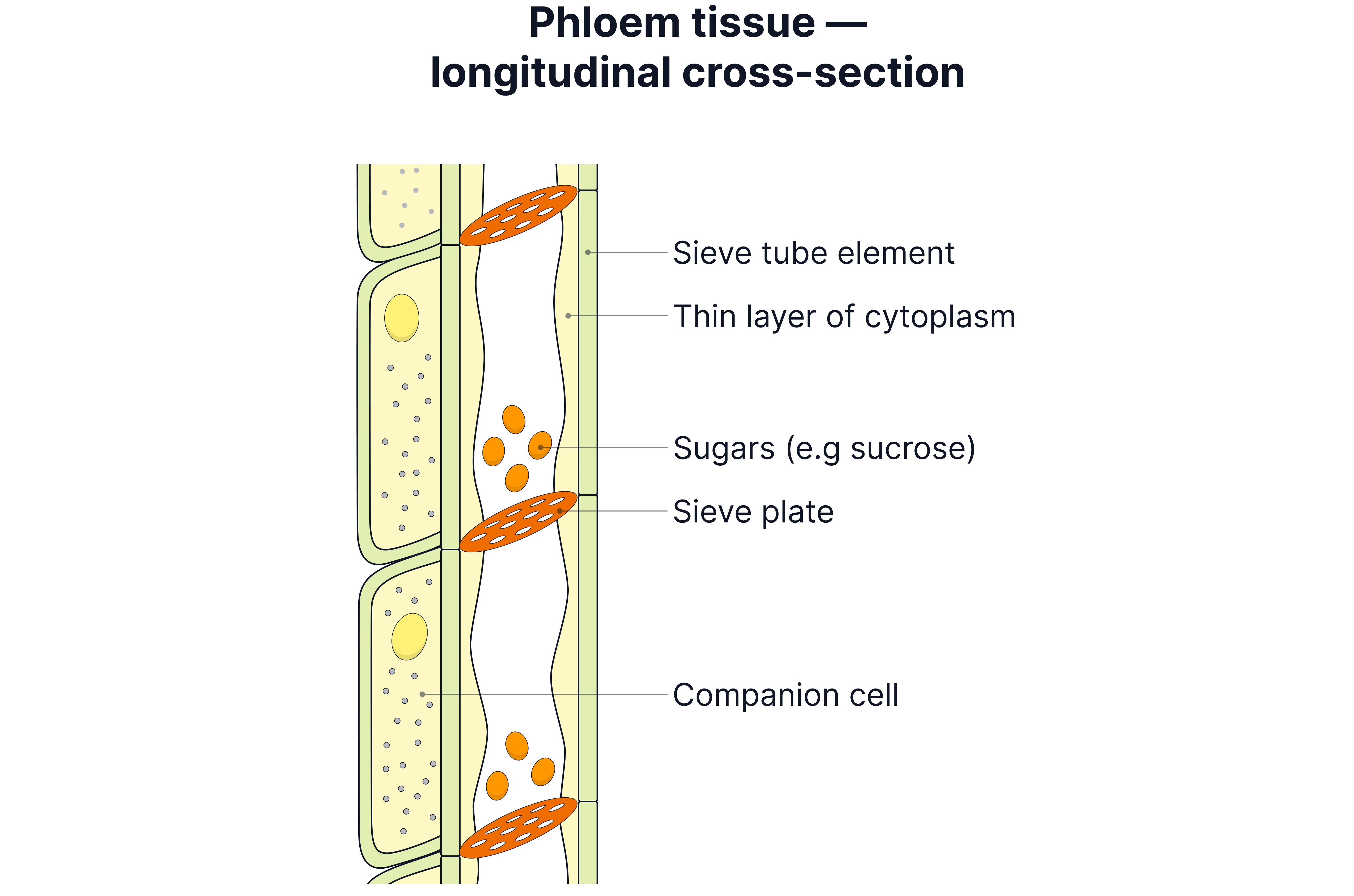
Describe the sieve tube elements.
Living cells, no nucleus, no organelles.
What is the function of companion cells in the phloem?
They provide ATP for the active transport of substances (as the sieve tube elements cannot do this).
What is the name for the site of production of sucrose in a plant?
The source.
What is the name for the site of use of sucrose in a plant?
The sink.
Give an example of a source cell in a plant.
A leaf cell.
Give an example of a sink cell in a plant.
A respiring cell.
Talk me through the process of translocation in less detail.
1) Higher concentration of sucrose in the source cell, so decreased water potential.
2) Decreased water potential causes water to enter the source cell via osmosis (increasing hydrostatic pressure in source cell).
3) Lower concentration of sucrose in the sink cell as it is being used up in respiration, so increased water potential.
4) Increased water potential causes water to leave the sink cell via osmosis (decreasing hydrostatic pressure in sink cell).
5) The higher hydrostatic pressure in the source cell forces the fluid containing sucrose into the phloem where it moves to areas of lower hydrostatic pressure like the sink cell.
Talk me through the process of translocation in more detail.
1) Sucrose is actively transported (so against concentration gradient) out of the source cell and into the sieve tube elements of the phloem by companion cells (companion cells use ATP to do so).
2) Movement of sucrose into sieve tube elements decreases water potential of phloem, so water enters the phloem via osmosis from adjacent xylem vessel.
3) Influx of water increases hydrostatic pressure in the phloem.
4) At the sink sucrose moves into the sink cells via active transport and facilitated diffusion, this increases water potential of phloem.
5) This causes water to move via osmosis out of the phloem and back into the xylem vessel, lowering the hydrostatic pressure in the phloem.
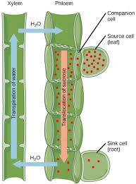
Diagram showing process above.
Explain how tracers support the fact that sugars are transported in the phloem.
When plants are provided with only radioactively labelled carbon dioxide, the plant will make sugars containing radioactive carbon over time. Areas where sugars are transported (the phloem) go black under an x-ray when radioactive carbon is present in a stem slice.
Explain how ringing experiments prove that sugars are transported in the phloem.
When bark is removed from a tree (which contains the phloem, so the phloem is removed from the tree), the trunk swells above the removed section. The liquid in the swelling is analysed to contain sugar. This means that when the phloem is removed, sugars cannot be transported and therefore the phloem transports sugars.