Albinism & Retinotopy - Visual Neurophysiology and Perception Spring 2026
1/196
There's no tags or description
Looks like no tags are added yet.
Name | Mastery | Learn | Test | Matching | Spaced | Call with Kai |
|---|
No analytics yet
Send a link to your students to track their progress
197 Terms
genetic
Albinism is a _____ disorder
a generalized lack of melanin in the body
What is albinism characterized by?
can be either
Is Albinism Tyrosinase + or -?
the lack of melanin is NOT absolute
What is Tyrosinase + Albinism?
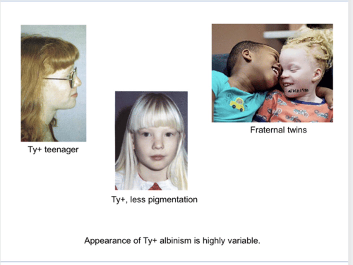
the signs & symptoms will be less severe than Tyrosinase -
What are the visual signs and symptoms of Tyrosinase + Albinism?
where melanin is completely missing
What is Tyrosinase - Albinism?
the signs & symptoms will be more severe than Tyrosinase +
What are the visual signs and symptoms of Tyrosinase -Albinism?
an enzyme that plays key role in multi-step process of producing melanin
What is Tyrosinase?
No -- resembles a newborn retina
Is the fovea in an individual with albinism well-differentiated from the rest of the retina?
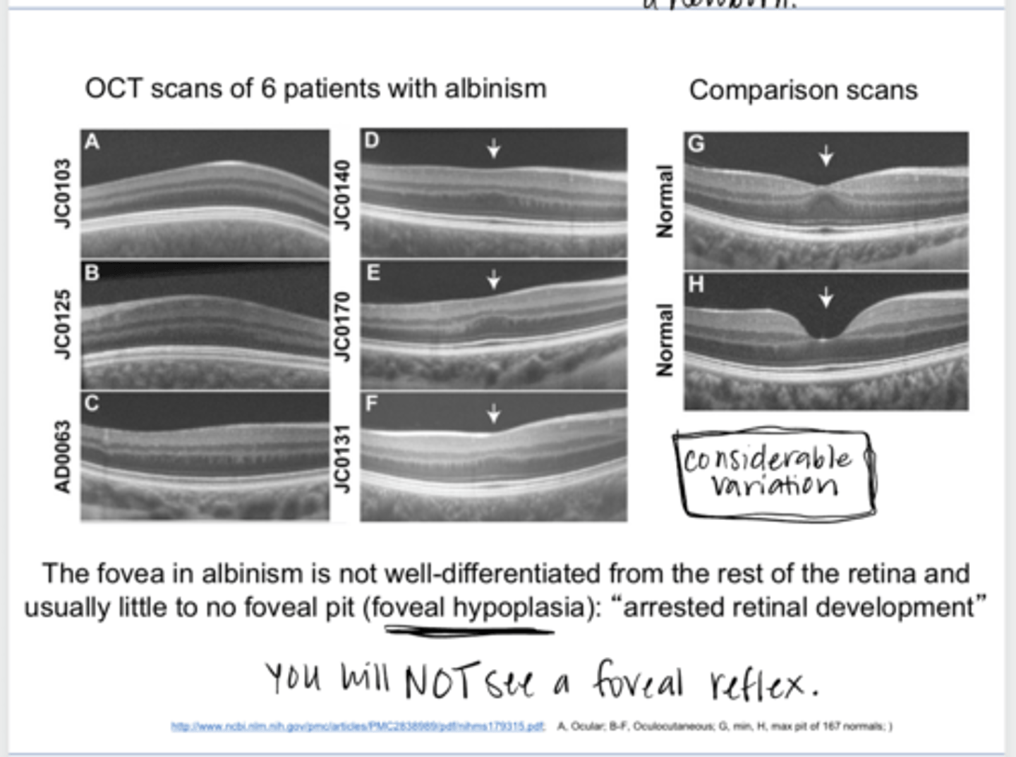
No -- termed foveal hypoplasia
Does the fovea in an individual with albinism show a foveal pit?
true
True or False:
Multiple genetic mutations can cause albinism
Defect of tyrosinase
What is the most common genetic mutation can cause albinism?
Tyrosinase
REVIEW: What is necessary to make melanin?
wiring development of the visual system
What else is tyrosinase important for besides making melanin?
-light iris, which trans-illuminates
-poor foveal development
-reduced VA
-large refractive errors (hyperopia, myopia, astigmatism)
-Difficulty w/ emmetropization
-Nystagmus
Albinism phenotypes often show what?
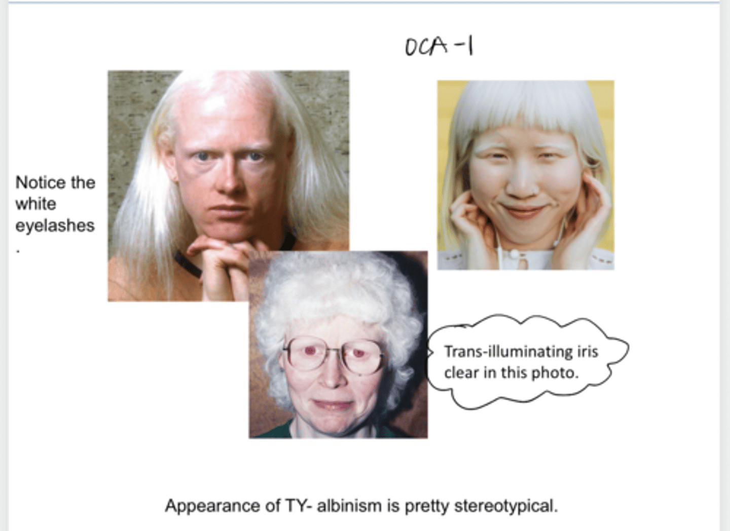
OCA1
What is the most common type of oculo-cutaneous albinism?
eyes, skin, hair
What does oculo-cutaneous albinism affect?
Tyrosinase (-); absolute lack of melanin all throughout the body
Describe OCA1 oculo-cutaneous albinism
-white hair
-pale skin
-white eyelashes/eyebrows
-light iris that transilluminates
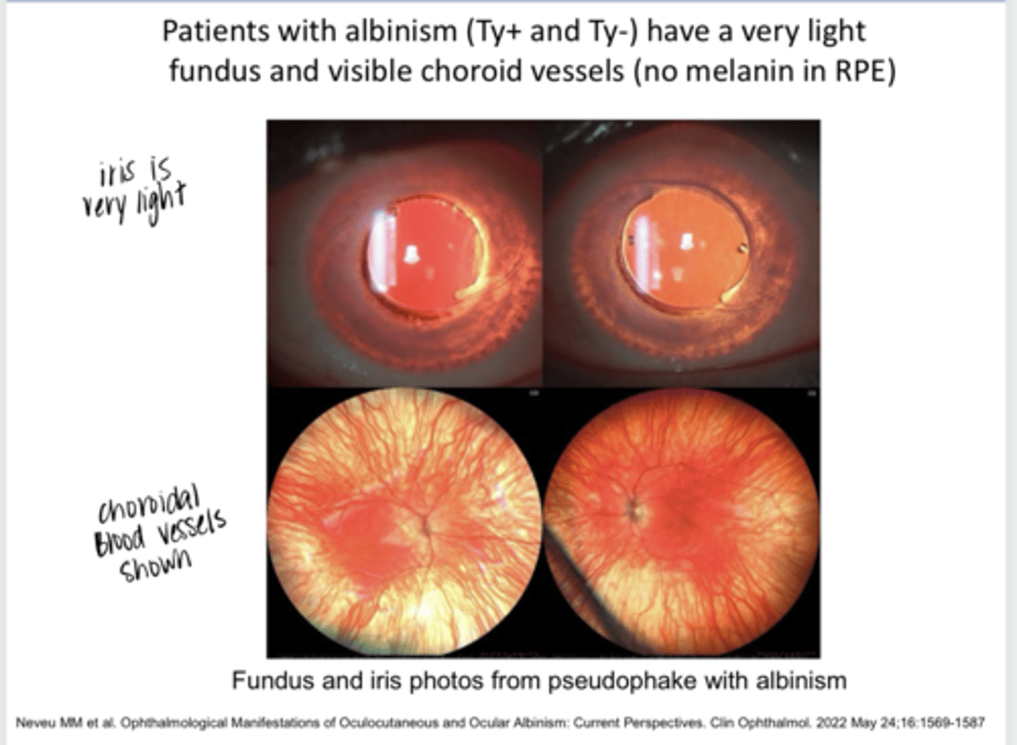
-no melanin in the RPE; hyper-reflective fundus; visible choroid vessels
What are the features of OCA1 oculo-cutaneous albinism?
20/100-20/400
What is the typical VA of an individual with OCA1 oculo-cutaneous albinism?
-some pigmentation of skin and hair, either from melanin or related pigment
-pigmentation may increase over time
-iris has variable pigment
-RPE no pigment
Tyrosinase (+) oculo-cutaneous albinism features
20/60-20/200
What is the typical VA of an individual with tyrosinase (+) oculo-cutaneous albinism?
Tyrosinase (-) Albinism Appearance (Pic)
Tyrosinase (-) Albinism Appearance (Pic)

Tyrosinase (+) Albinism Appearance -- Variable (Pic)
Tyrosinase (+) Albinism Appearance -- Variable (Pic)
Patients with Albinism Ty(+) or Ty(-) Have a Very Light Fundus & Visible Choroid Vessels D/t No Melanin in the RPE (Pic)
Patients with Albinism Ty(+) or Ty(-) Have a Very Light Fundus & Visible Choroid Vessels D/t No Melanin in the RPE (Pic)
Maybe; maybe not
When an individual has Ty(+) ocular albinism, will their skin/hair be obviosuly hypopigmented?
sex linked recessive
Inheritance pattern of ocular albinism
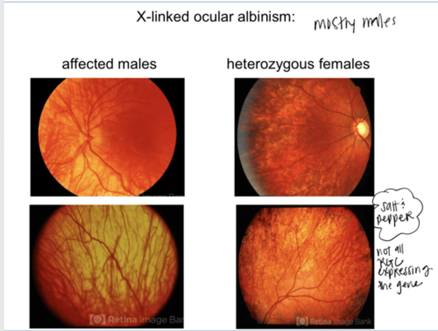
males
Ocular albinism is mostly seen in (females/males)

true
True or False:
Some forms of ocular albinism have associated hearing loss
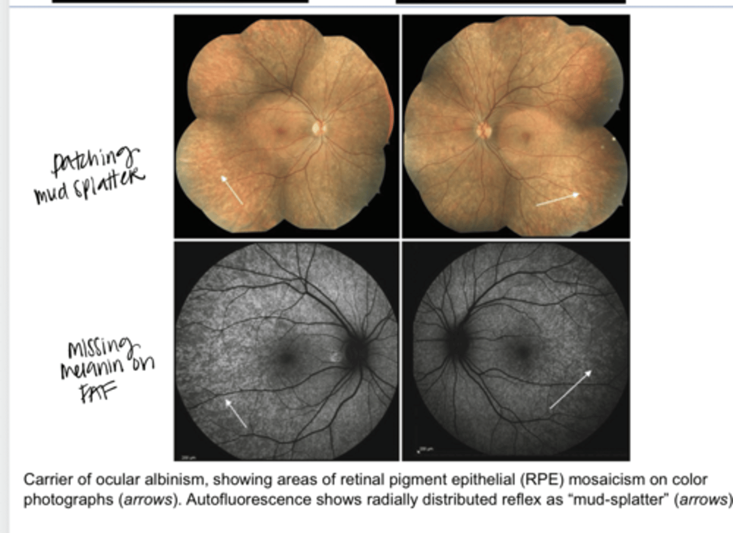
salt and pepper "mud splattered" retina
In females who are heterozygous (carriers) of sex-linked ocular albinism, what will their fundus look like?

X chromosome inactivation in some cells but not others
What is genetic mosaicism as found in ocular albinism?

X Linked Ocular Albinism in Males/Female Carriers (Pic)
X Linked Ocular Albinism in Males/Female Carriers (Pic)

Carrier of Ocular Albinism -- Showing Areas of RPE Mosaicism & Mud Splatter Appearance (Pic)
Carrier of Ocular Albinism -- Showing Areas of RPE Mosaicism & Mud Splatter Appearance (Pic)
-infant like fovea
-more glare d/t lack of melanin in RPE
-projections from the retina to the thalamus are abnormal
What are some consequences of albinism on the retina?
No or little foveal pit (no foveal reflex will be present); this leads to reduced acuity
Describe the fovea in an individual with albinism & the consequence of this
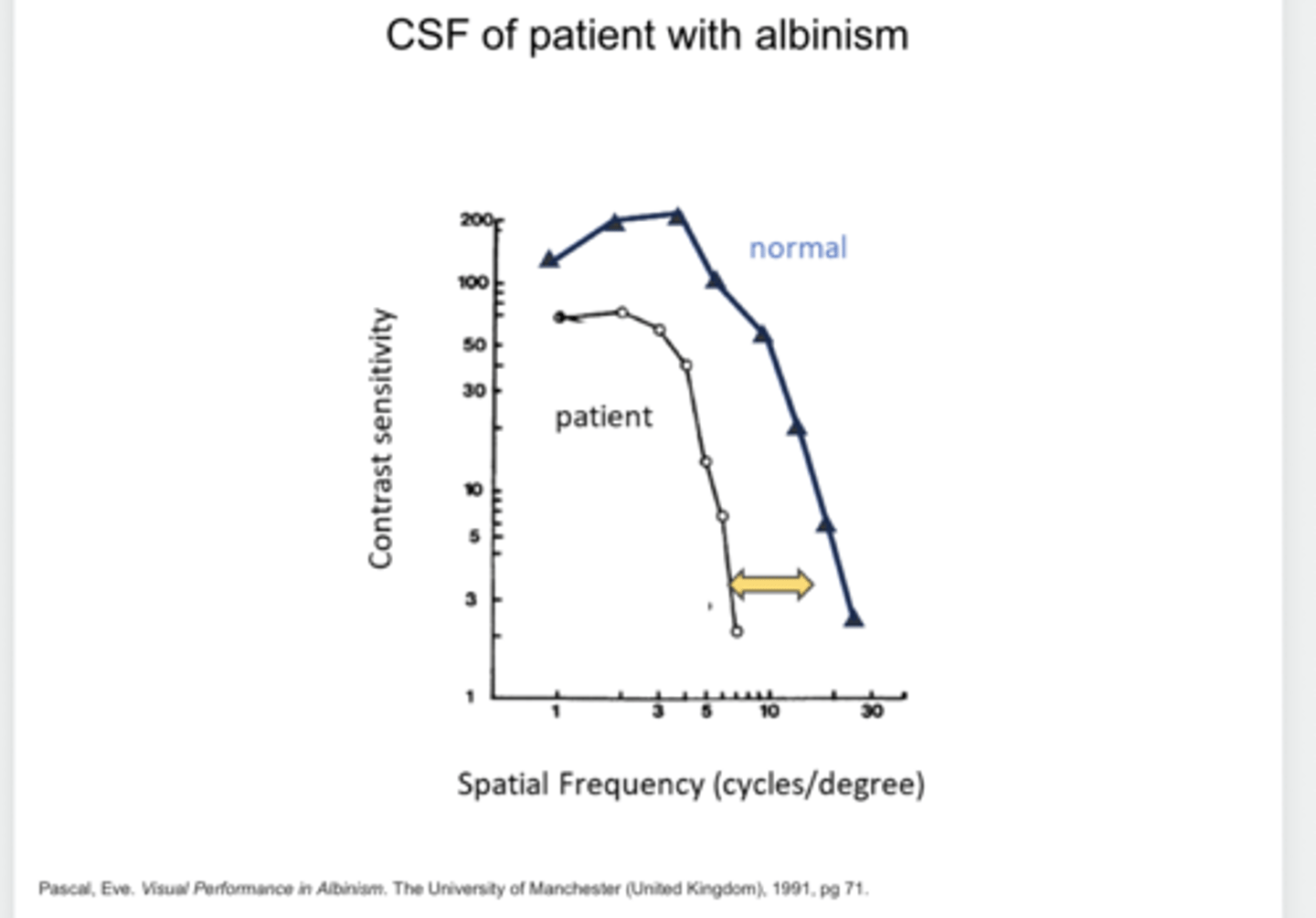
Hypopigmented fundus --> Scattered light --> Reduced Contrast Sensitivity Beyond the Loss of VA
Why would an individual with albinism experience MORE glare? What is the consequence of this?
Abnormal decussation at the optic chiasm --> projections to the LGN don't show the temporal/nasal RGC axon crossing/not crossing that is typically seen
Describe the abnormal projections from the retina to the thalamus in an individual with albinism..
Contrast Sensitivity Function in an Individual with Albinism -- Reduced from Normal (Pic)
Contrast Sensitivity Function in an Individual with Albinism -- Reduced from Normal (Pic)
tyrosine kinases
The Eph molecules expressed in the RGCs are ___________
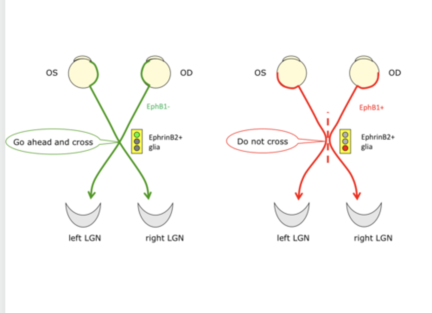
No
Are Eph molecules present in Tyrosinase (-) albinism?

They can be reduced or absent (not rlly understood rn)
Are Eph molecules present in Tyrosinase (+) albinism?

development in determining whether an RGC axon will cross at the chiasm to the contralateral side of the brain OR stay ipsilateral
What do the Eph molecules play a key role in?

Not much
Is EphrinB1 expressed in the temporal RGCs of patients with ocular albinism?

The ipsilateral projection does NOT occur during embryological development in patients with albinism
What is the problem with EphrinB1 not being expressed in temporal RGCs of individuals with ocular albinism?

Crossing/Not Crossing at the Optic Chiasm (Pic)
Crossing/Not Crossing at the Optic Chiasm (Pic)

Normal Crossings v Albinism Crossings at the Optic Chiasm (Pic)
Normal Crossings v Albinism Crossings at the Optic Chiasm (Pic)

left
In a normal left eye, the right visual field will project to what hemisphere of the occipital lobe?

right
In a normal left eye, the left visual field will project to what hemisphere of the occipital lobe?
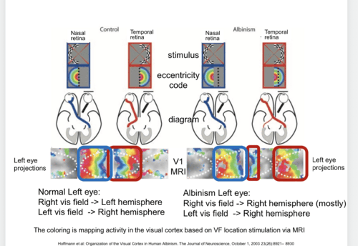
mostly to the right hemisphere
In an albinism left eye, the right visual field will project to what hemisphere of the occipital lobe?

to the right hemisphere
In an albinism left eye, the left visual field will project to what hemisphere of the occipital lobe?

Optic Radiations (Pic)
Optic Radiations (Pic)
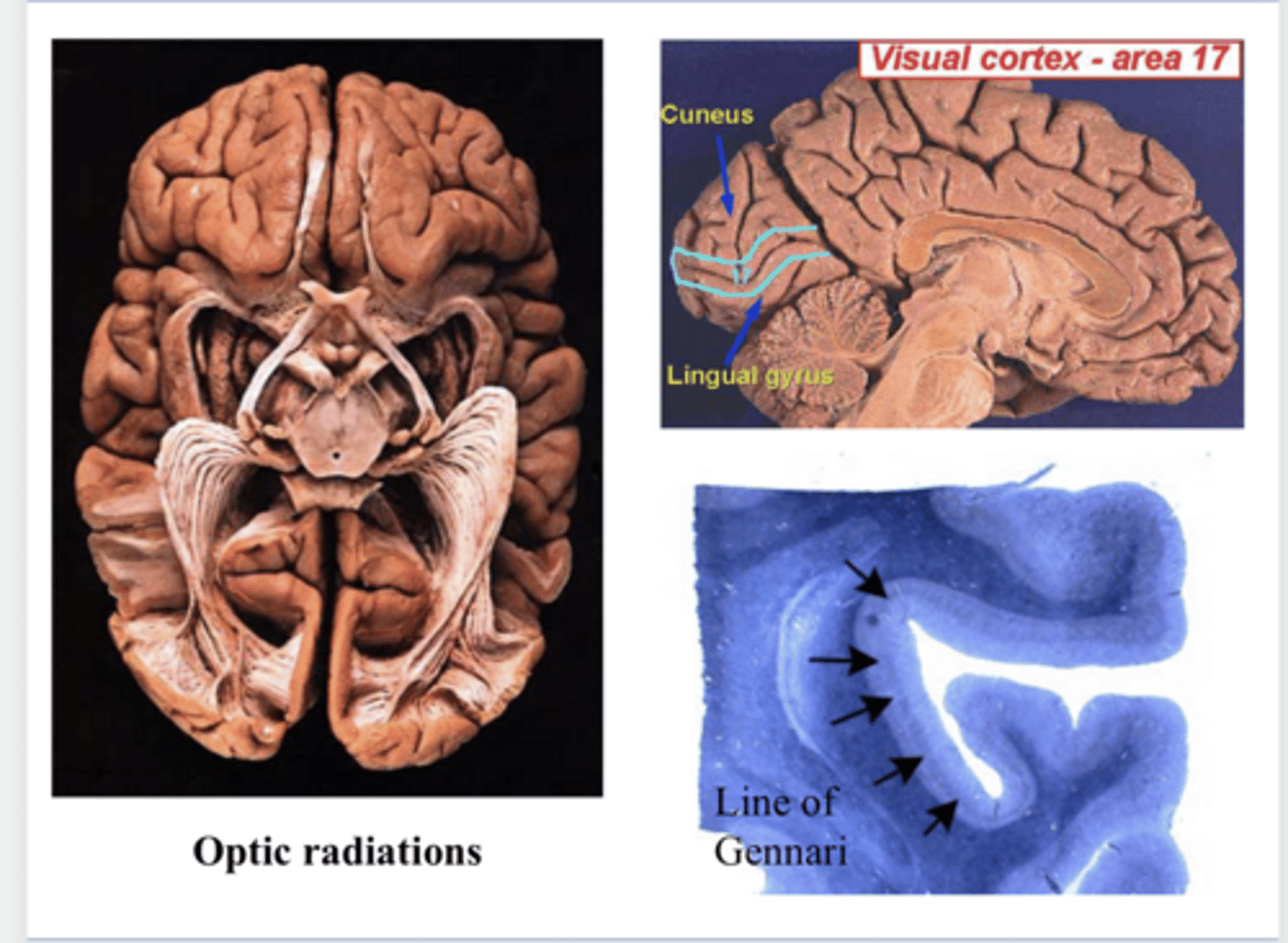
Brodmann Area - V1 - Primary Visual Cortex (Pic)
Brodmann Area - V1 - Primary Visual Cortex (Pic)
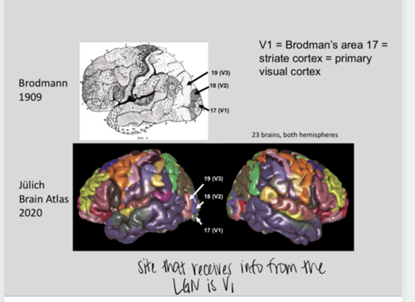
V1
What is the primary area that receives info from the LGN?

true
True or False:
The retinal image is "retinotopically" related to the real world stimulus because of the optics of the eye
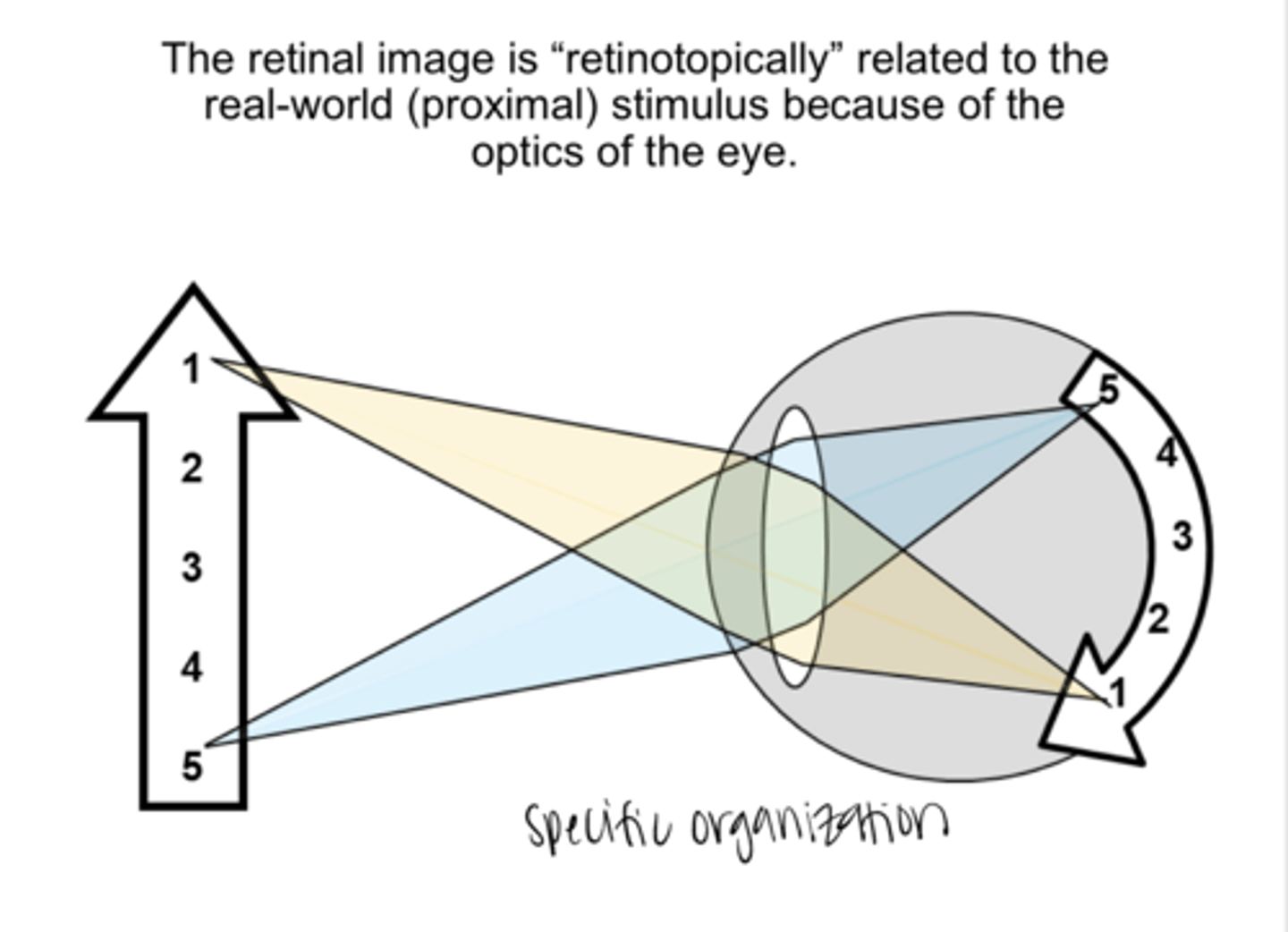
the mapping of the visual stimulus as one moves from the retina to the brain (first LGN and then V1) 00 the order in the retina is maintained in these brain regions
What is the meaning of Retinotopic?
If a stimulus excites retinal patch A and patch B, which are beside each other, the RGCs in these patches are going to project to neurons in the LGN that are close to each other
Example of Retinotopic Mapping
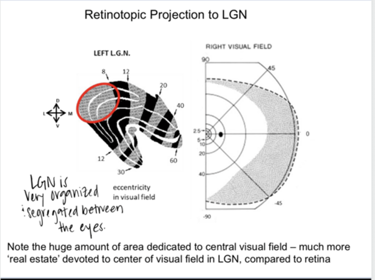
Retinotopic Projection to the LGN (Pic)
Retinotopic Projection to the LGN (Pic)

yes -- much more "real estate" dedicated to center of visual field in the LGN compared to the retina
Is there a huge amount of area in the LGN dedicated to central vision?
yes
Is the primary visual cortex (V1) retinotopic?
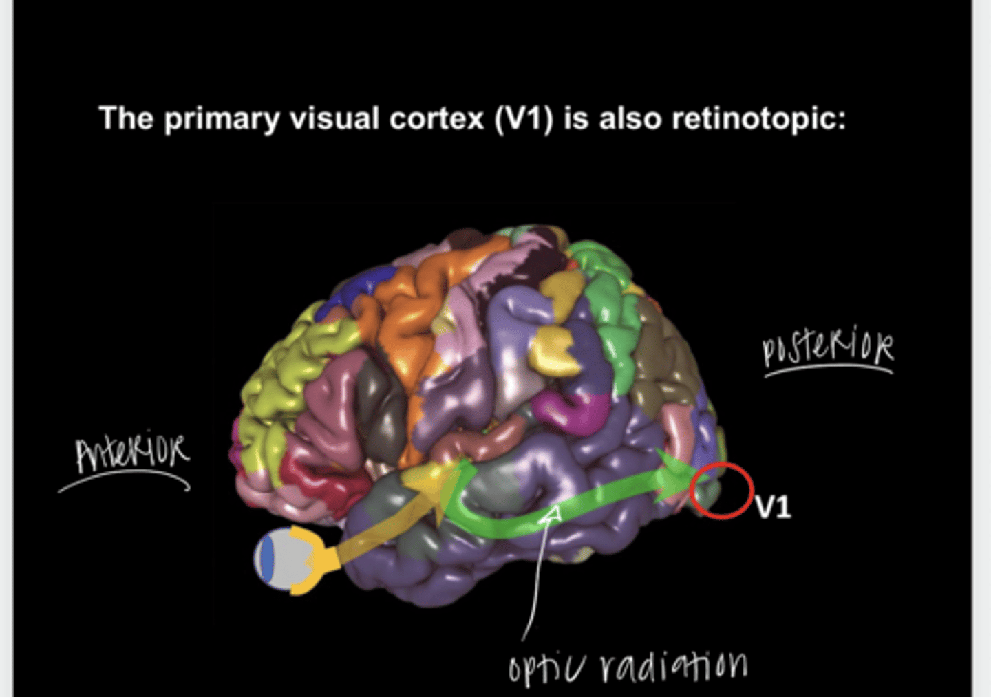
inferior
Neurons from the superior VF will end up in the (inferior/superior) V1

superior
Neurons from the inferior VF will end up in the (inferior/superior) V1

fovea/central vision
What vision is located at the very posterior of V1?
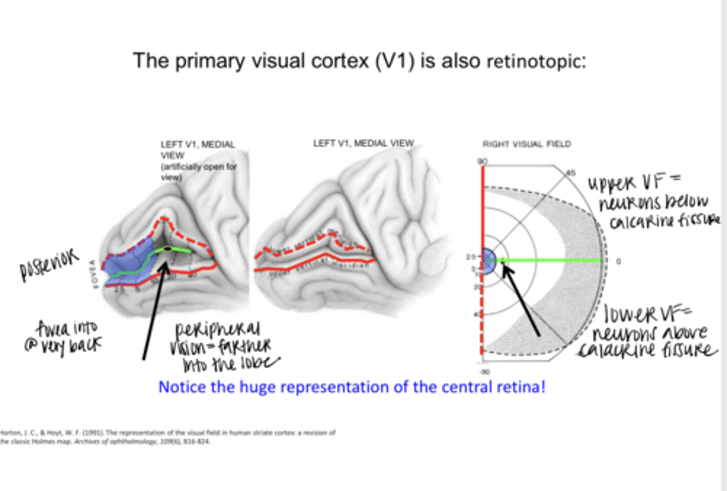
farther INTO the lobe
Where does the peripheral vision project to in V1?

Yes
Is there a huge representation of the central retina in the primary visual cortex?

convergence
6 million cones; 120 million rods; 1 million RGCs
From photoreceptors --> RGC there is a lot of (divergence/convergence) overall
1:1
Ratio of LGN output axons to optic nerve axons
divergence -- image processing will occur here
From axons leaving the LGN --> cells in the striate nucleus there is a lot of (divergence/convergence) overall
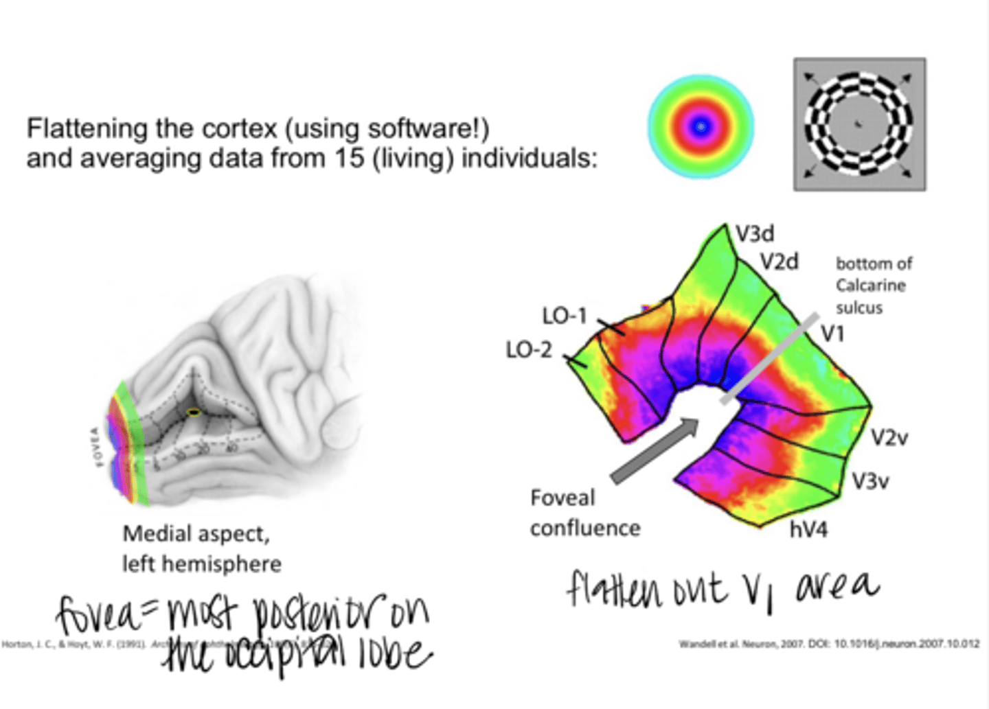
"busy" parts of the brain have increased blood flow (hemodynamic response) and particularly the amount of oxygenated blood increases
What is shown in BOLD (Blood Oxygenation Level Dependent) imaging?
visual stimuli
_____ can make certain parts of the brain busy
what parts of the brain respond to various stimuli
The variation in the BOLD signal as the stimulus changes can tell us what?
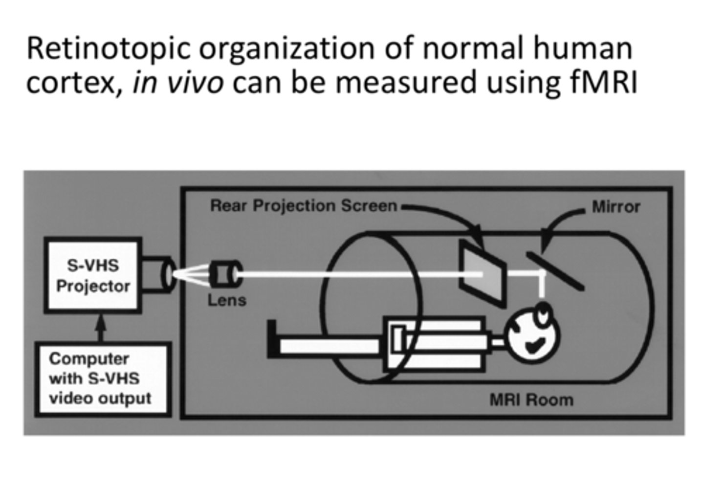
fMRI
Retinotopic organization of normal human cortex, in vivo, can be measured using _____
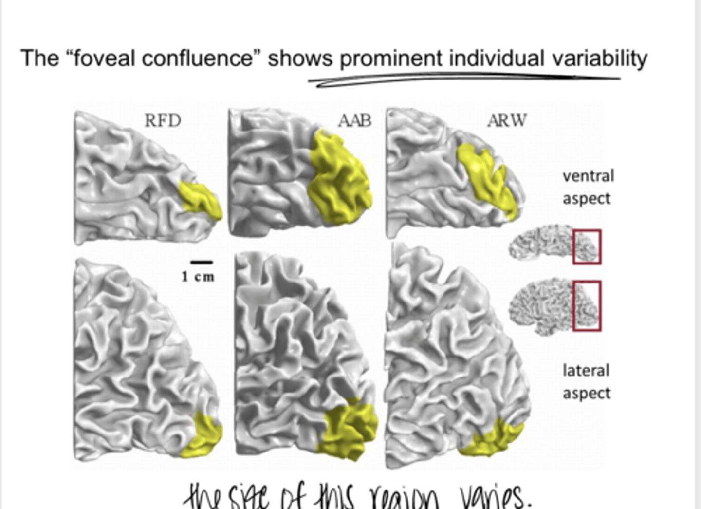
Flattening the Cortex & Mapping of the Foveal Confluence of V1 (Pic)
Flattening the Cortex & Mapping of the Foveal Confluence of V1 (Pic)
variability
The foveal confluence shows prominent individual _____
retina
SUMMARY: The relative amount of visual cortex devoted to each area of the visual field is set at the ______
fovea
SUMMARY: A large fraction of V1 is devoted to the ____
fMRI
SUMMARY: Scientists can measure the retinotopic map in individual people using a _____, which reveals a lot of individual variability
foveal confluence
SUMMARY: The representation of the fovea is in a merged _______
why damage to the brain has different effects on different people
SUMMARY: The individual variability of the retinotopic map explains what?
-Projecting neurites growth cones follow chemical signposts up the concentration gradient
Cell nuclei are born locally and axons must join distant brain regions. How do the axons know where to go?
amblyopia
Understanding how the visual system is wired up in a retinotopic fashion helps to understand the development of ______
Role of EphrinB1 in Determining Whether RGCs Cross or Not at the Chiasm (Pic)
Role of EphrinB1 in Determining Whether RGCs Cross or Not at the Chiasm (Pic)

true
True or False:
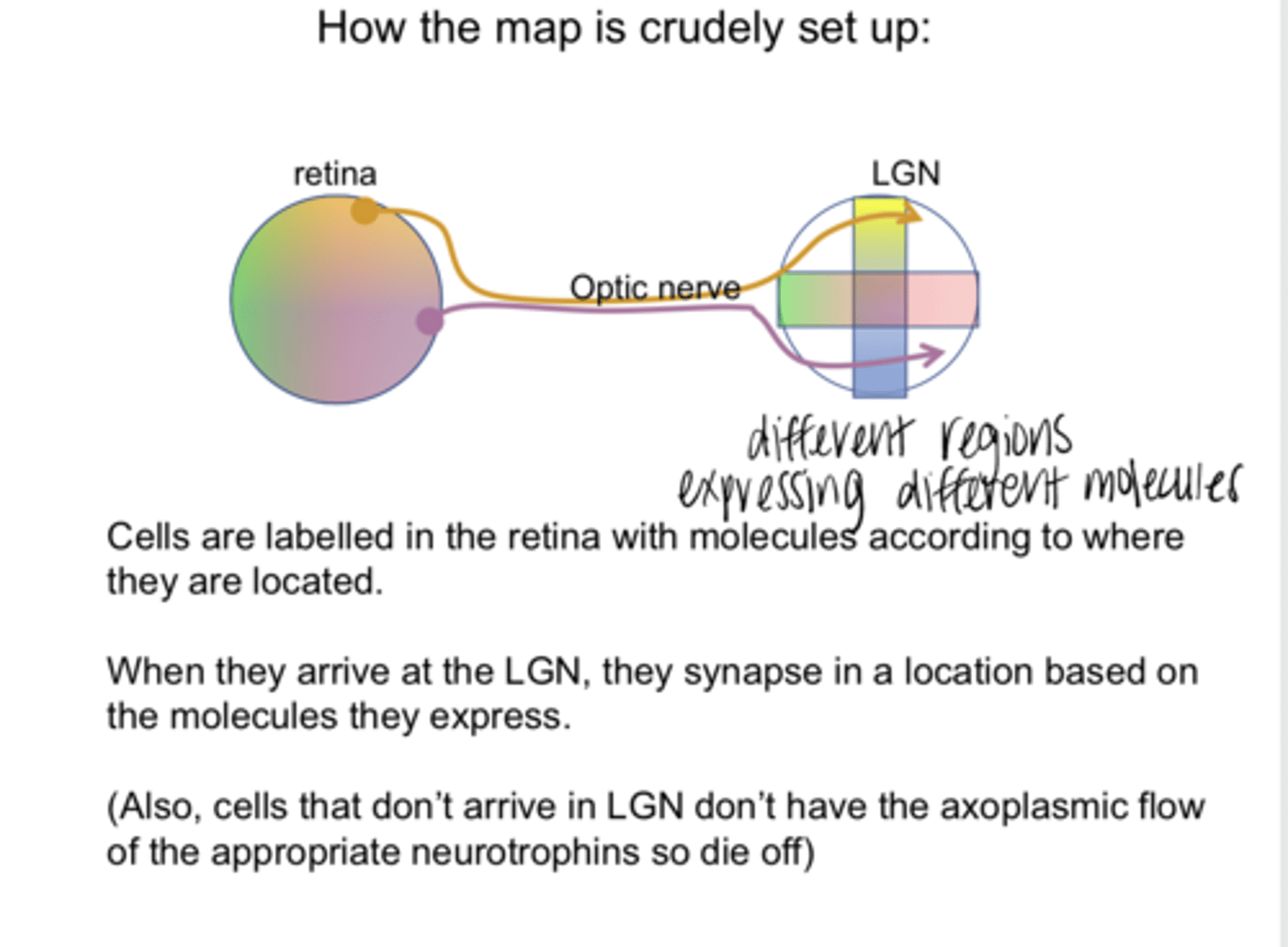
Cells are labelled in the retina with molecules according to where they are located
the molecules they express
When cells arrive at the LGN, they synapse in a location based on what?

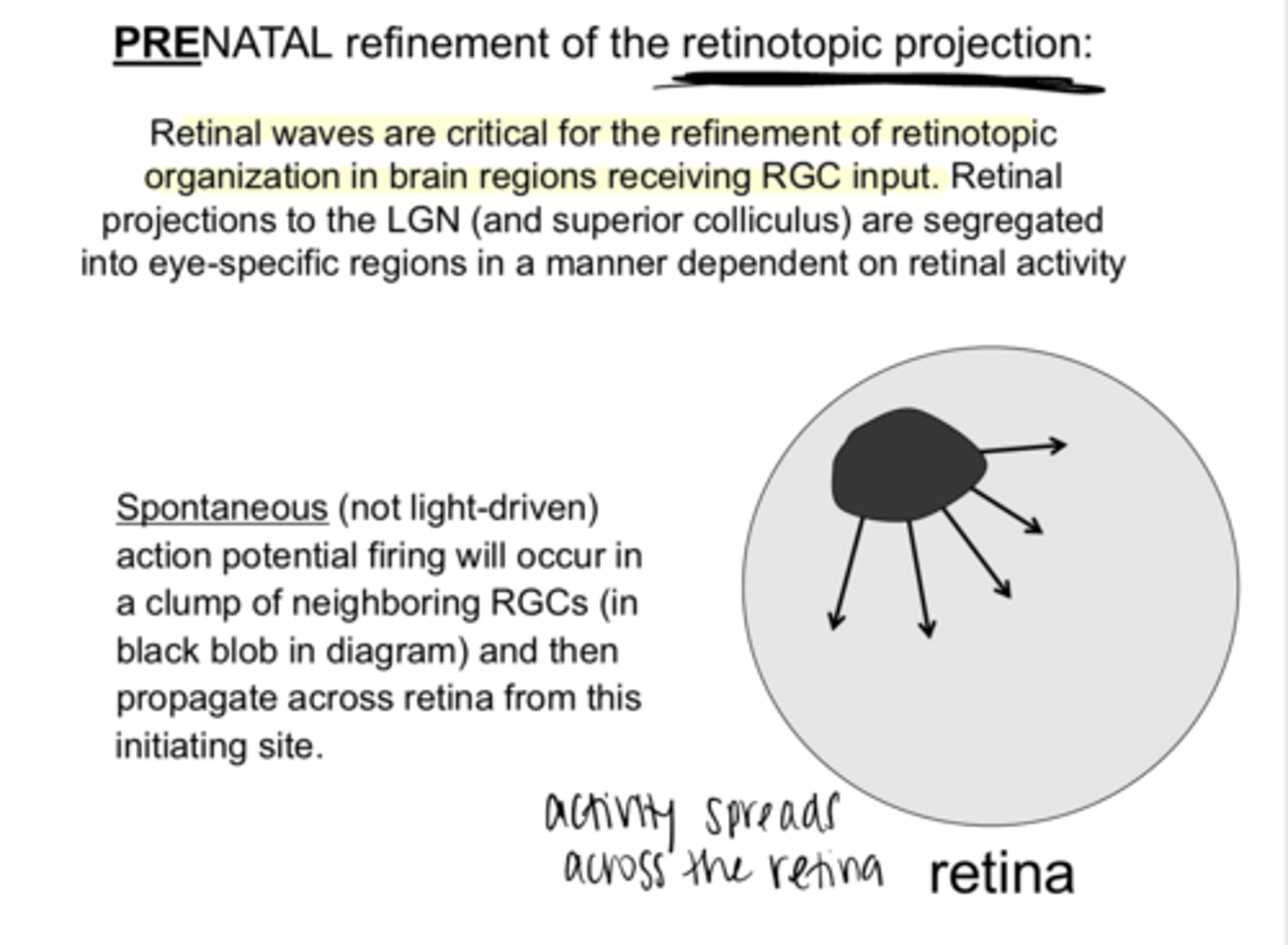
Retinal waves
______ are critical for the refinement of retinotopic organization in brain regions receiving RGC input
true
True or False:
Retinal projections to the LGN are segregated into eye-specific regions in a manner dependent on retinal activity

A clump of neighboring RGCs will fire and then propagate across the retina from this initiation site
Spontaneous (not light driven) action potential firing will occur how in retina?

the same
Neighboring retinal cells tend to fire at (the same/different) times
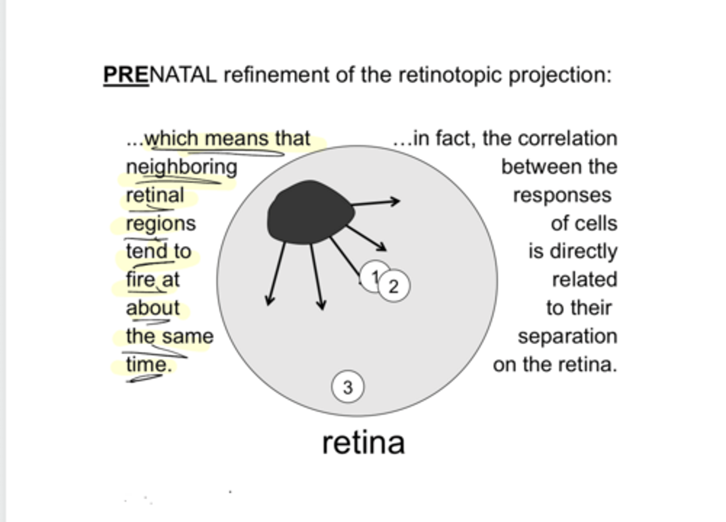
separation
The correlation between the responses of cells is directly related to their ____ on the retina

Multi-Electrode Array of Spontaneous Waves Travelling Across Neonatal Rat Retina (Pic)
Multi-Electrode Array of Spontaneous Waves Travelling Across Neonatal Rat Retina (Pic)

wire together
Cells that fire together...
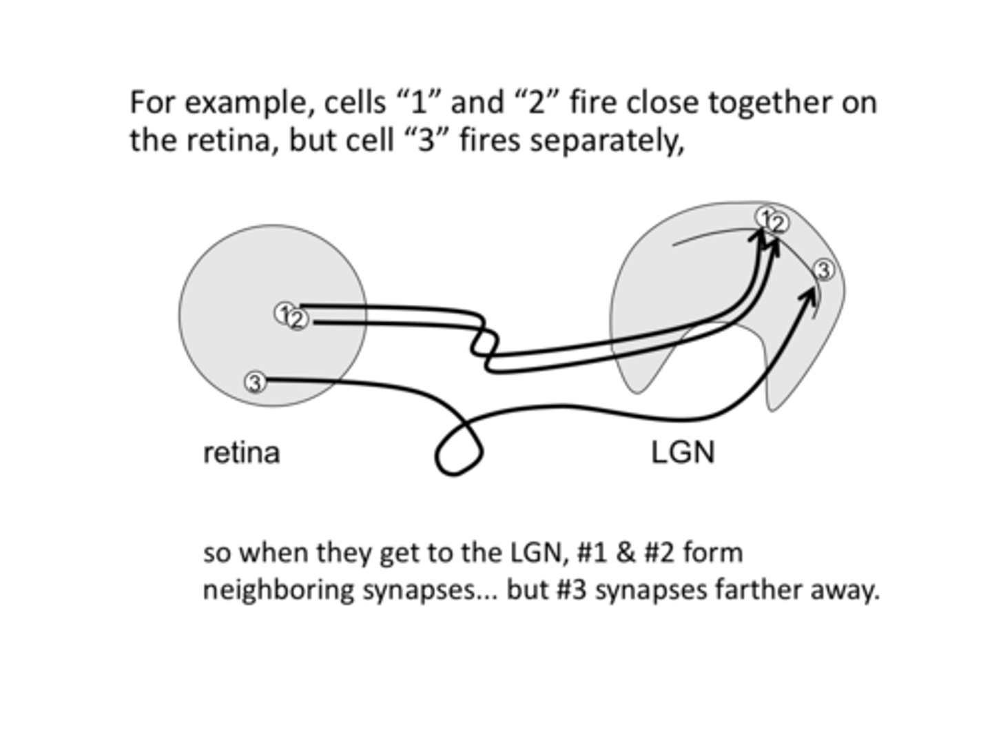
Cells "1" and "2" fire close together on the retina, but cell "3" fires separately. So, when they get to the LGN, "1" and "2" form neighboring synapses but "3" synapses farther away.
Explain this Pic
1) Molecular control
2) Functional refinement
Two ways in which the brain is retinotopically wired-up at birth
-Growth cones on neurites follow pathways marked with molecular signals
-Synapses form in approx the right places based on molecular markers distributed in a gradient across the tissue
What is the molecular control that controls how the brain in retinotopically wired-up at birth?
-The retinotopic map is refined by correlation in the responses of RGCs that are neighbors on the retina
-All of these happen also in V1 and other retinotopic cortical areas
What is the functional refinement that controls how the brain in retinotopically wired-up at birth?
large
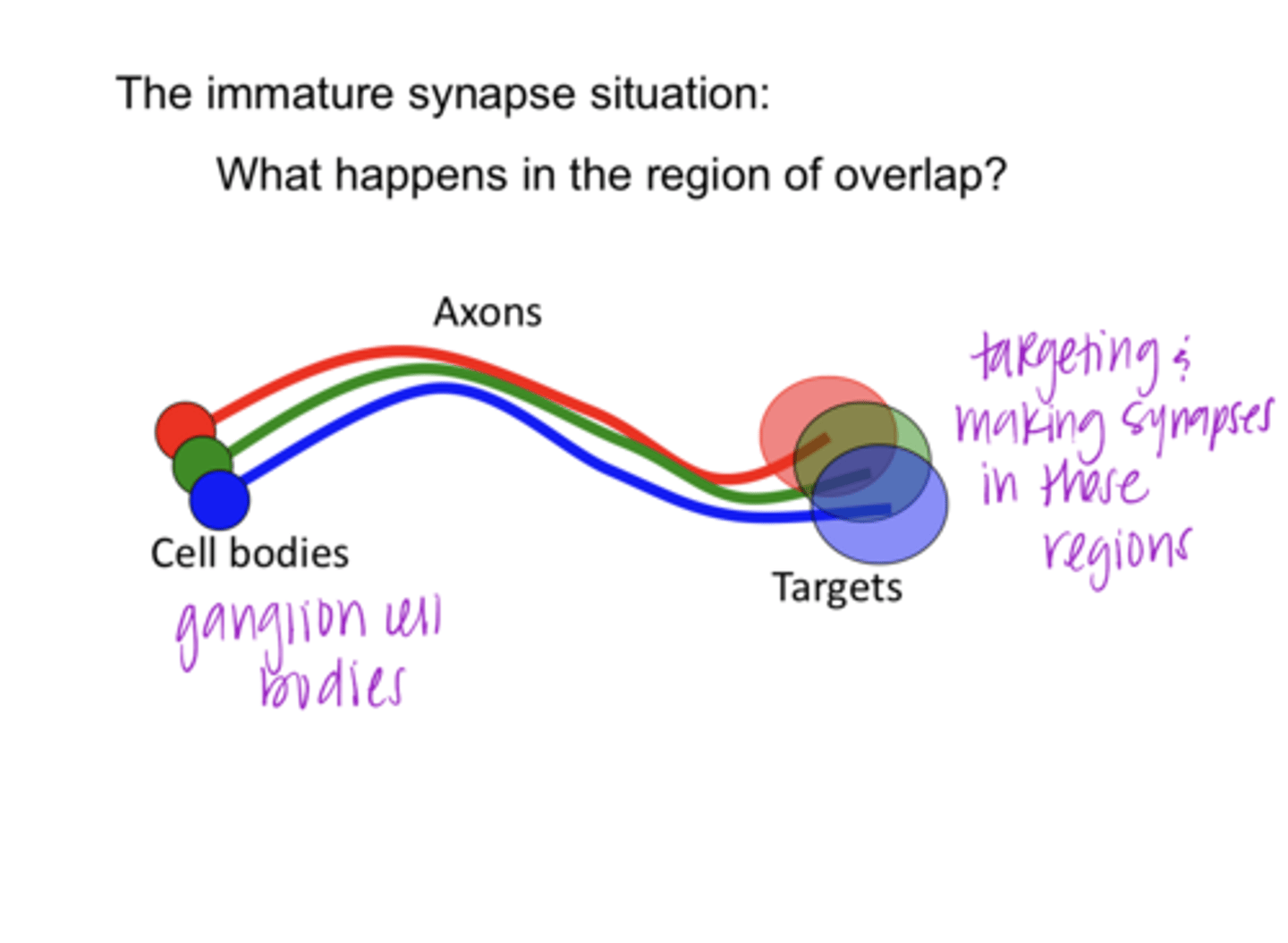
When they arrive at their destinations, each axon terminal synapses over a relatively (small/large) area
true
True or False:
Target cells have a lot of cells synapsing onto them

Birth
When will there be more synapses? Birth or adult
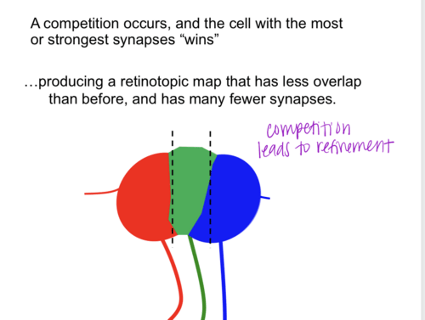
a competition occurs and the cell with the most or strongest synapses wins producing a retinotopic map that has less overlap than before w/ fewer synapses
Immature Synapse: What happens in the region of overlap?
refinement
competition leads to ______
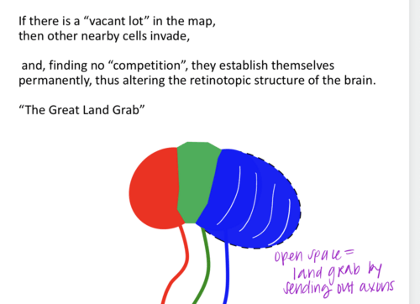
Nearby cells will invade when they find no competition. They will Permanently alter the retinotopic structure of the brain
**land grab
If there is a "vacant lot" in the map, what happens?