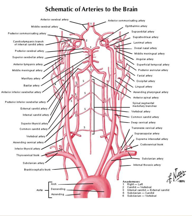
Cerebral Blood Supply
1/40
There's no tags or description
Looks like no tags are added yet.
Name | Mastery | Learn | Test | Matching | Spaced | Call with Kai |
|---|
No analytics yet
Send a link to your students to track their progress
41 Terms
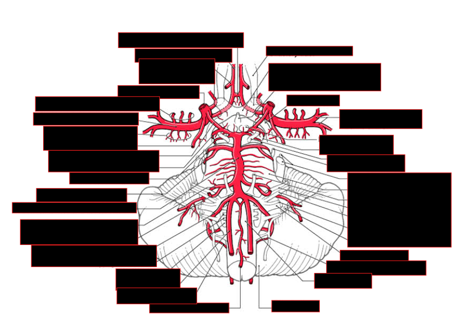
Describe the Sources of blood to the brain
Blood Supply to Brain:
Two Sources: Internal Carotid + vertebrobasilar system.
joins together via communicating arteries in circle of Willis on base of brain.
allow collateral circulation
Describe the pathway of the ICA
Describe its first branches
Pathway of ICA:
ascends from bifurcation → petrous portion via carotid canal → cartilage filled foramen lacerum → Cavernous sinus → travels anteriorly alongside sphenoid → ascends medial to anterior clinoid process → pierces through dural roof of sinus to become cerebral part → arches back below optic nerve → bifurcates @ anterior perforated substance → forms ant./mid cerebral arteries
Branches:
No major branches are given off from petrous part
first branches associated w/ brain are in cavernous sinus to supply hypophysis and meninges.
Describe the hypophyseal arteries:
Branches of?
Supplies?
Branches?
Hypophyseal arteries:
Branches of ICA;
supplies pituitary gland and part of the infundibulum
Branches:
Inferior: arises in cavernous sinus
Superior: arises from the cerebral (supraclinoid) part
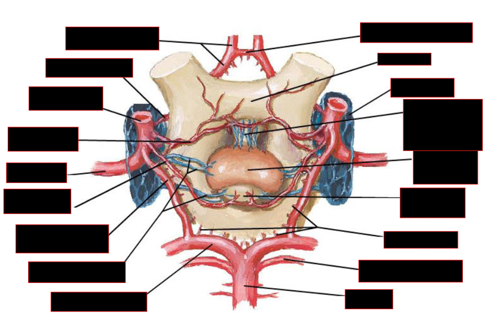
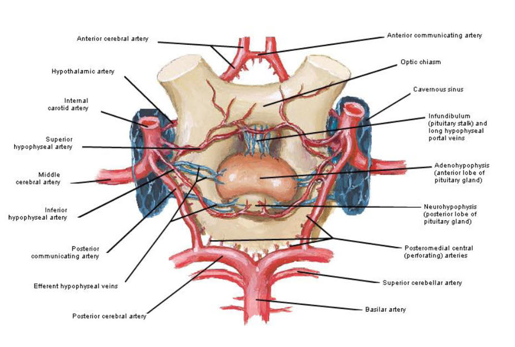
List the initial branches of the ICA
Initial Branches:
Ophthalmic artery
Posterior communicatinq artery
Anterior choroidal artery
Superior hypophyseal artery
Describe the ophthalmic artery
Pathway
Supply
Important Branch?
Clinical Importance?
ophthalmic artery
Pathway
enters orbit via optic canal → multiple branches
Supply:
eye + extraocular structures of orbit
Important Branch: Central artery of Retina:
critical for visual function
enters optic nerve as it approaches eyeball and supplies the retina.
Clinical Importance:
can carry small particles (microemboli) from carotid →
ransient or permanent visual loss
appearance of small ischemic areas visualized on retina
This is b/c ophthalmic artery arises early from ICA
Describe the posterior communicating artery
Pathway
STATs
PCA:
Pathway:
travels over oculomotor nerve in posteromedial direction → joins w/ posterior cerebral artery,
connects carotid w/ vertebrobasilar system
STATs:
size of this artery varies greatly
may be absent in small percentage of individuals
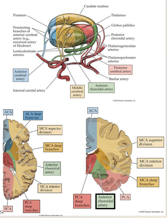
describe the anterior chordal artery
Pathway/Supply
Branches
Clinical Importance
AchA:
Pathway/Supply:
follows optic tract in caudal/lateral direction around crus cerebri → @ lateral geniculate body, branches → branches enter inferior horn of lateral ventricle via choroidal fissure → supply choroid plexus.
Branches:
to optic tract
crus cerebri,
amygdala,
anterior hippocampus,
tail of the caudate,
medial globus pallidus,
posterior limb of the internal capsule (ventral portion),
retrolenticular limb of intemal capsule.
lateral thalamus (including LGB),
CLINICAL IMPORTANCE:
damage = consistant w/ damage to posterior limb of int. capsule:
Hemiplegia
Hemianesthesia
Hemianopsia
NOTE: visual field defects are a variable feature and if present, may be transient.
List the terminal branches of the ICA
Terminal Branches:
Anterior Cerebral
Middle Cerebral
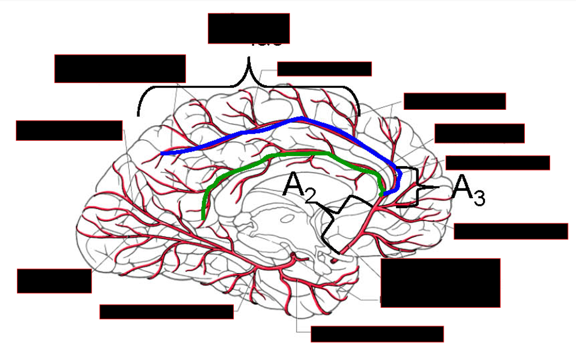
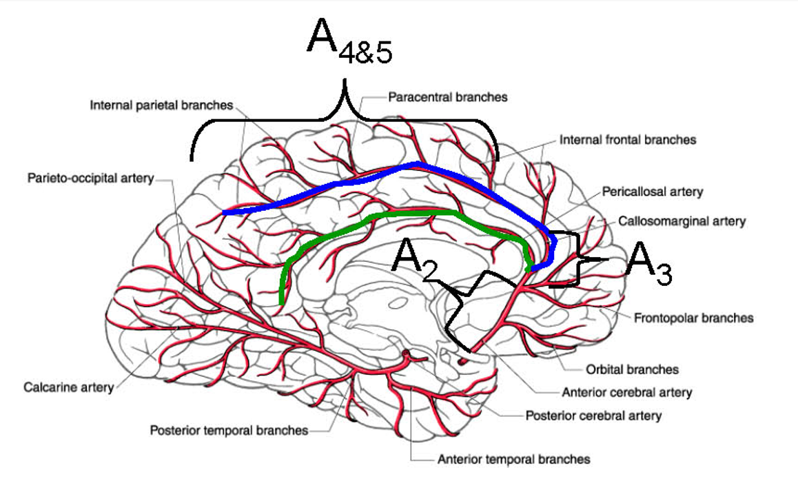
Describe the anterior cerebral artery
Pathway
Branches
Supplies
Clinical Importance
Anterior Cerebral Artery
Pathway:
runs medially over optic chiasm to near midline position → joined by contralateral counterpart via anterior communicatinq artery near cistern of lamina terminalis
Branches extend dorsolaterally onto lateral surface of the hemisphere
Branches:
orbitofrontal,
frontopolar,
callosomarqinal
pericallosal artery.
Supplies:
Paracentral Lobule
Contains cortical centers for movement and sensation of the lower limb
Ventromedial Prefrontal Cortex
Important for executive functions including decision making and planning
Clinical Importance:
symptoms depends on which branch is occluded:
Paralysis of opposite foot and leg — UMN lesion
Sensory deficits over toes, foot, and leg
Cognitive impairments
planning and decision making;
loss of initiative;
memory and emotional disturbances
Transcortical motor aphasia may also be present if in left (dominant) hemisphere
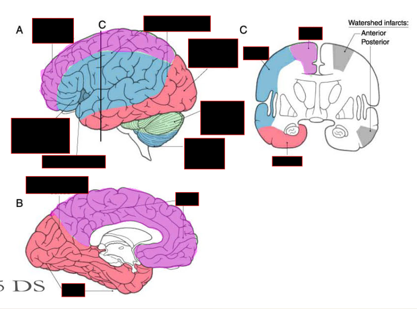
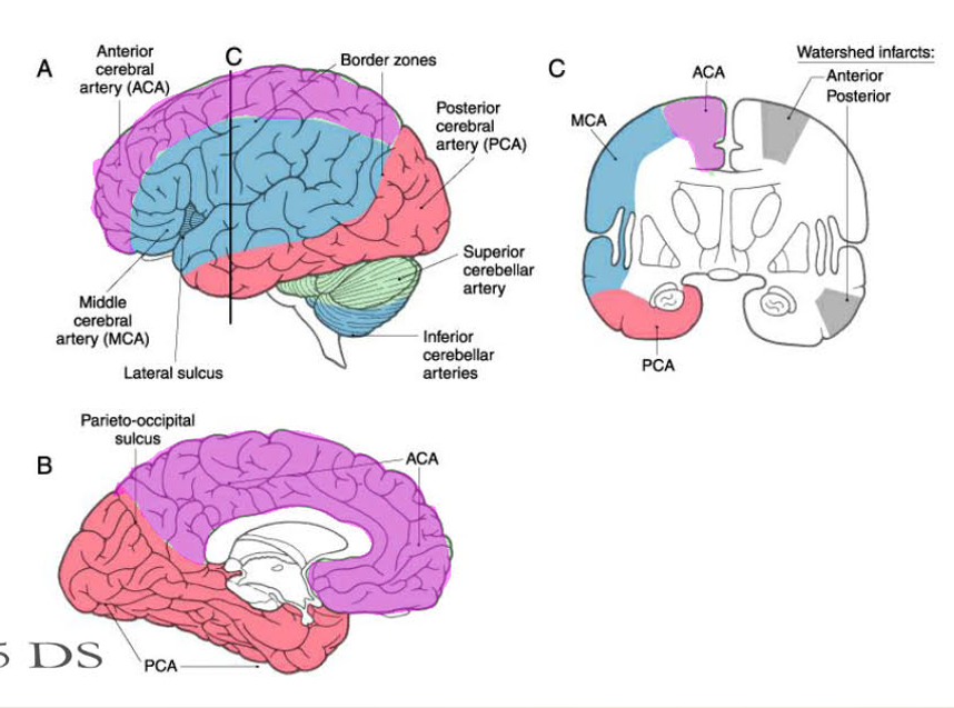
NOTE: should watch recording when he was talking about watershed infarcts, b/c information spoken wasn’t on the slide
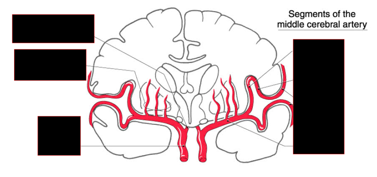
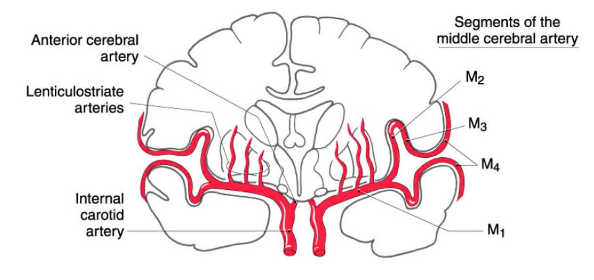
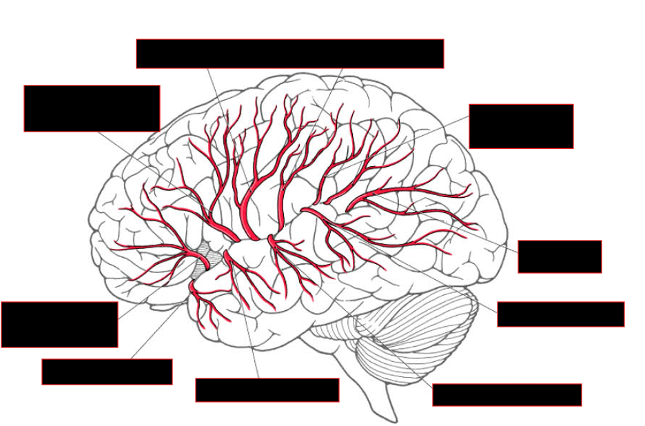
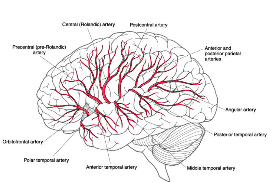
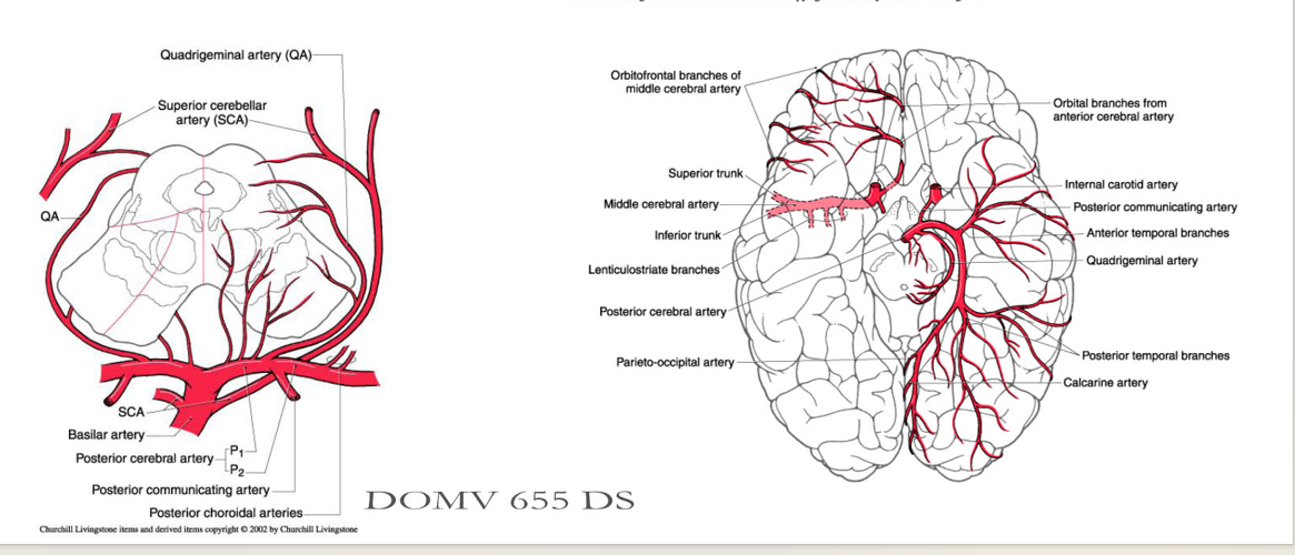
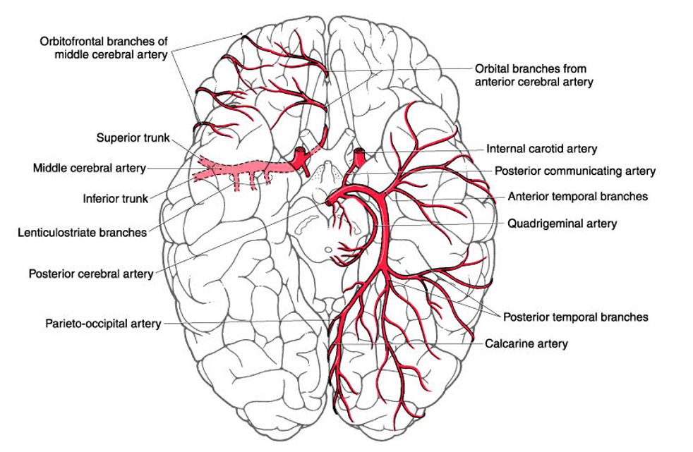
Describe the Middle Cerebral Artery
STATs
Pathways
Branches of Lat. Hemisphere
Lat vs Med hemisphere
Supplies
Clinical Importance
middle cerebral artery
STATs:
usually (70% of the time) the larger of the terminal branches of the internal carotid
Pathway/Branches :
moves laterally through Sylvian cistern → gives off lenticulostriate arteries to basal ganglia and internal capsule → splits into Sup/Inf. Trunks as it enters Insula → supply inner aspects of the opercula and the lateral surface of the cerebral hemisphere
Branches of Lateral Hemisphere = variable:
Includes branches adj to central sulcus
(Precentral, Central and Postcentral (Rolandic) arteries).
Lat vs Med hemisphere:
Lat:
MCA supplies central area that is surrounded by Ant/post. cerebral Artery
Medial:
does NOT supply the medial aspect of the hemisphere.
Supplies:
Primary sensory and motor areas for face and arm
Left Side: Broca's/Wernicke's area
Optic radiation
Parietal association cortex
Frontal Eye Field
CLINICAL IMPORTANCE:
Symptoms = depends on which divisions/branch
Paralysis of opposite face and arm
Sensory deficits over opposite face and arm
Aphasia if on left side
Visual field defects
Inattention and neglect of contralateral side of body or space and denial of illness if on right side
Transient paralysis of conjugate gaze to the contralateral side.
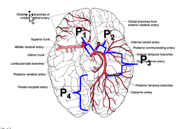
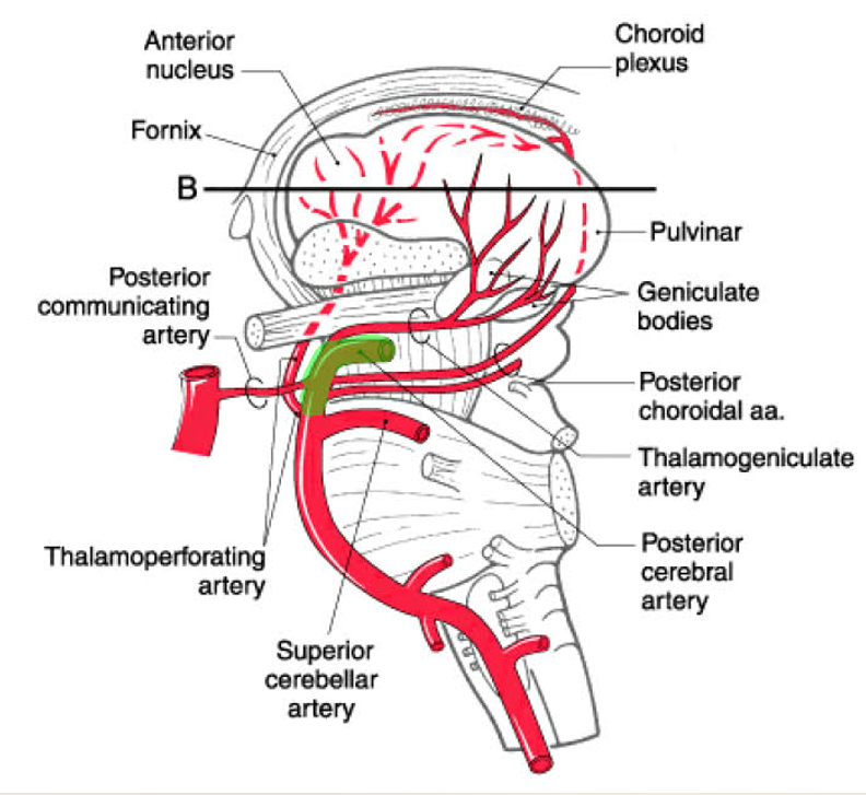
Describe the posterior cerebral artery
Pathway
Early Branches
Supplies
Clinical Presentation
PCA:
Pathway:
Arise from bifurcation of basilar A @ level of interpeduncular cistern → travels laterally over oculomotor nerve → wraps around midbrain in ambient cistern → runs along ventral/medial surface of temporal lobe → continues post. to supply ventral/medial surfaces of the occipital lobe
NOTE: includes the calcarine artery. which supplies primary visual cortex.
Note: considered part of vertebrobasilar system; but supplies post. area of hemisphere
Early Branches: Collective supply midbrain, thalamus and choroid plexus.
quadriqeminal artery,
thalamoperforatinq arteries
medial and lateral posterior choroidal arteries
thalamoqeniculate artery
Supplies:
Visual cortex
Medial surface of occiptal lobe (primary and association cortices)
Temporal lobe, caudal parietal lobe, and splenium of corpus callosum
Part of thalamus:
thalamogeniculate branch — ventral lateral thalamus;
thalamoperforating branches to anterior and medial thalamus;
posterior choroidal arteries — posterior thalamus, choroid plexus of 3rd and lateral ventricles
Clinical Presentation:
Homonymous Hemianopia (with macular sparing)
MCA provides collateral supply to occipital pole- macular vision
Pure Alexia (inability to read)
w/ involvement of posterior corpus callosum + left visual cortex;
color agnosia may also be present.
Sensory deficits with deep territory involvement
Describe the segments of the PCA
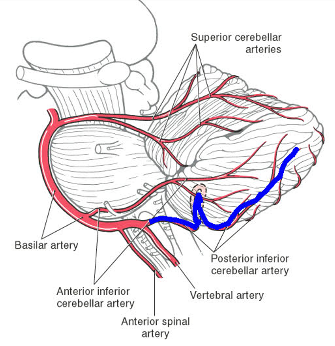
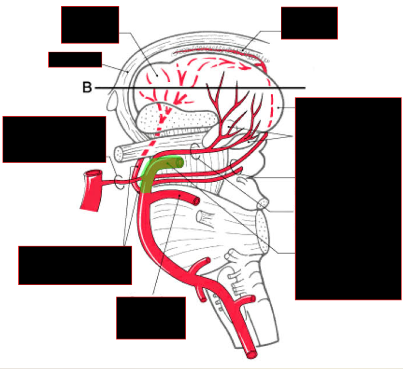
Describe the vertebral Artery
Pathway
Branches supplies
VA:
Pathway:
enter lateral cerebellomedullary cistern after penetrating atlanto-occipital membrane.
Branches Supplies:
part of the medulla oblongata,
cerebellum
dura of the posterior cranial fossa.
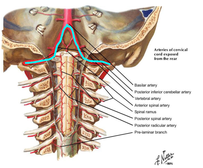
Describe the Posterior Inferior Cerebellar Artery
Pathway/Supplies
PICA:
first major branch of the vertebral artery
Pathway/Supplies:
supplies dorsolateral medulla as it wraps around medulla → emerges in cisterna magna where it supplies the choroid plexus of the fourth ventricle and then the posterior and medial parts of the inferior cerebellar surface.
Differentiate between PSA and ASA:
Origins
Supplies
posterior spinal artery
Origins:
most often arises from PICA.
However, in about 25% of cases, it arises from the vertebral artery.
Supplies:
dorsal part of the spinal cord
dorsolateral region of the medulla oblongata
caudal to the area served by PICA.
Anterior Spinal Artery:
Origin:
Prior to joining → basilar artery; each vertebral artery gives rise to an ASA → 2 ASA joins to form one; seen on ventral surface of the spinal cord.
Supplies:
anterior and medial areas of the medulla and spinal cord.
Describe the AICA
Origins
Pathway
Supplies
Branches
Anterior Inferior Cerebellar Artery
Origins: First large branch of Basilar artery
basilar artery lies on ventral surface of pons in prepontine cistern
Pathway:
wraps around caudal middle cerebellar peduncle → inferior surface of the cerebellum.
Supplies:
ventral and lateral surfaces of cerebellum
portion of pons
small part of the choroid plexus that extends out of the fourth ventricle.
Branches:
labyrinthine artery
arises near CN 7/8 → enters internal acoustic meatus w/ them
supply portions of the inner ear
paramedian, short and Ionq circumferential branches
supply pons
Describe the superior cerebellar artery
Origins
Pathway
Supplies
SCA:
Origins: basilar artery below it distal bifurcation.
Pathway:
travels caudal to CN III around crus cerebri in ambient cistern → divides into medial + lateral branches.
Supplies:
superior portion of cerebellum
most of deep cerebellar nuclei
lateral tegmentum of rostral pons.
caudal portions of midbrain.
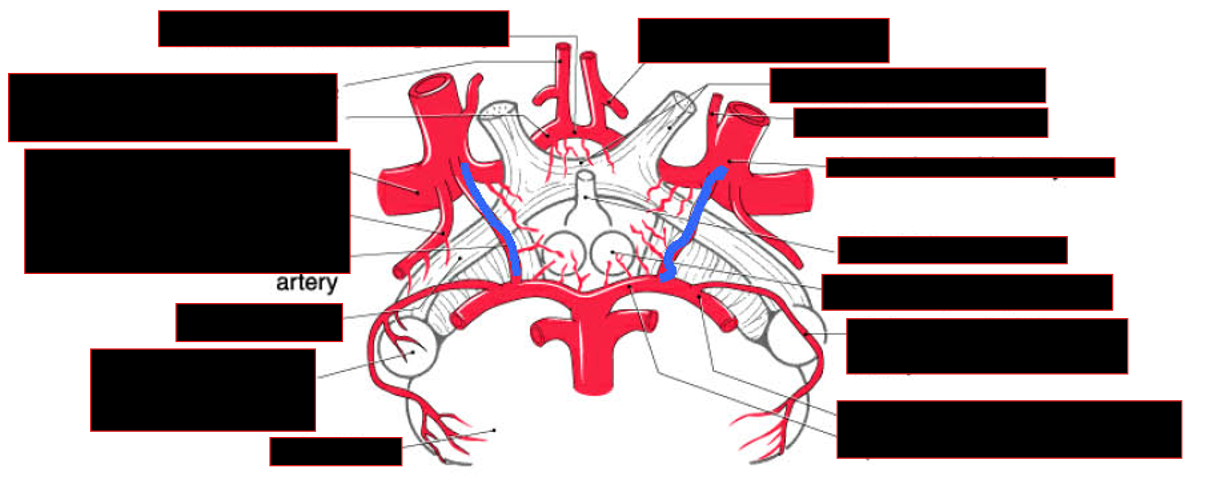
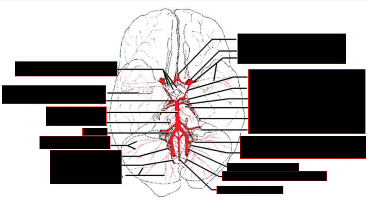
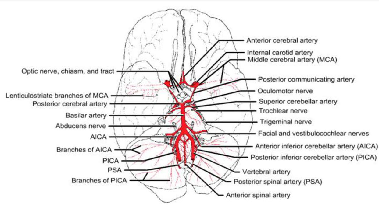
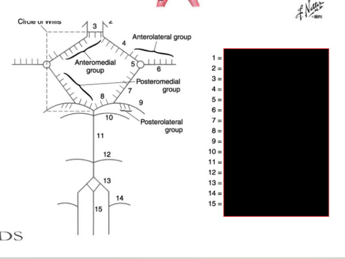
Describe the Circle of Willis
Perforating groups
Divisions
Supplies (ant vs post group)
Circle of Willis
Perforating Groups:
arteries forming circle of Willis give rise to numerous small ( perforating, central, ganglionic) branches that supply the ventromedial structures of the brain.
Divisions:
anteromedial group
anterolateral group
posteromedial group
posterolateral group
Supplies:
Anterior Group
optic chiasm
anterior hypothalamus
anterior perforated substance
NOTE: lateral striate (lenticulostriate) arteries are sometimes included.
Posterior Group
crus cerebri
middle + posterior parts of hypothalamus
Forms the post. perforated substance as they enter the interpeduncular fossa
NOTE: thalamoperforating, tuberothalamic, thalamogeniculate, and medial and lateral posterior choroidal arteries may be included
Differentiate between the med/lat striate artery:
Origins
Supply
Medial vs Lateral Striate Arteries:
Origins:
Medial: Ant. Cerebral Artery (A2)
Lat: Middle Cerebral Artery (MI)
Supply:
Medial (recurrent artery of Heubner):
rostral part of the head of the caudate & lenticular n
anterior limb of the internal capsule.
Lat (lenticulostriate):
head and body of the caudate
most of the lenticular n
dorsal part of anterior limb
genu
posterior limb of the internal capsule
Describe the blood supply to the thalamus
Primarily by?
Ant. Thalamus
Post. Thalamus
Posteriolat.
Blood Supply to Thalamus:
Primarily by branches of posterior cerebral artery
thalamoperforating (Pl area),
thalamogeniculate
medial and lateral posterior choroidal arteries
arise from beyond the branching of the posterior communicating artery (P2 area) on the posterior cerebral artery.
Ant. Thalamus:
Via tuberothalamic (polar) artery
branch of posterior communicating artery.
WHEN ABSENT (33%):
thalamoperforatinq arteries supply this area in addition to the medial thalamus
Posterior thalamus:
posterior choroidal arteries
Posteriolat:
thalamoqeniculate artery
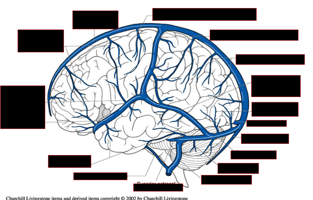
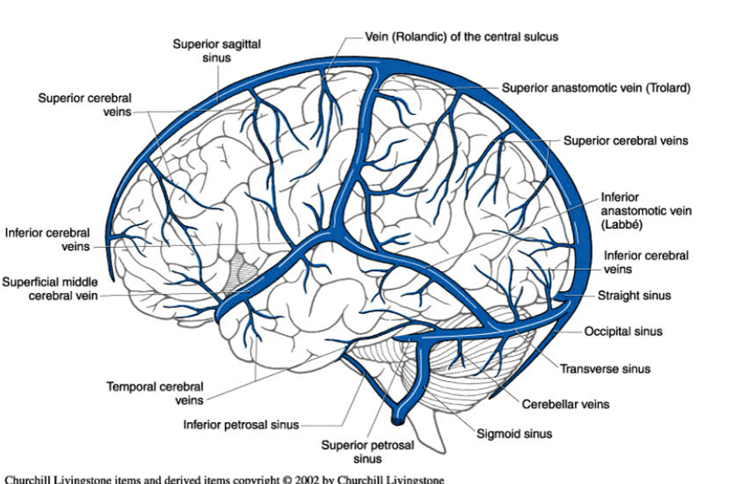
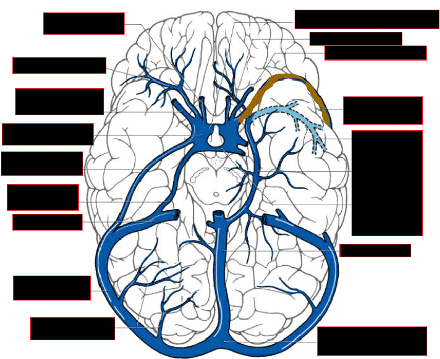
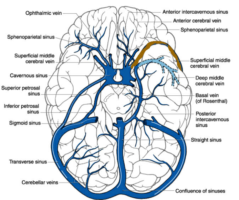
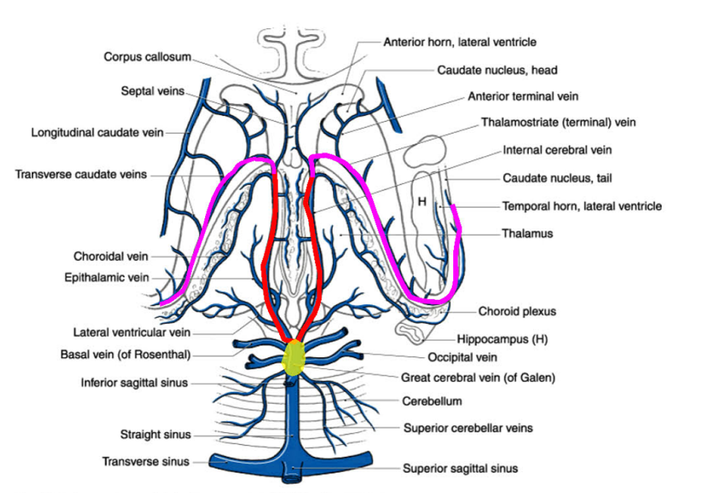
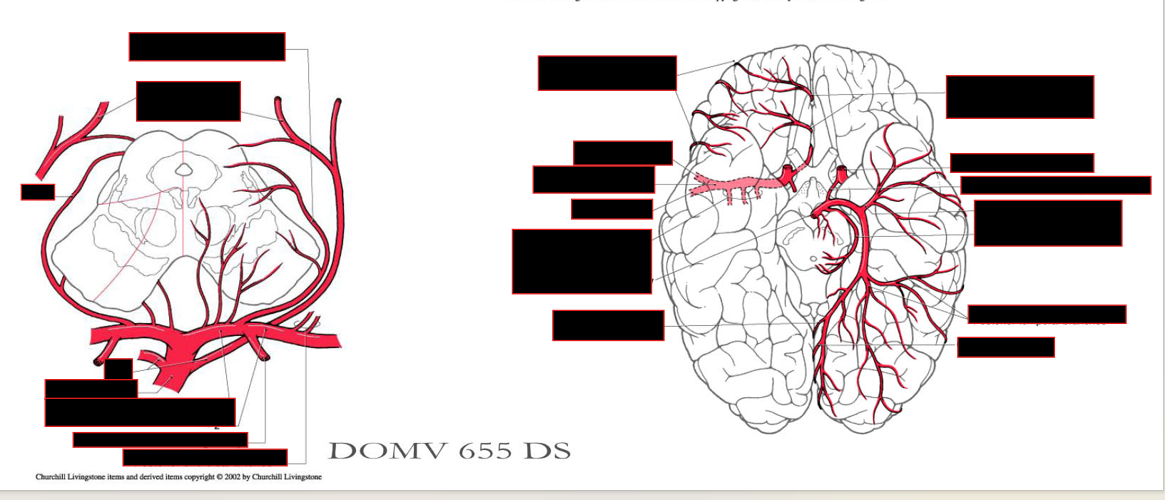
Describe the cerebral venous system
Lat. Surface:
Vessels found
Vessel Connections
Pathway
Midsagittal Surface:
Pathway
Base of Brain:
Which sinuses are found
Internal Cerebral Veins
What are they?
Pathway?
Consists of
Lateral Surface of hemisphere:
Vessels Found:
superior anastomotic vein (of Trolard) + inferior anastomotic vein (of Labbe)
Vessel Connections :
w/ superior sagittal + transverse sinuses + superficial middle cerebral vein
Pathway:
vessel courses medially around temporal pole → cavemous sinus.
Midsagittal Surface:
Pathway:
vessels drain into sagittal sinus.
Others from lower medial surface near corpus callosum + medial temporal lobe (the basal vein of Rosenthal) → intemal cerebral veins → qreat cerebral vein (of Galen) → straiqht sinus
Base of Brain:
superior + inferior petrosal sinuses link cavernous sinus to transverse sinus and origin of IJV, respectively.
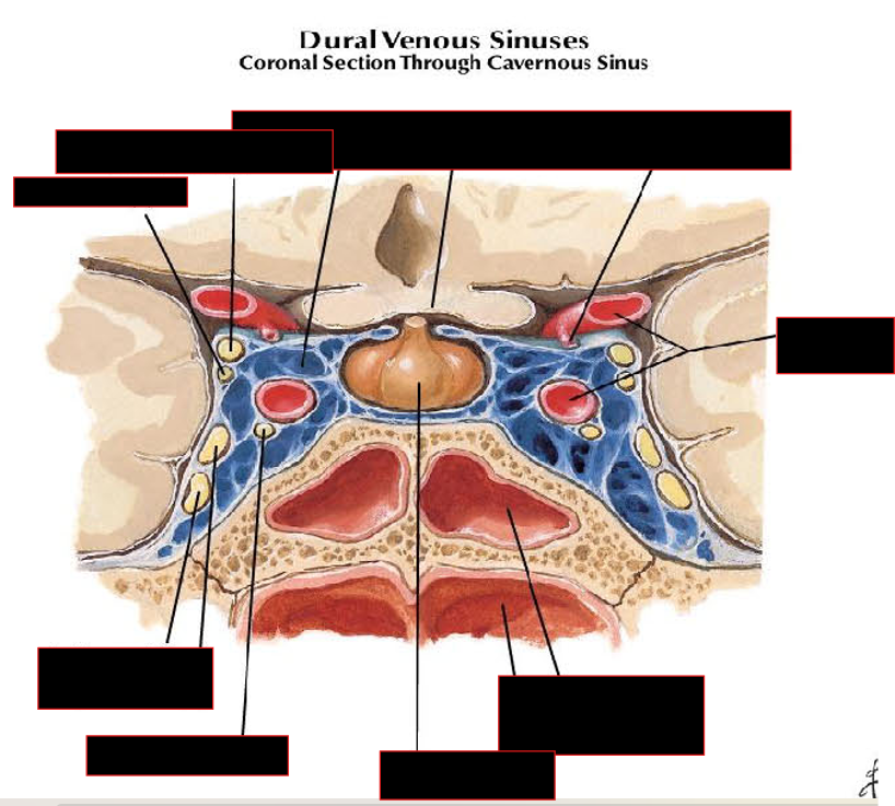
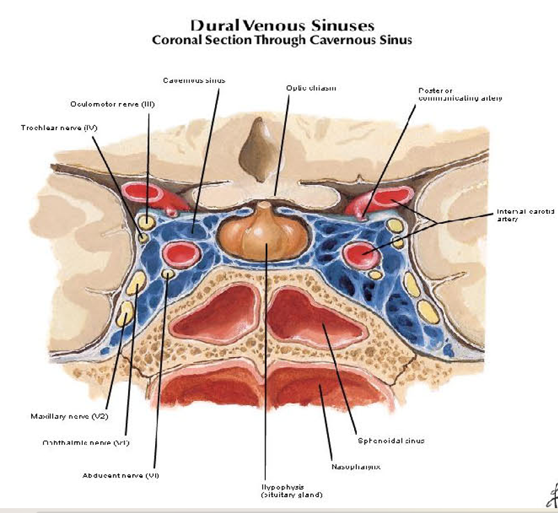
[REVIEW]: The internal carotid artery passes forward through the lumen of the cavemous sinus along with the abducens nerve (CN VI). The oculomotor n., trochlear n., and the ophthalmic and maxillary branches of the trigeminal n. lie in the lateral wall.
Internal Cerebral Veins:
What are they?
veins draining the internal structures of the cerebral hemispheres
Pathway:
join @ great cerebral vein and empty into straight sinus.
Consist of:
choroidal veins
traveling from choroid plexus
thalamostriate (terminal vein) vein
runs adj to the caudate nucleus.