DH 228: FINAL IDENTIFICATION
1/50
There's no tags or description
Looks like no tags are added yet.
Name | Mastery | Learn | Test | Matching | Spaced | Call with Kai |
|---|
No analytics yet
Send a link to your students to track their progress
51 Terms
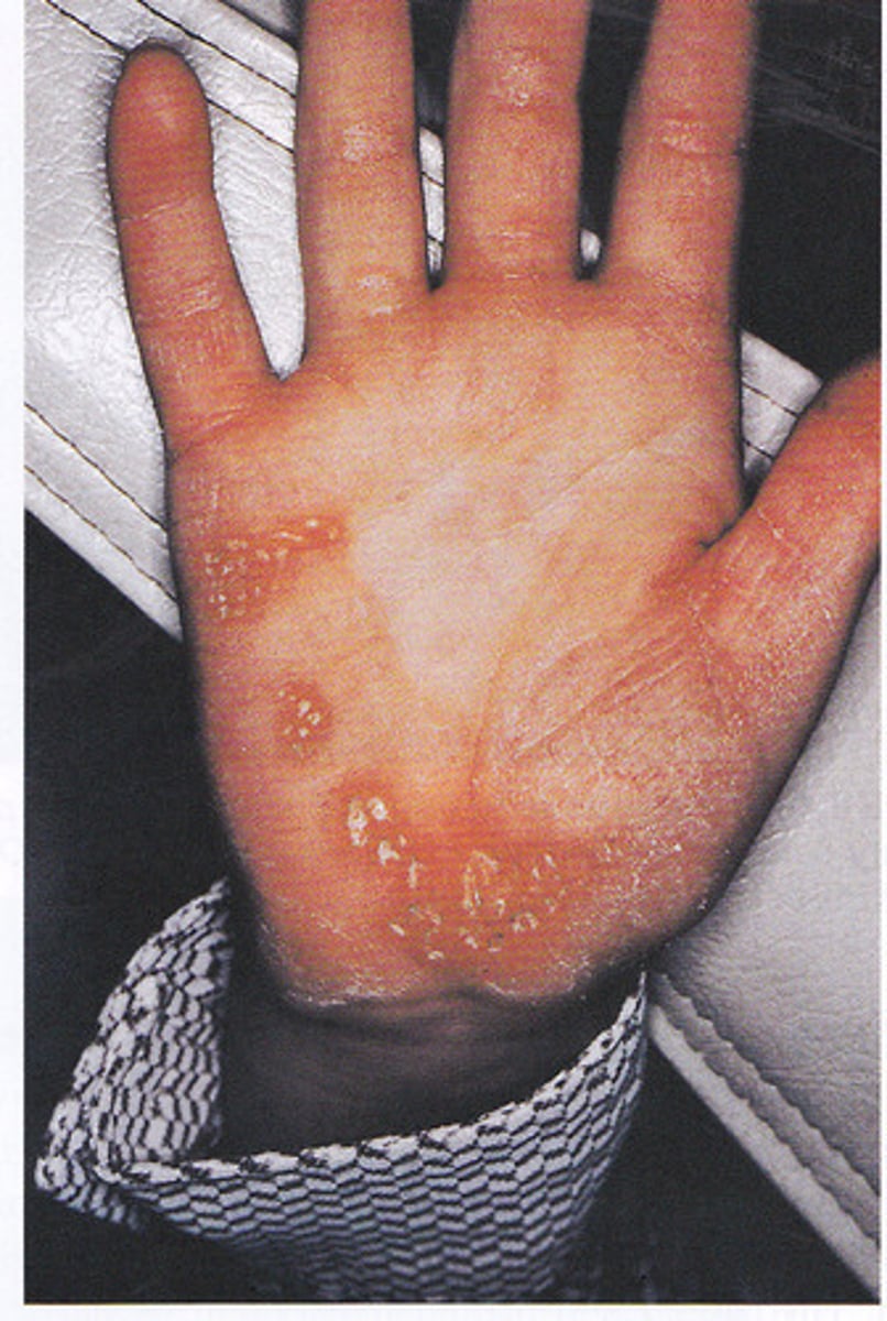
Papillon-Lefevre syndrome
-Associated with Periodontitis associated with genetic disorders (V)
-Severe periodontal destruction
-Premature tooth loss
-Hyperkeratosis of the palms of hands & soles of feet
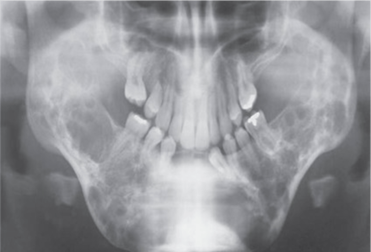
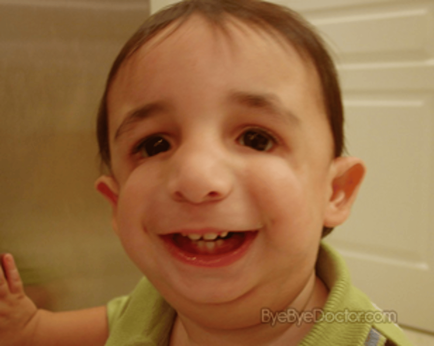
Cherubism
-progressive bilateral facial swelling at 1-4 years of age, most commonly involves the mandible
-increased distance between eyes
-"soap bubble" appearance
-pseudodontia
Gardner Syndrome
Presence of osteomas in various bones of the skull
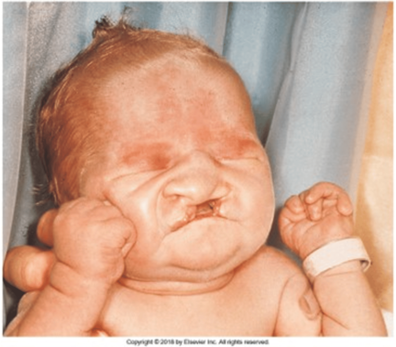
Trisomy 13 (Patau Syndrome)
bilateral cleft lip and palate, microphthalmia (small eyes) or anophthalmia (no nose), superficial hemangioma of the forehead or nape of the neck, growth retardation, severe mental handicap.
-75% of live born infants die within the first 7 months
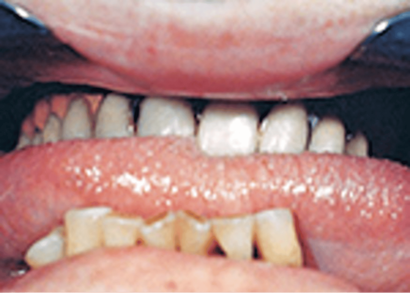
dentinogenesis imperfecta
incomplete or improper development of dentin tissue, between enamel and cementum
-short roots and almost complete lack of pulp chambers
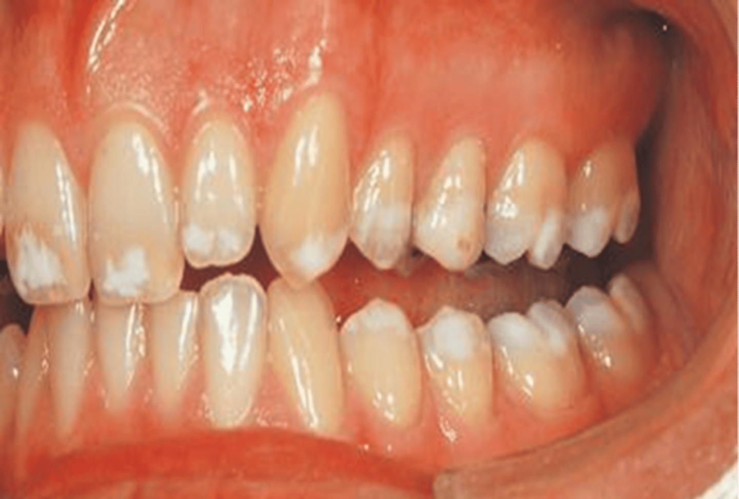
Type III amelogenesis imperfecta
incomplete or improper development of the enamel tissue
-Uniform white incisal edges and occlusal cusps
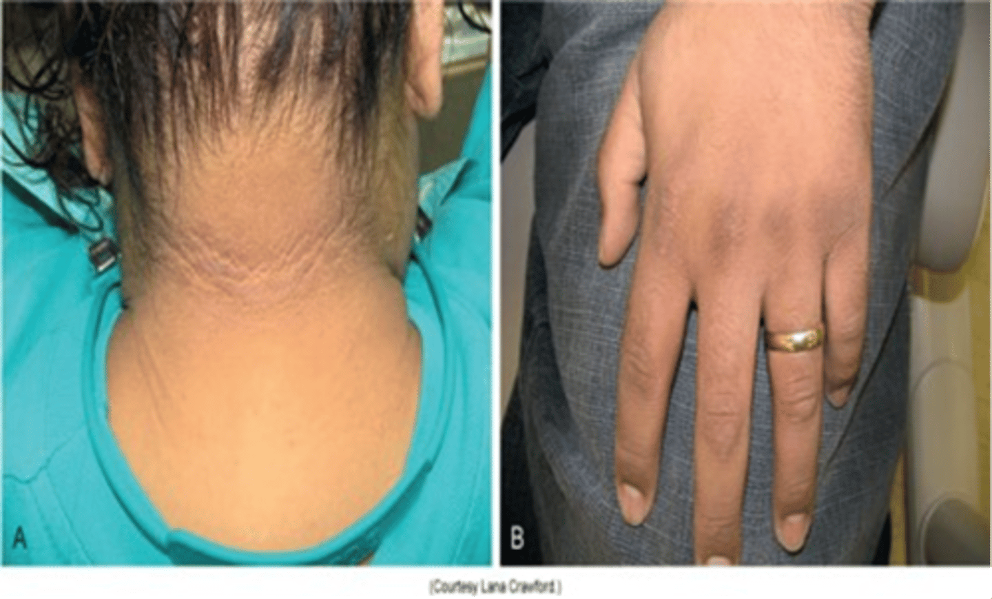
Acanthosis nigricans
thickening and darkening of skin near axillary region, associated with Diabetes Type II and gastric carcinoma
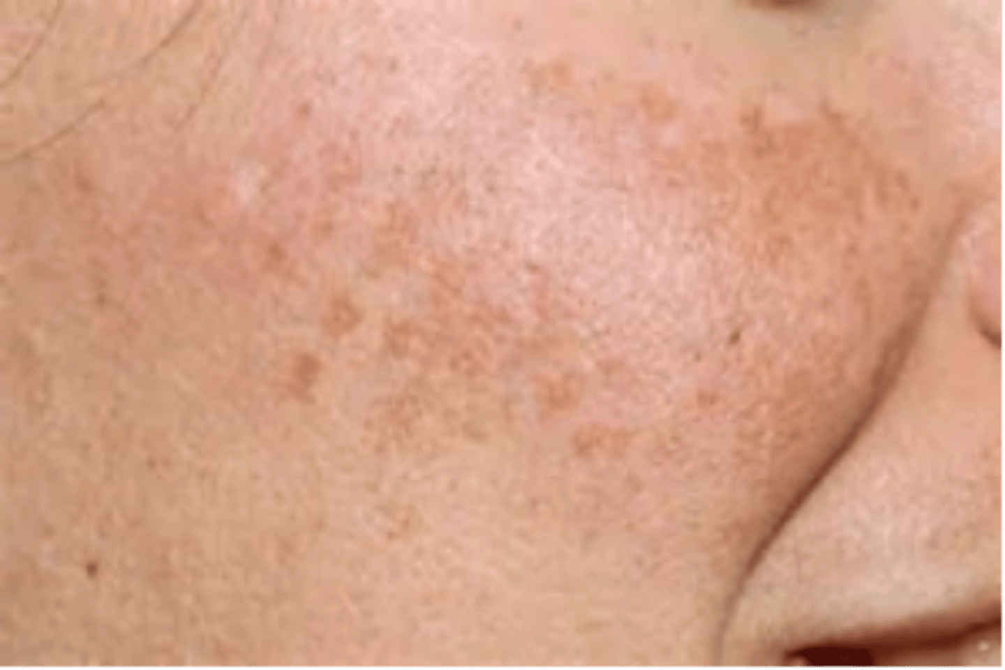
Peutz-Jeghers syndrome
mucosal pigmentation, associated with gastrointestinal polyposis.
periapical cemento-osseous dysplasia (cementoma)
Asymptomatic fibro-osseous lesion
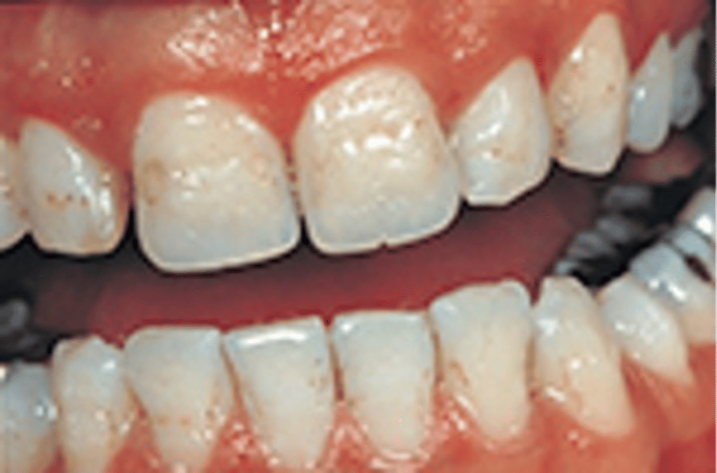
Type I Amelogenesis Imperfecta
incomplete or improper development of the enamel tissue, this picture shows the pitting on the labial surface
Paget diseaseEnlargement of the Maxilla with
chronic metabolic bone disease
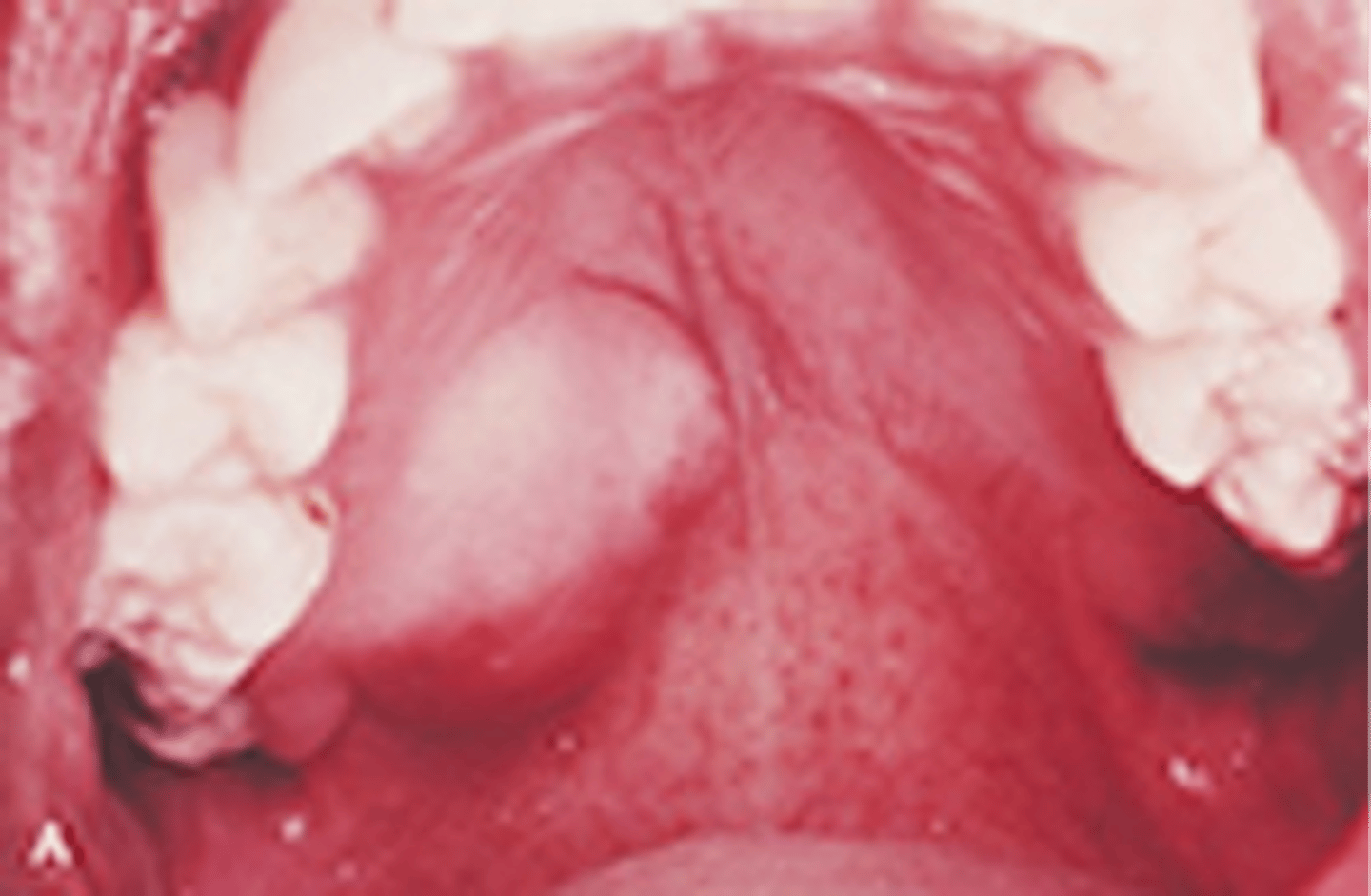
Pleomorphic adenoma (benign mixed tumor)
-A benign salivary gland tumor
-90% of all salivary gland tumors
-The most common extraoral location is the parotid gland; the most common intraoral location is the palate
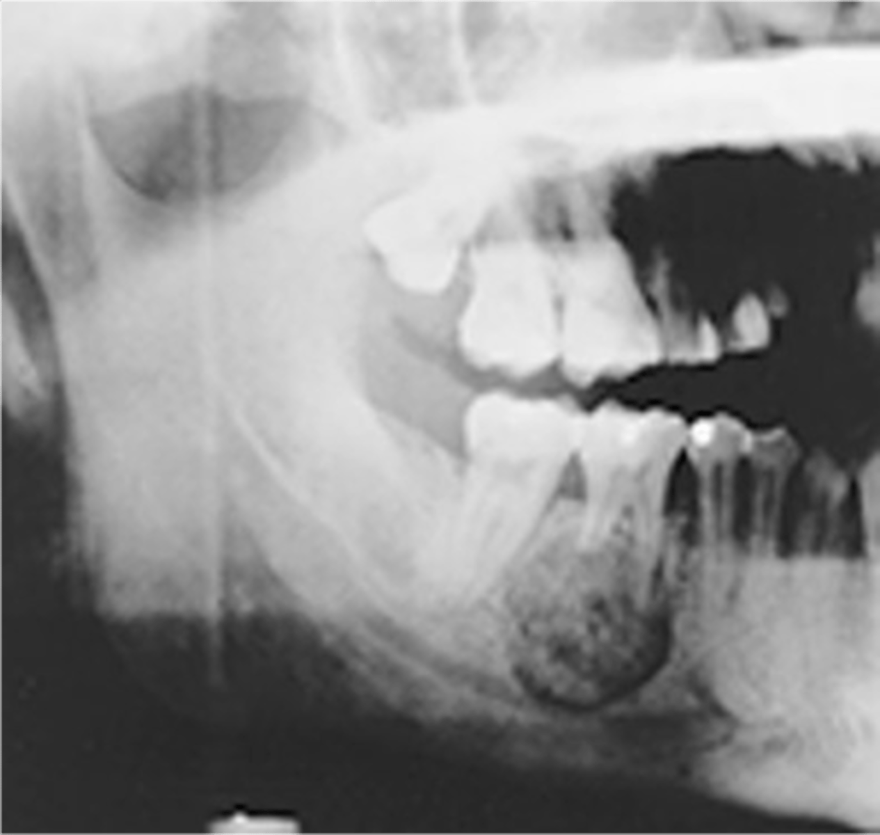
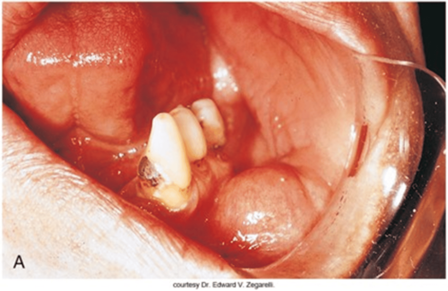
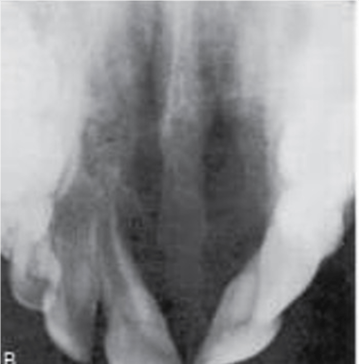
Cementoblastoma
Odontogenic tumor of cementoblasts that resembles another odontogenic tumor but has direct attachment to a tooth
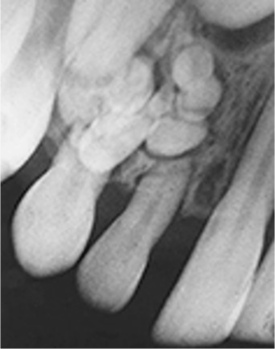
compound odontoma
- Small radiopaque masses
- Usually in the anterior jaw
- Resemble an accumulation of small teeth
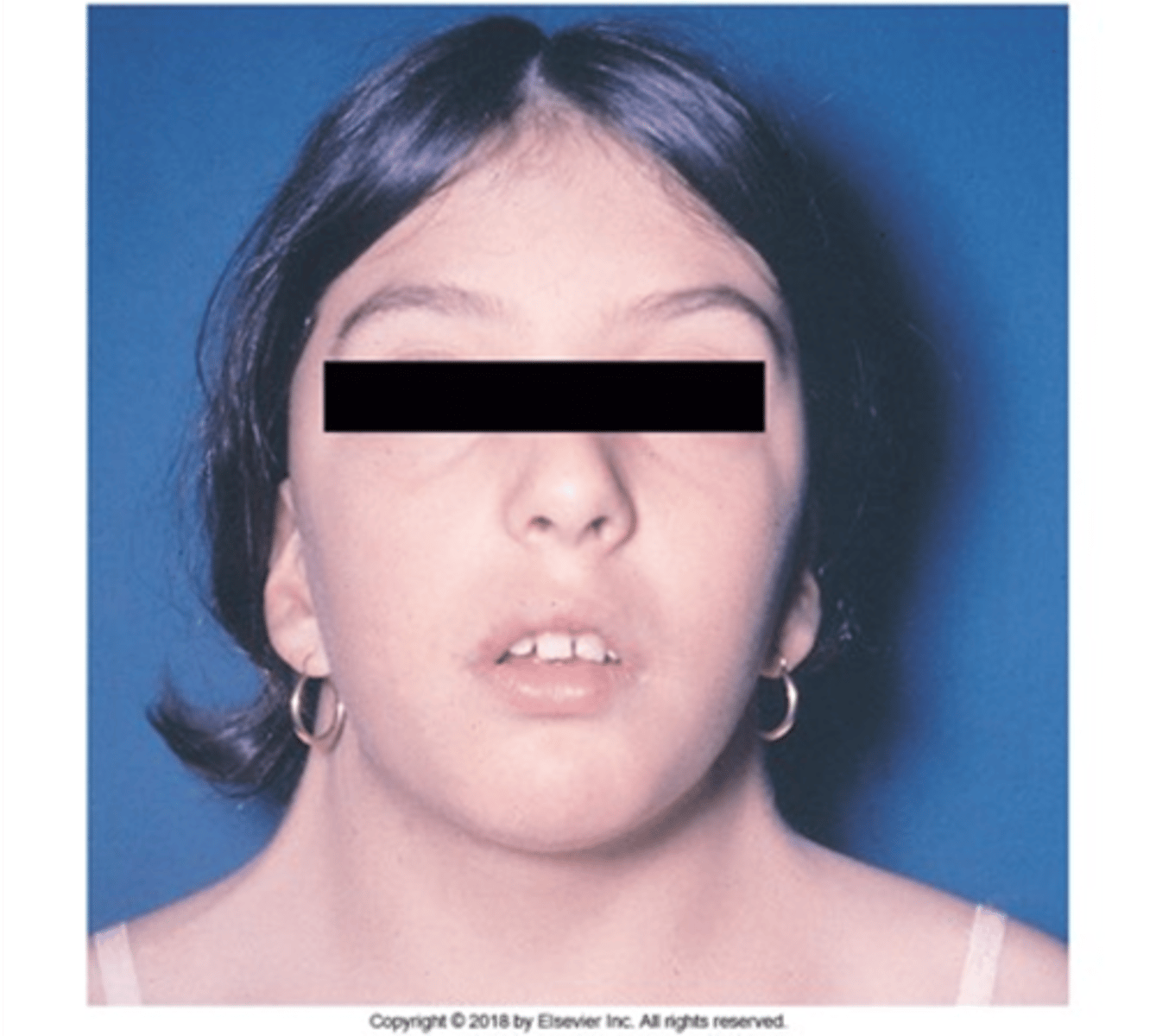
Mandibulofacial Dysostosis
Also called Treacher Collins Syndrome, it is a genetic disorder that affects the development of the facial structure. Characterized by a fish-like mouth (high palate, downward sloping of the lip commissures, and an open-bite). Deafness results from a lack of otic ossicles.
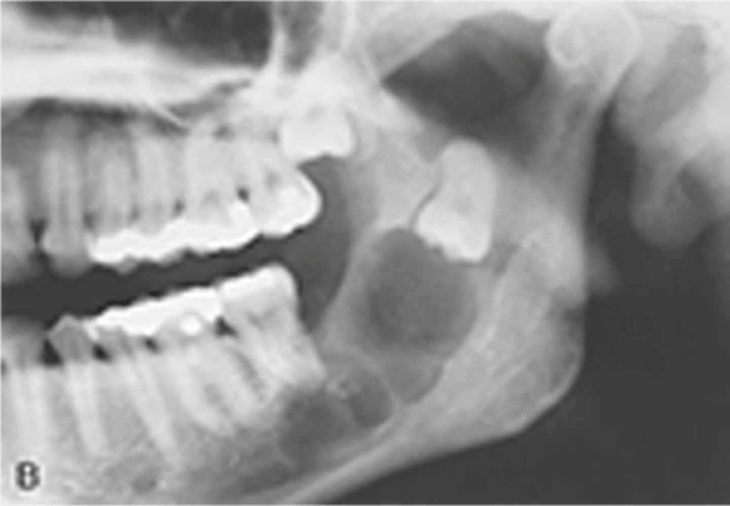
Ameloblastoma
Benign but aggressive tumor w sig recurrence
Most common in post mand but can be in the ant
Most common true odontogenic tumor
multilocular radiolucency (soap bubble)
often assoc w/ impacted tooth
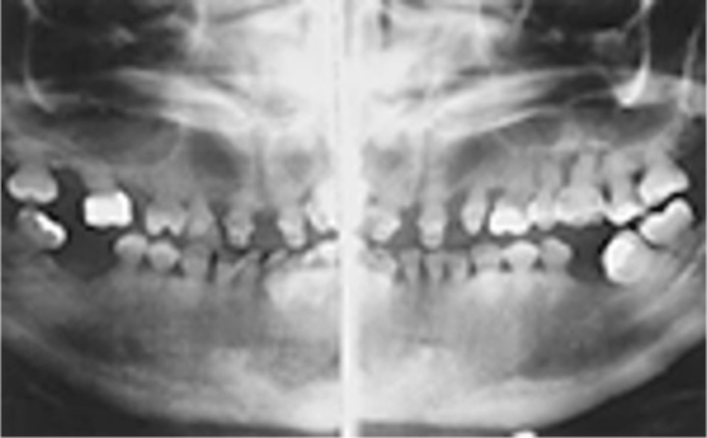
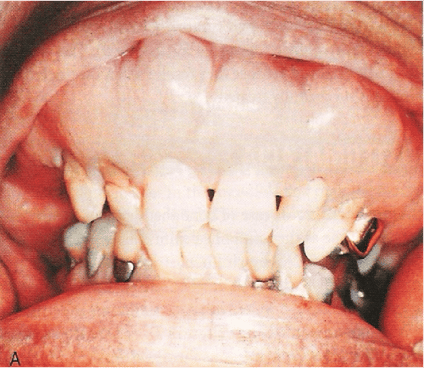
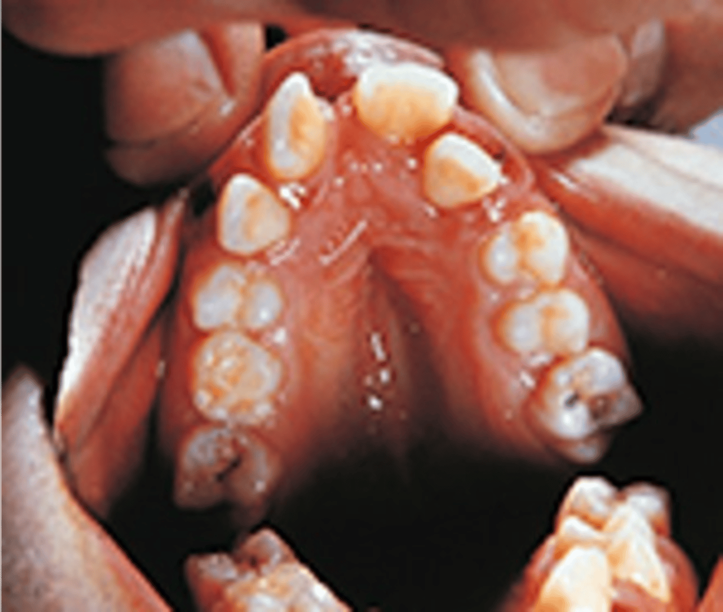
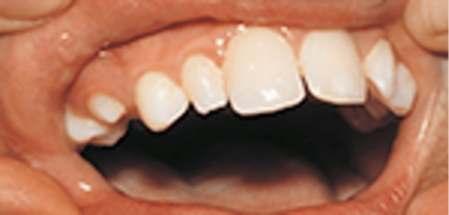
Cleidocranial dysplasia
supernumerary teeth extracted resulting from the cranium developing into a mushroom shape because the fontanelles remain open
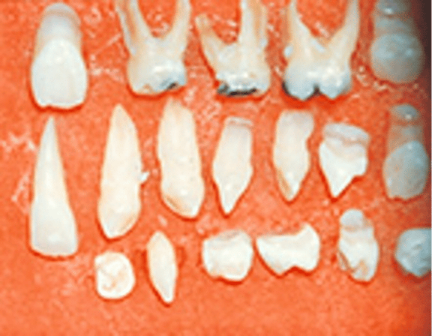
pegged maxillary lateral incisors
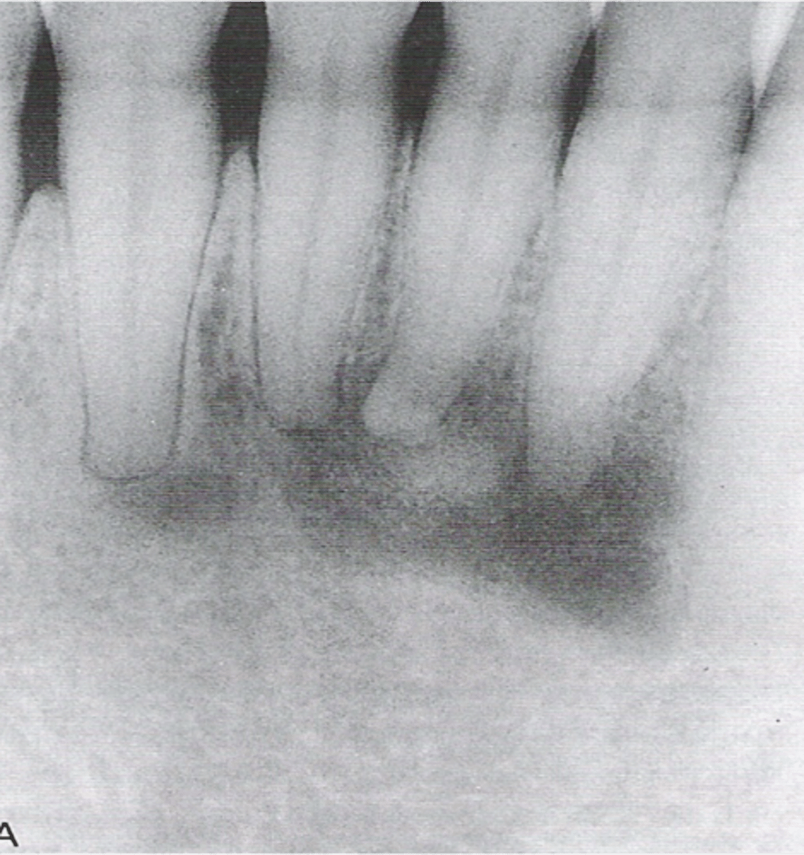
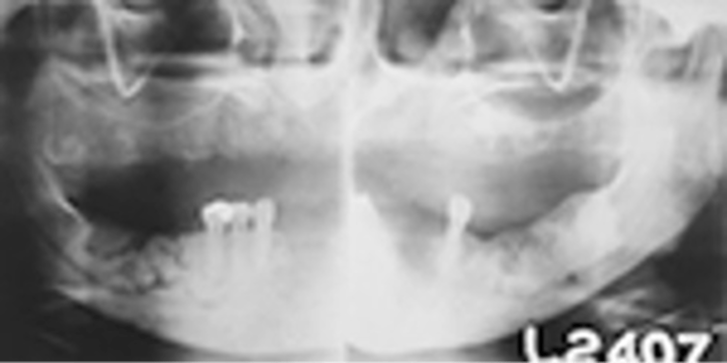
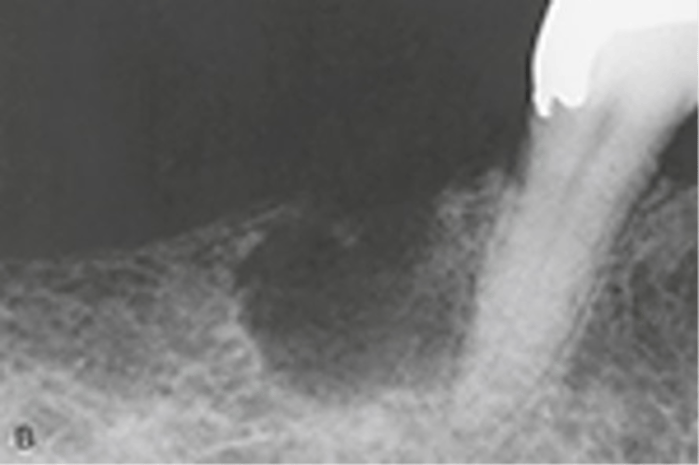
Mandibular lesion
seen radiographically in a patient with hyperparathyroidism
-radiolucencies also called brown tumors
osteonecrosis
death of bone tissue associated with bisphosphonate therapy
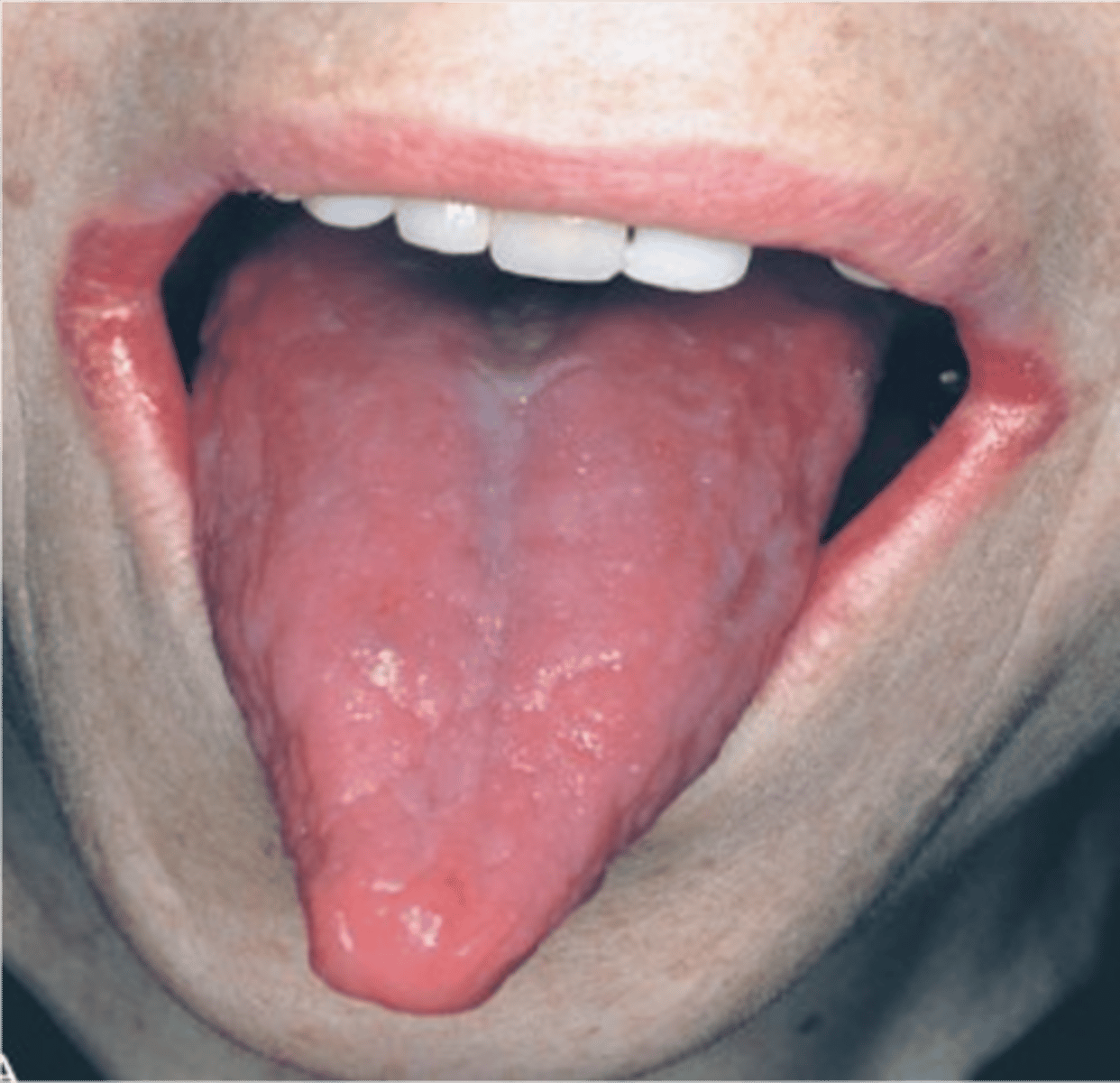
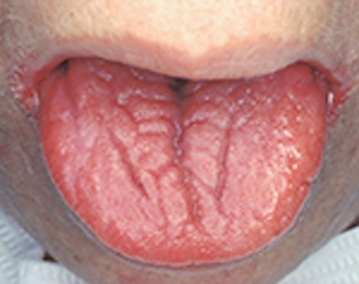
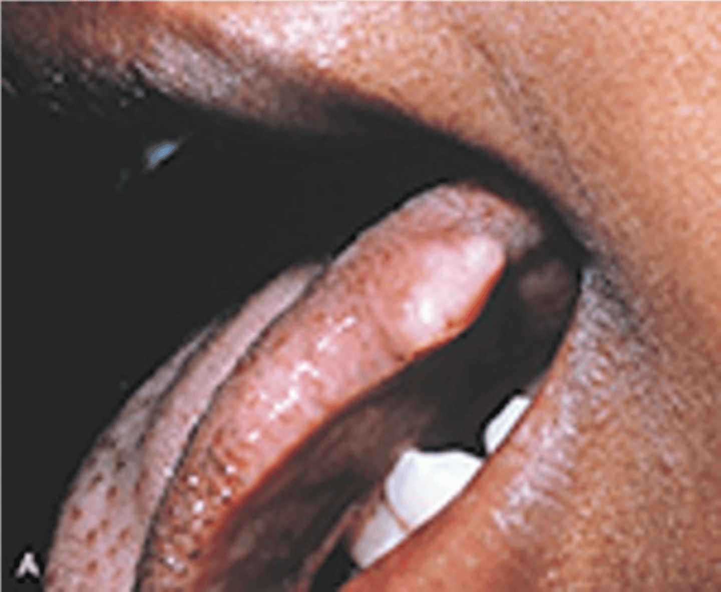
Macroglossia
Enlarged tongue; associated with acromegaly
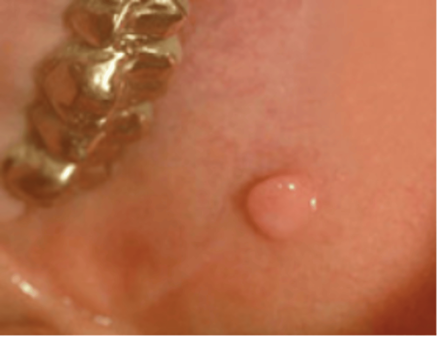
Papilloma
a benign, superficial wart-like growth on the epithelial tissue or elsewhere in the body, such as in the bladder
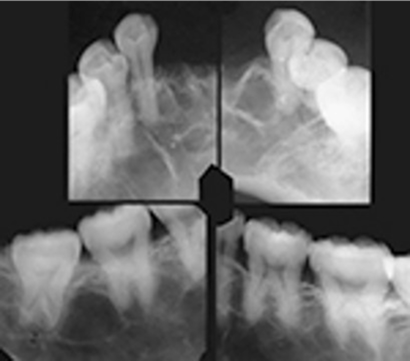
Florid Osseous Dysplasia
exuberant form of periapical cemento-osseous dysplasia, may involve entire jaw, African American middle aged women
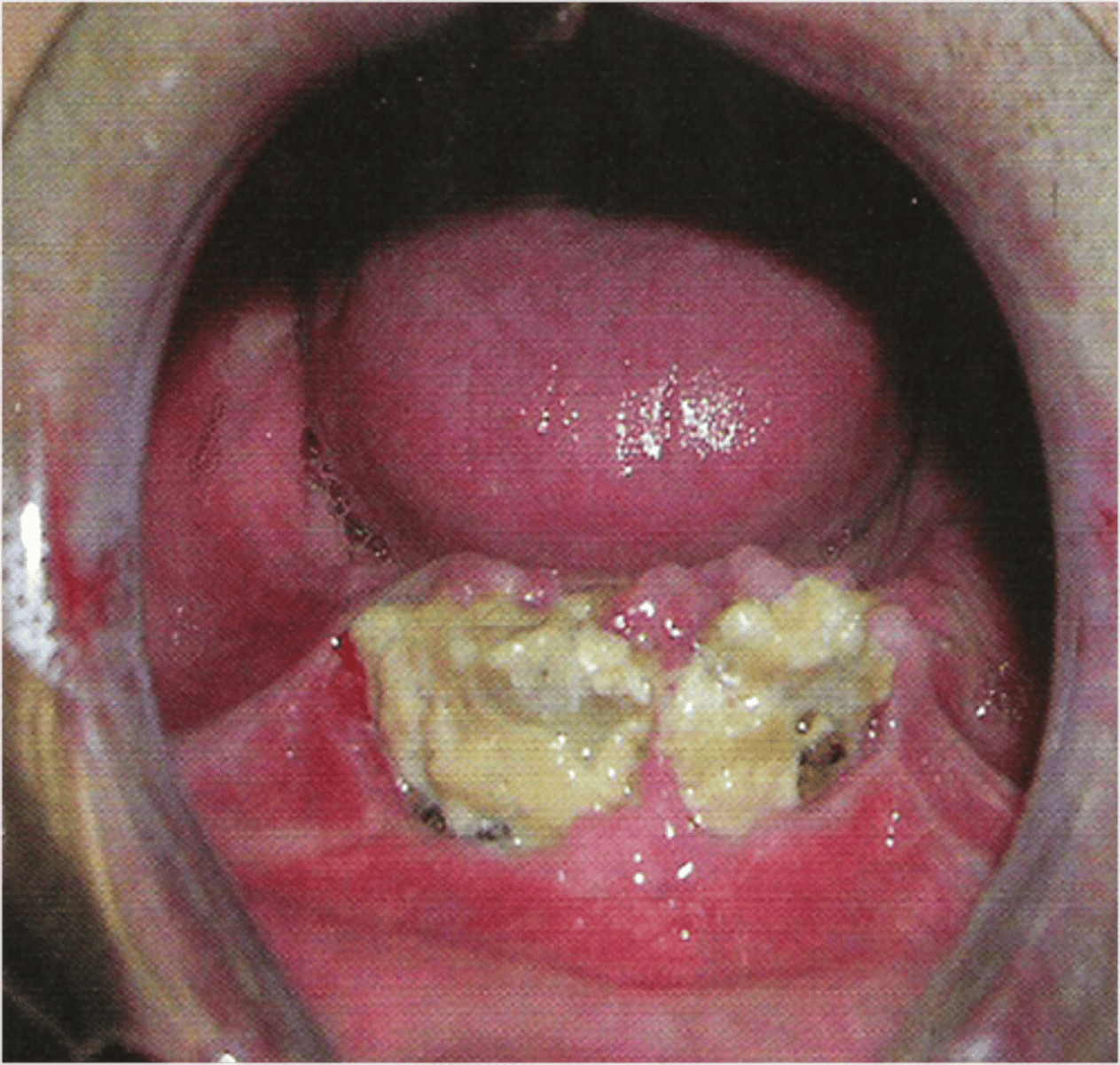
Cyclic Neutropenia
ulcerated gingivitis is characterized by a decrease in the number of circulating neutrophilic leukocytes
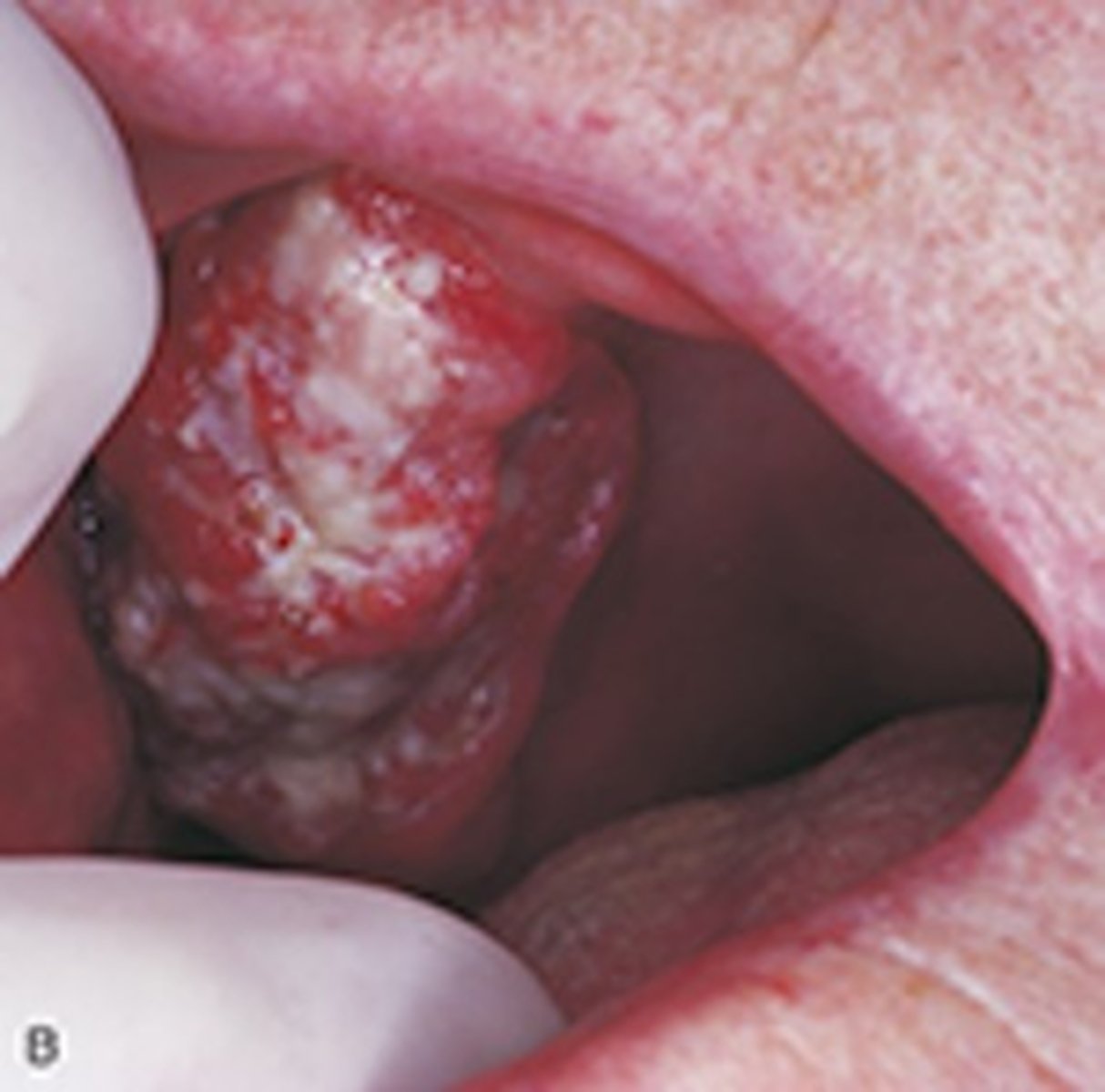
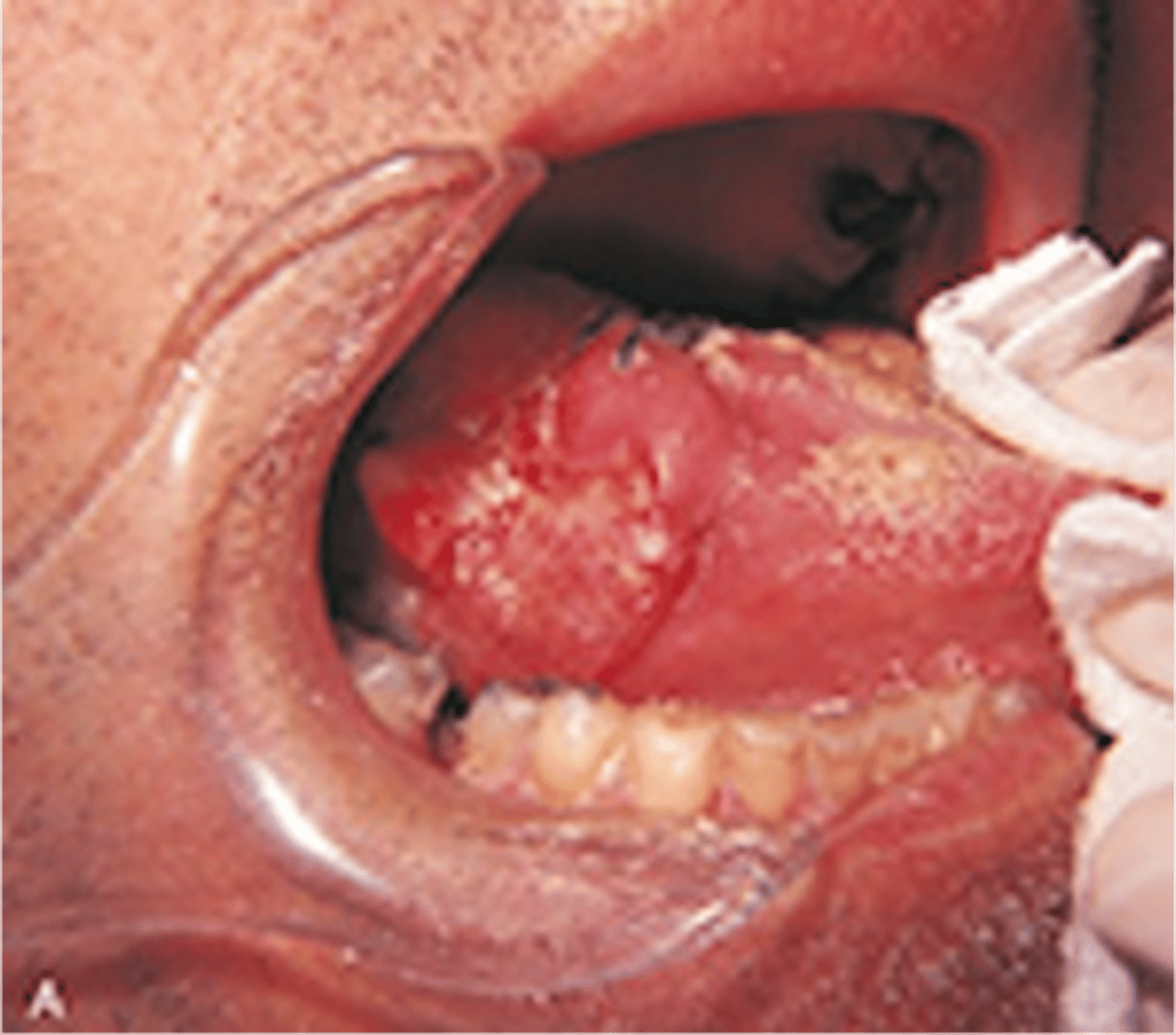
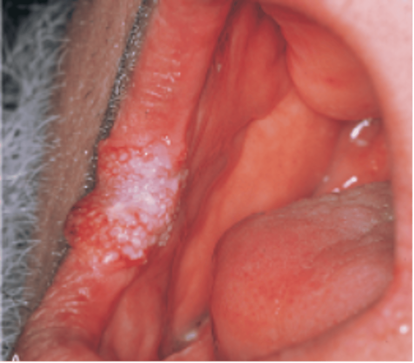
Verrucous Carcinoma
Specific type of squamous cell carcinoma that is separated from other squamous cell carcinomas because it has a much better prognosis
Central Giant Cell Granuloma
May present as a multilocular radiolucency, most common on the anterior segments
pernicious anemia
lack of mature erythrocytes caused by inability to absorb vitamin B12 into the bloodstream
iron deficiency anemia
anemia resulting when there is not enough iron to build hemoglobin for red blood cells
sickle cell anemia
a genetic disorder in which erythroctyes take on an abnormal curved or "sickle" shape
Calcifing odontogenic cyst
most common >40
may have calcifications
squamous cell carcinoma
Lipoma
a benign, slow-growing fatty tumor located between the skin and the muscle layer
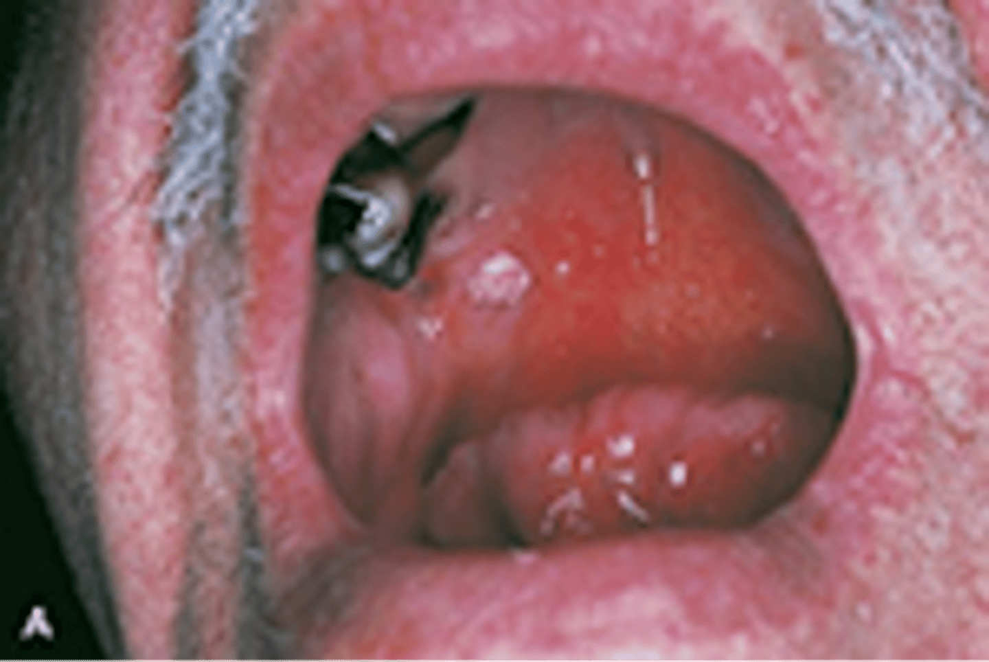
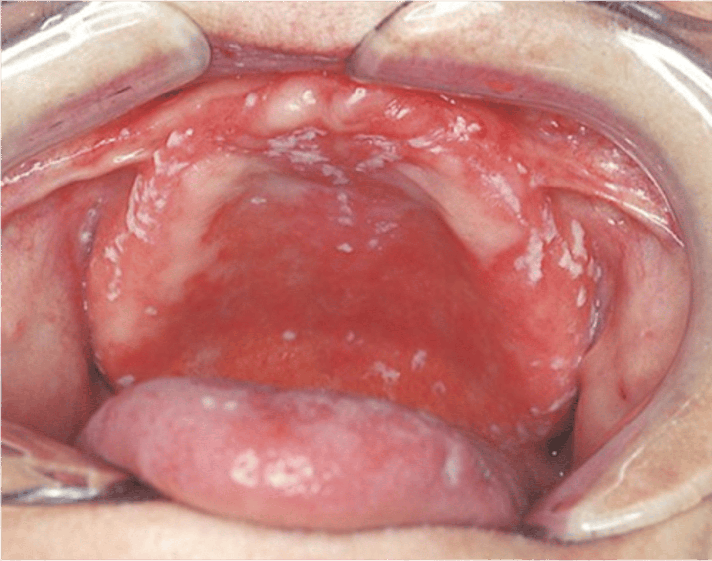
Candidiasis (Thrush)
Thick, white, raised patches in the mouth (this is probably caused from not cleaning the denture)
Central Giant Cell Granuloma (CGCG)
Non-neoplastic bone growth. Etiology unknown. Most commonly seen in females 30 years and younger. Treatment based on level of destruction. Corticosteroid injections reverse some CGCGs.
Schwannoma
Benign tumor of Schwann cells
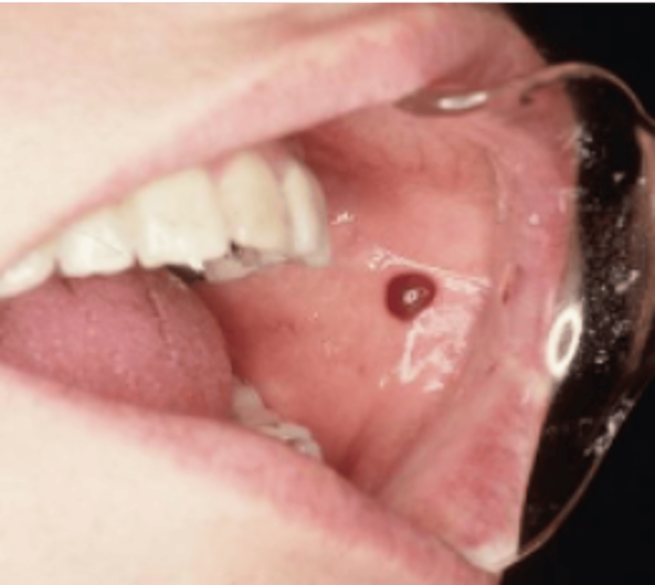
Hematoma
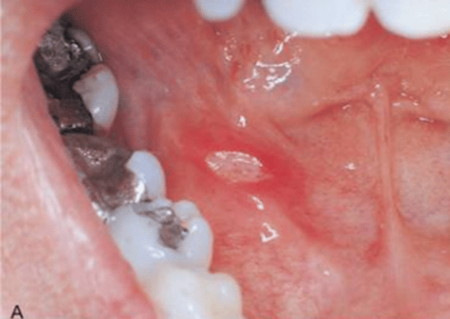
aphthous ulcer
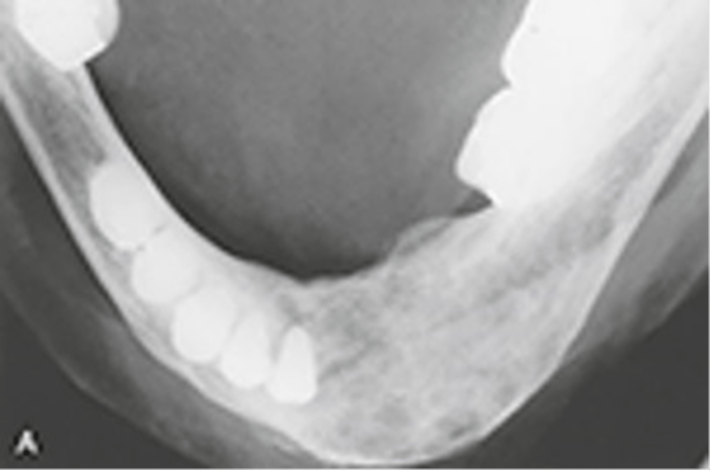
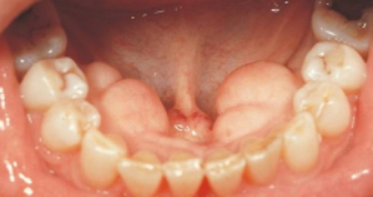
Mandibular tori
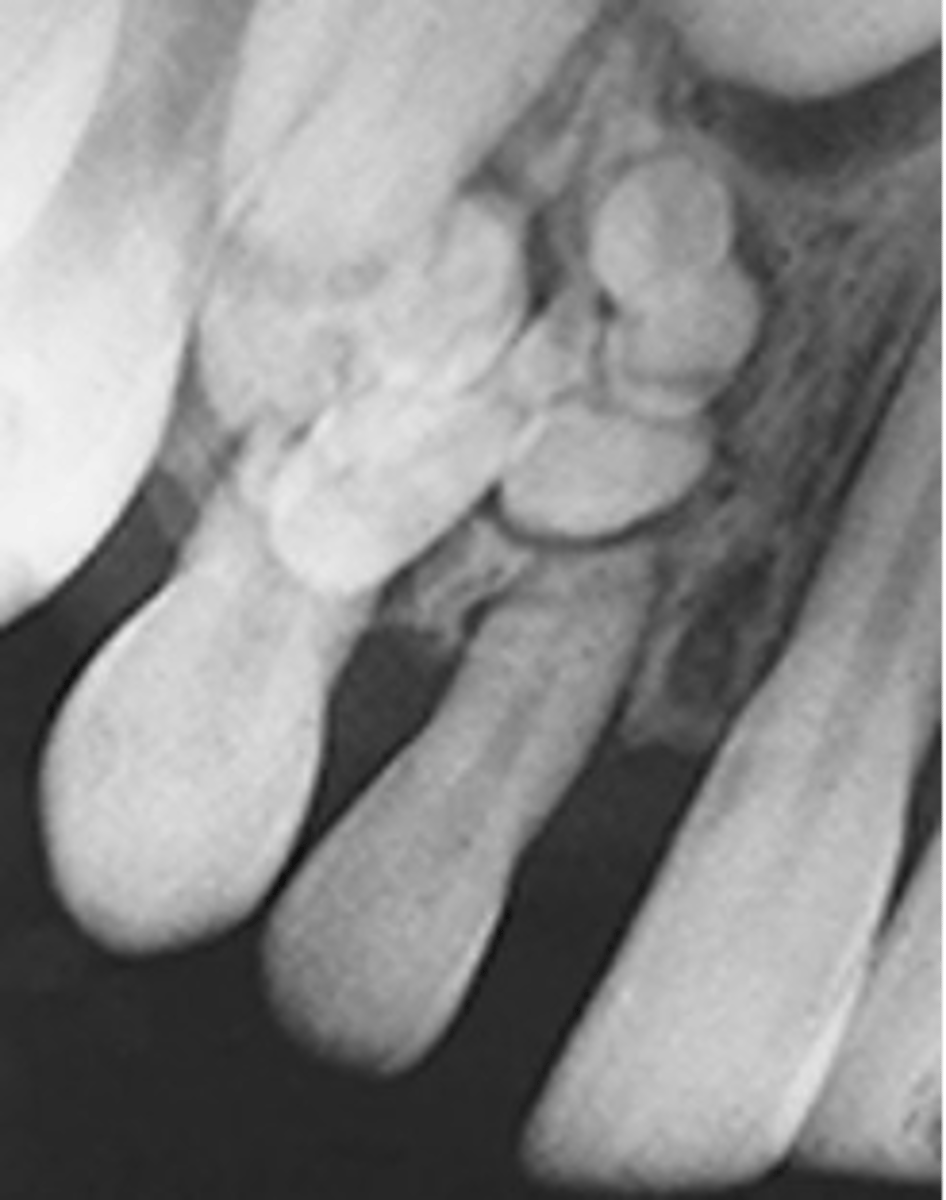
Compound odontoma
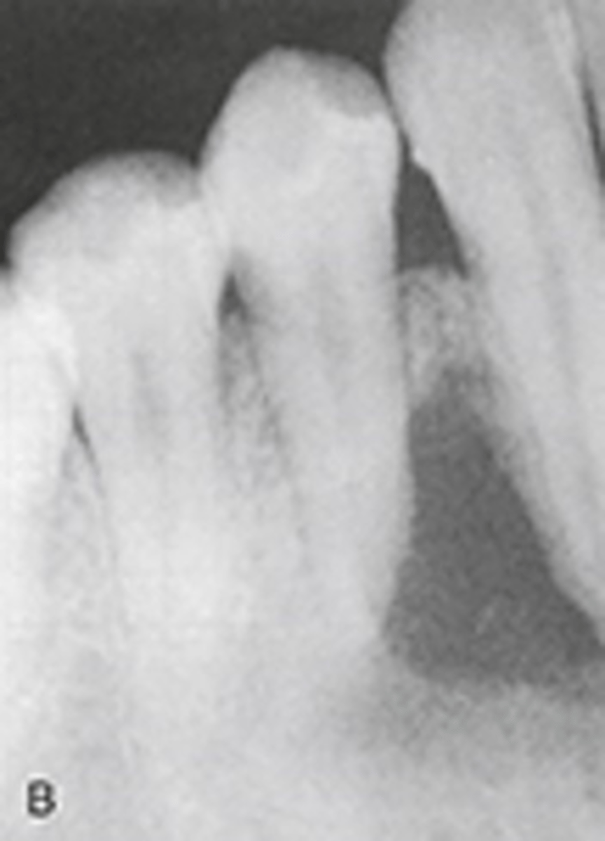
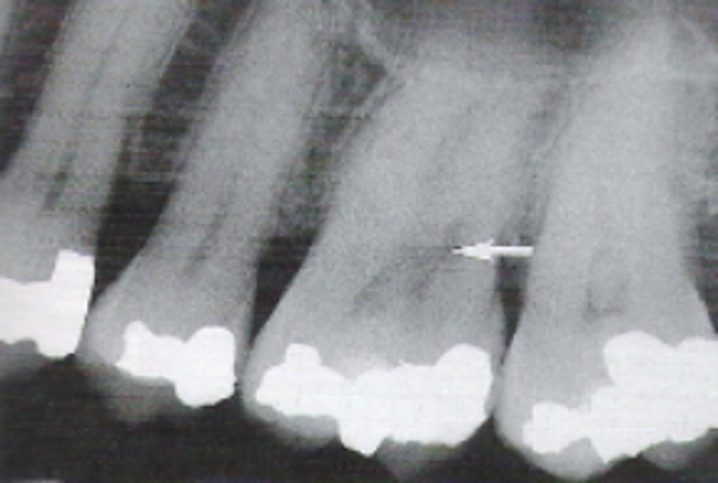
Internal root resorption
macule
verrucous carcinoma
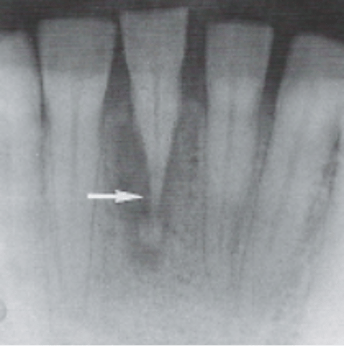
external resorption
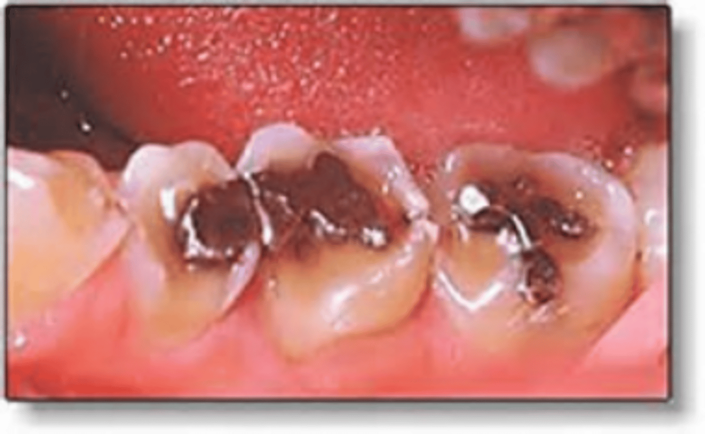
Erosion around Amalgam Restorations
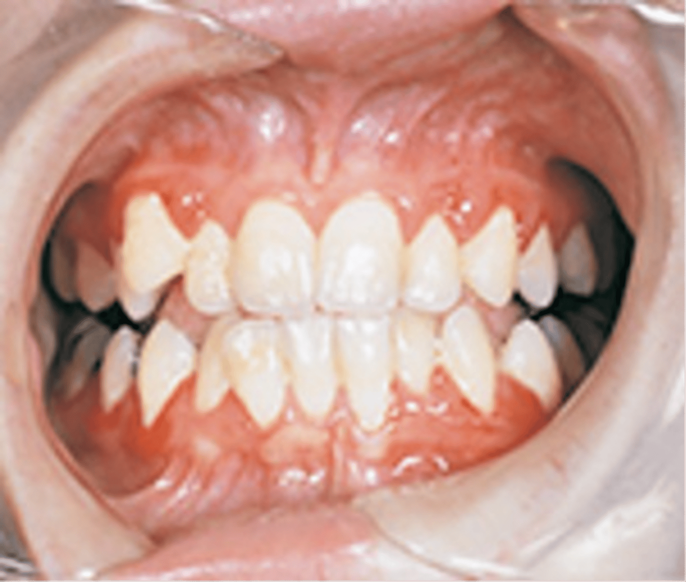
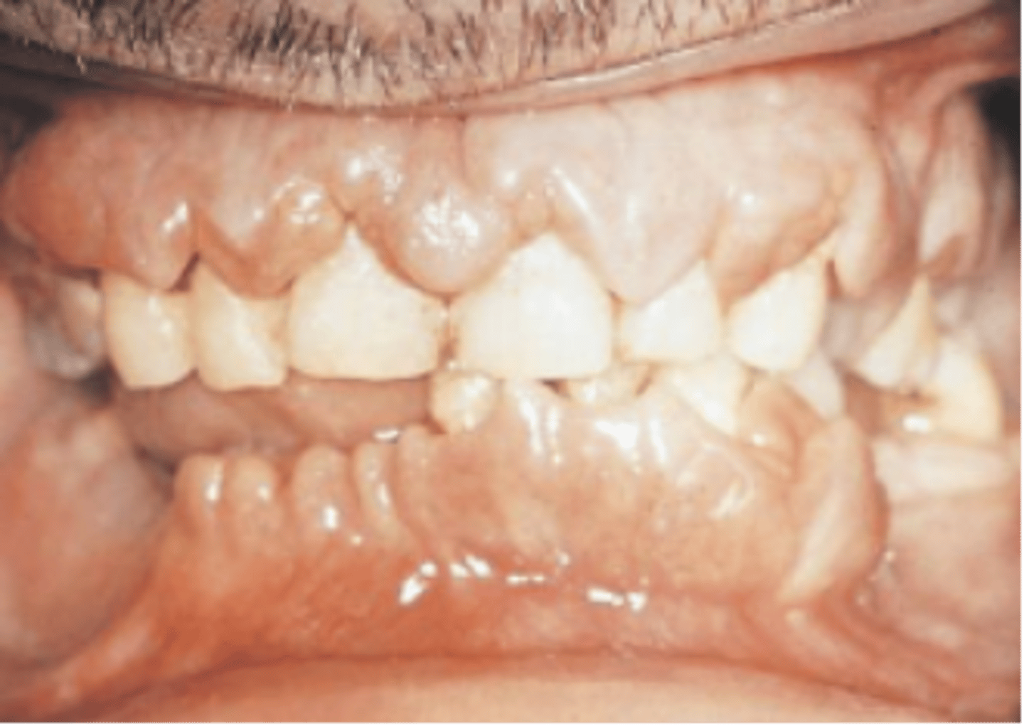
Leukemia: Gingival enlargement because of infiltration of leukemic cells
Treacher Collins syndrome
hearing loss and breathing (respiratory) difficulties. Cleft palate ,behavioral anomalies such as microcephaly and psychomotor delay, downward-slanting eyes, a very small jaw and chin, hearing loss, and vision loss
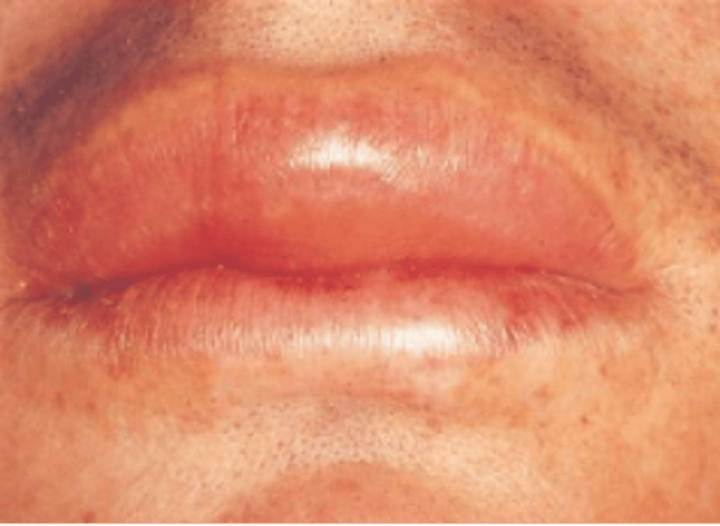
angioedema
The diffuse swelling caused by the increased permeability of deeper blood vessels depicted in the image below is called
Down Syndrome (Trisomy 21)

Turner syndrome
A chromosomal disorder in females in which either an X chromosome is missing, making the person XO instead of XX, or part of one X chromosome is deleted.
46 chromosone - normal
45 chromosone -> Turner syndrome
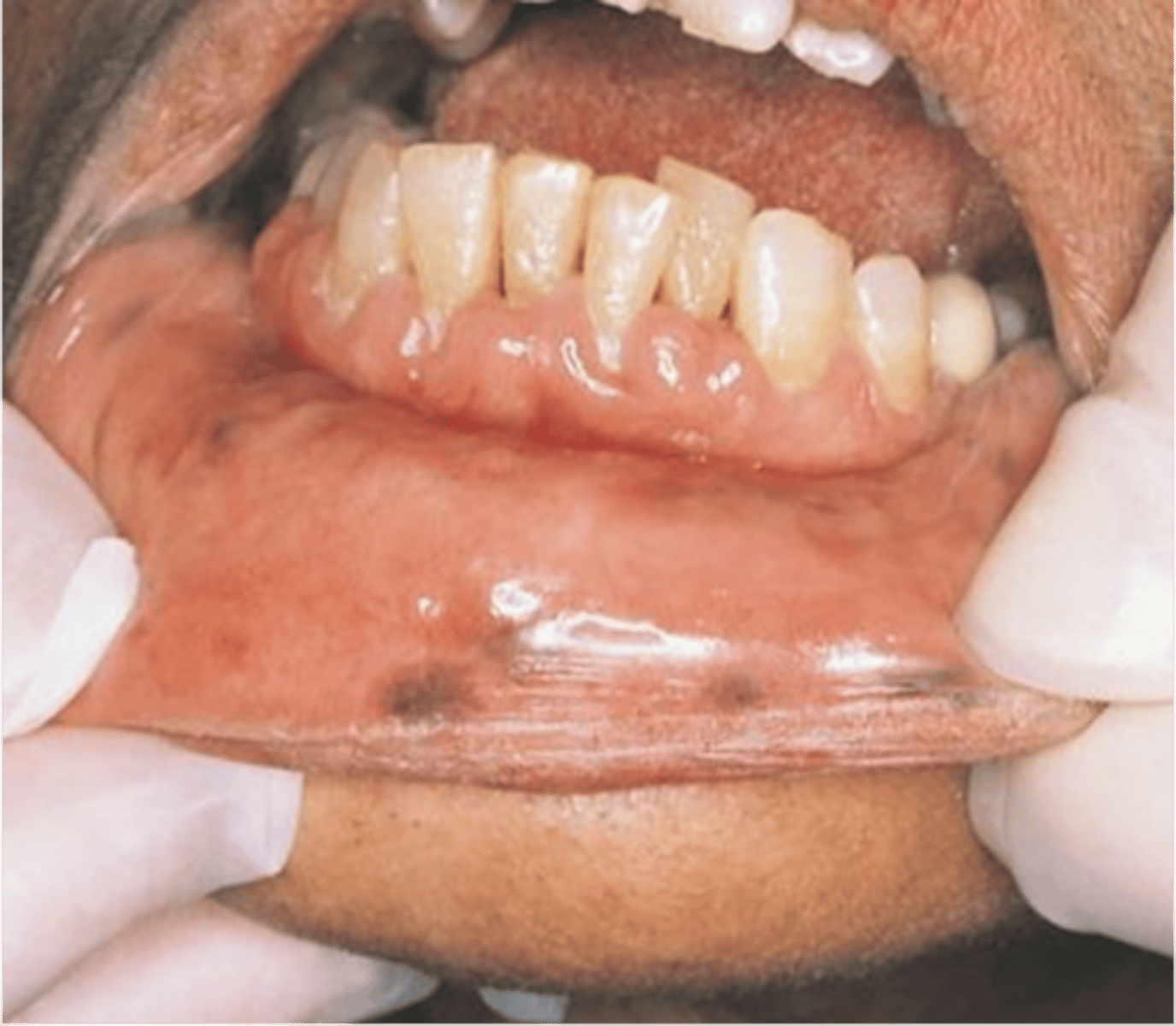
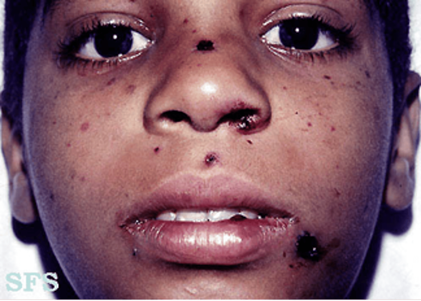
Thrombocytopenic Purpura
Spontaneous purpuric or hemorrhagic lesions on the skin.
Patients bruise easily
May have blood in urine
Frequent nosebleeds
Spontaneous gingival bleeding
Petechiae
Clusters of petechiae or purpuric spots
Ecchymosis
Nodule