Urinary and Reproductive Systems
1/69
There's no tags or description
Looks like no tags are added yet.
Name | Mastery | Learn | Test | Matching | Spaced | Call with Kai |
|---|
No analytics yet
Send a link to your students to track their progress
70 Terms
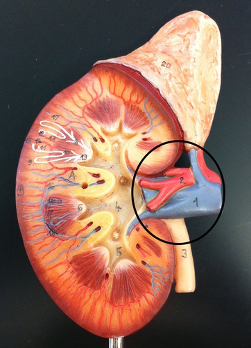
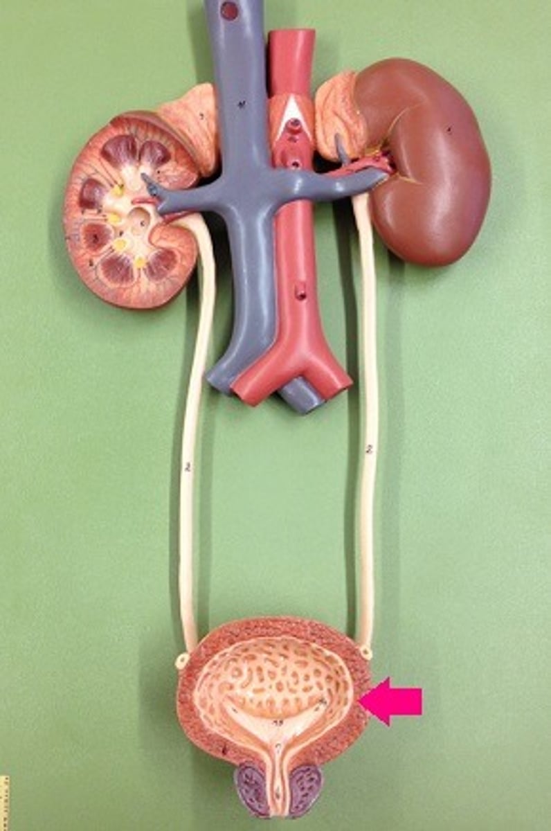
Hilus
depression where vessels and nerves enter an organ
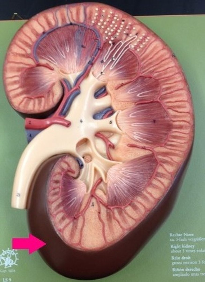
Fibrous Capsule of Kidney
the surrounding protective layer of the kidney
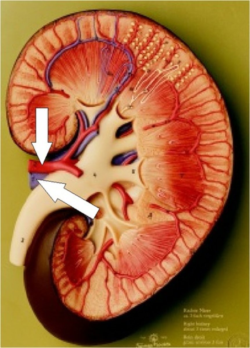
Renal artery and vein
blood supply to and from the kidneys
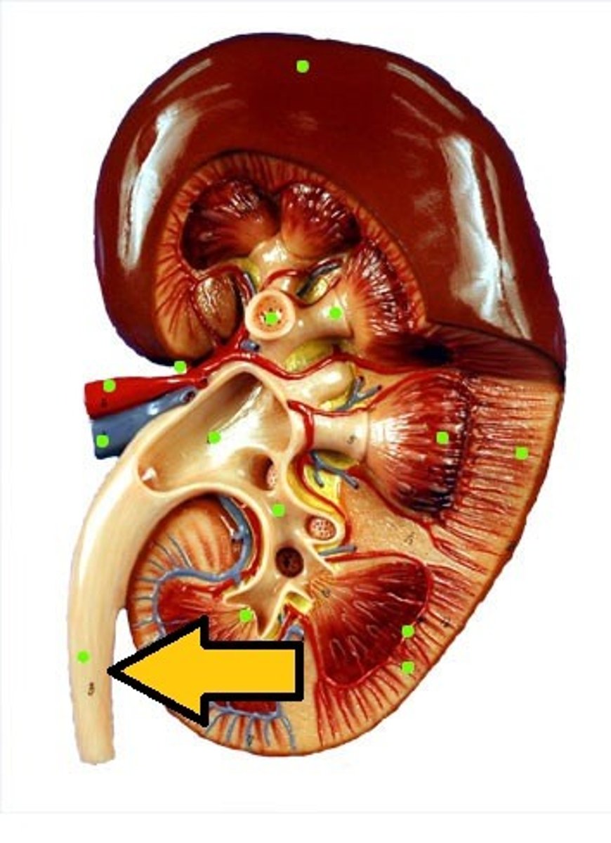
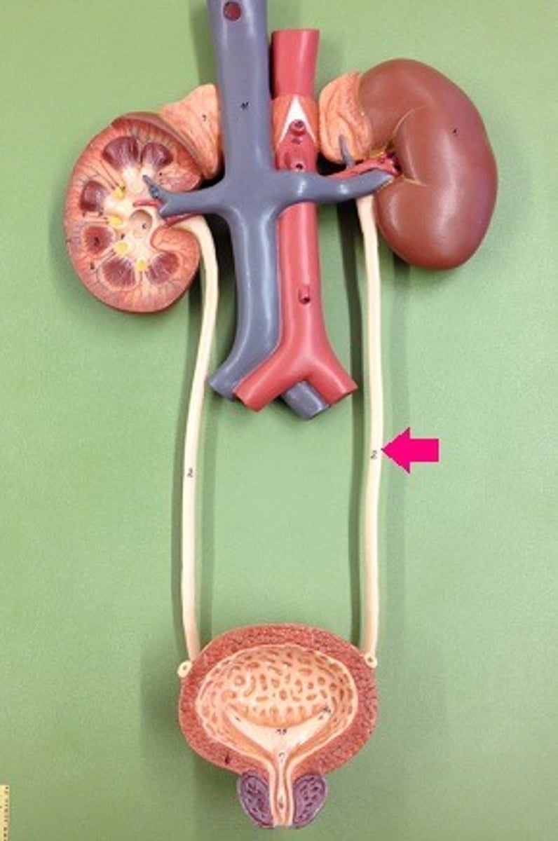
Ureter
A duct leading from the kidney to the urinary bladder.
the medulla of the kidney
inner portion of kidney, composed of collecting tubules that empty into the renal pelvis
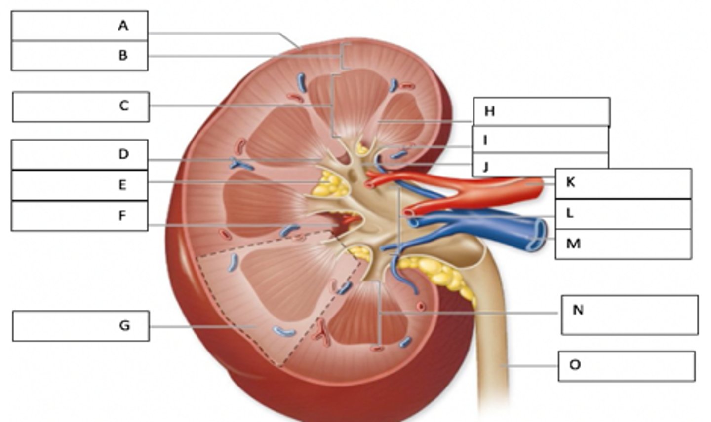
renal columns
Inward extensions of the cortex tissue separating the renal pyramids.
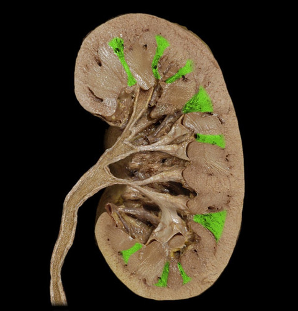
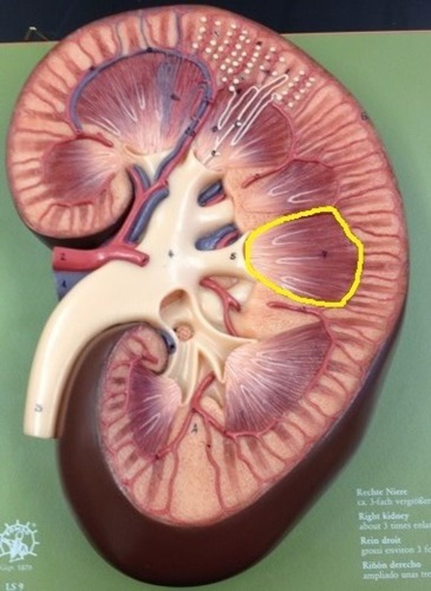
Renal Pyramids
triangular-shaped areas of tissue in the medulla of the kidney
The renal papillae
These are the tips at the base of the renal pyramids which project into a minor calyx. These structures drain urine from the pyramids into the minor calyxes.
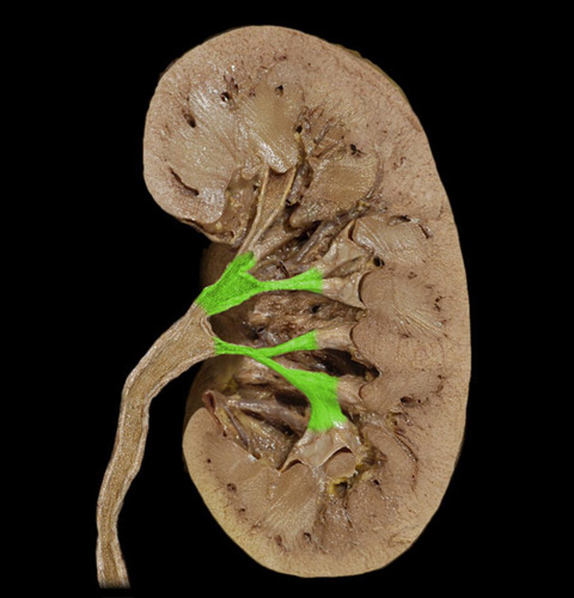
Major Calyx
The cavity formed by the convergence of several minor calyces, which drain urine from the minor calyxes into the renal pelvis
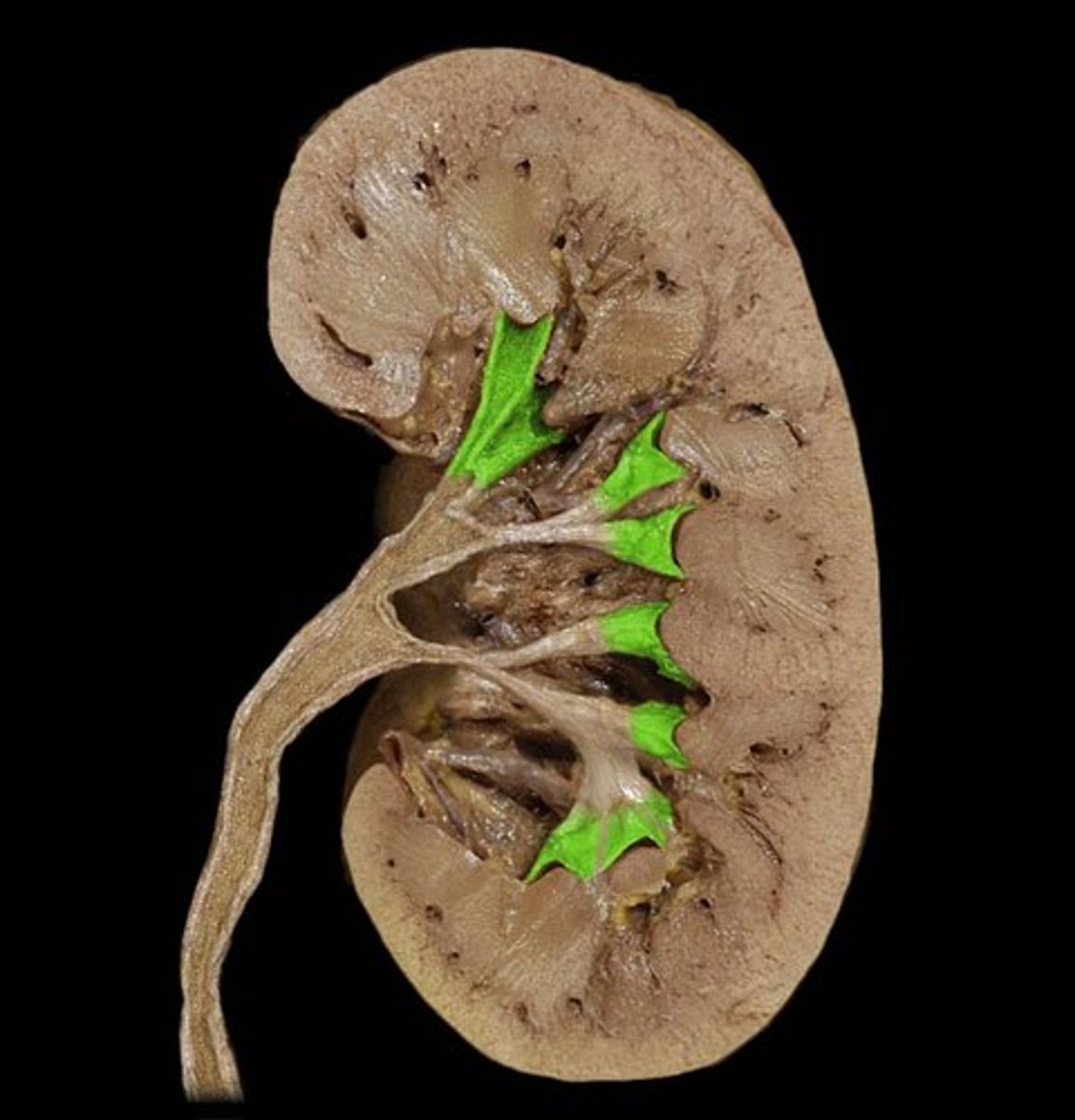
minor calyx
a cup-shaped extension of the pelvis that encircles the apex of a pyramid
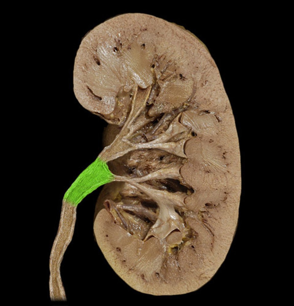
Renal pelvis
central collecting region in the kidney
Ureters
The tubes that carry urine from the kidneys to the bladder.
Urinary bladder
stores urine
Urethra
tube leading from the urinary bladder to the outside of the body
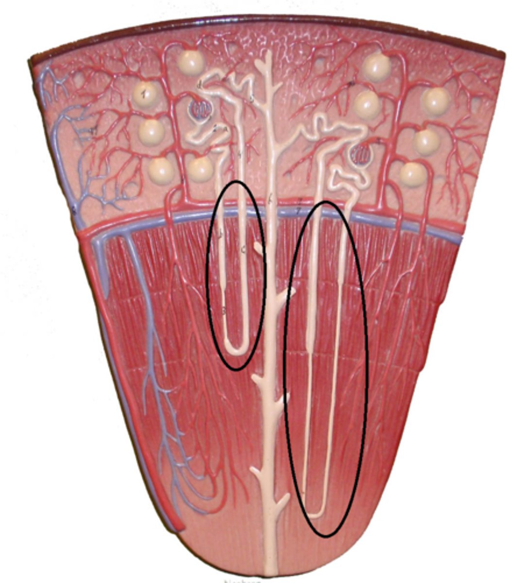
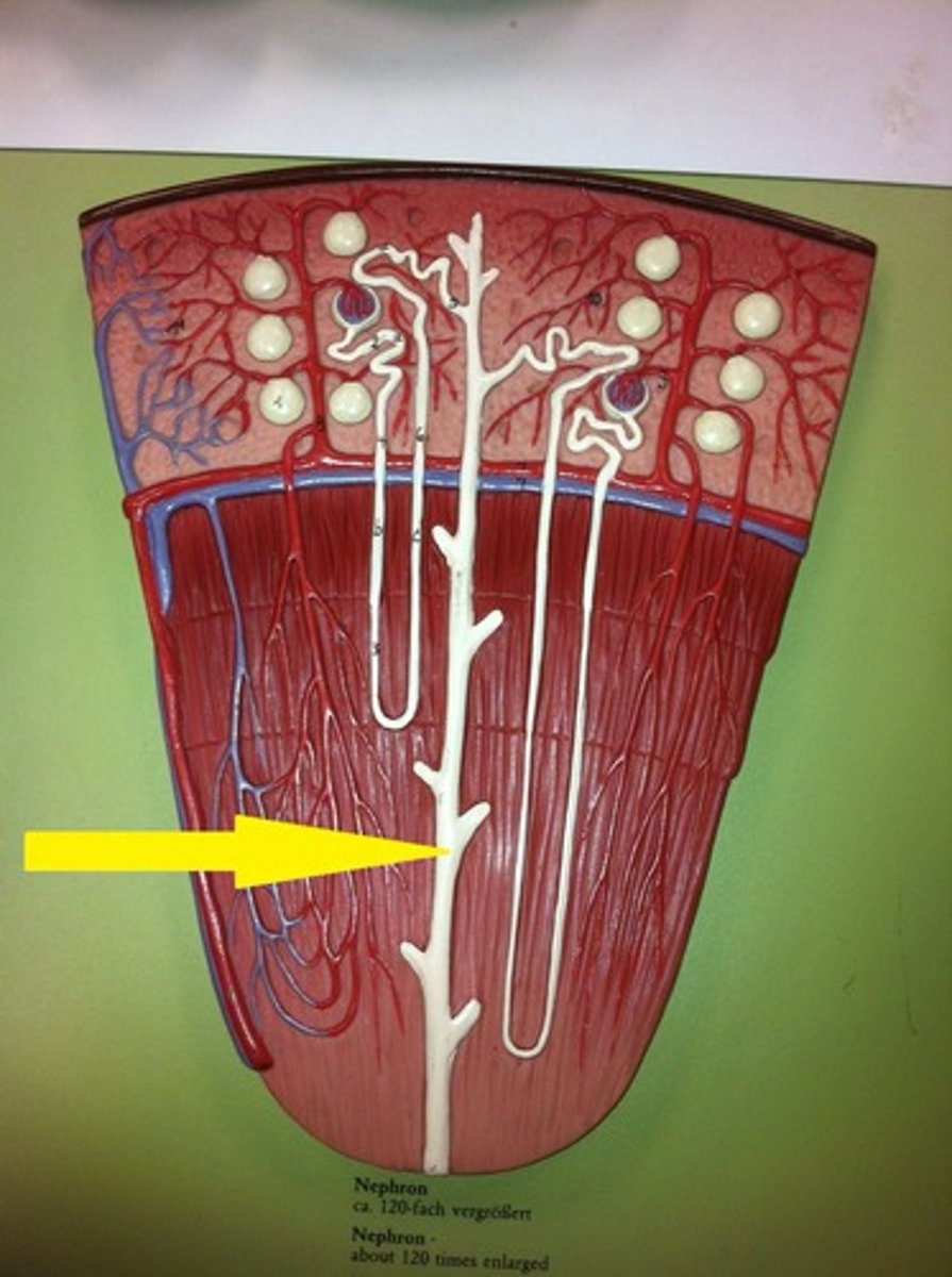
Nephron
functional unit of the kidney
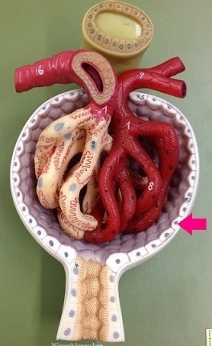
glomerulus
A ball of capillaries surrounded by Bowman's capsule in the nephron and serving as the site of filtration in the vertebrate kidney.
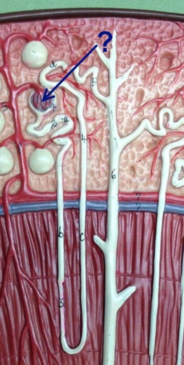
Glomerular Capsule/Bowman's Capsule
Cup-shaped, hollow structure surrounding glomerulus
Proximal Convoluting tubule
The first segment of the renal tubule where reabsorption of water, ions, and nutrients occurs.
F in diagram

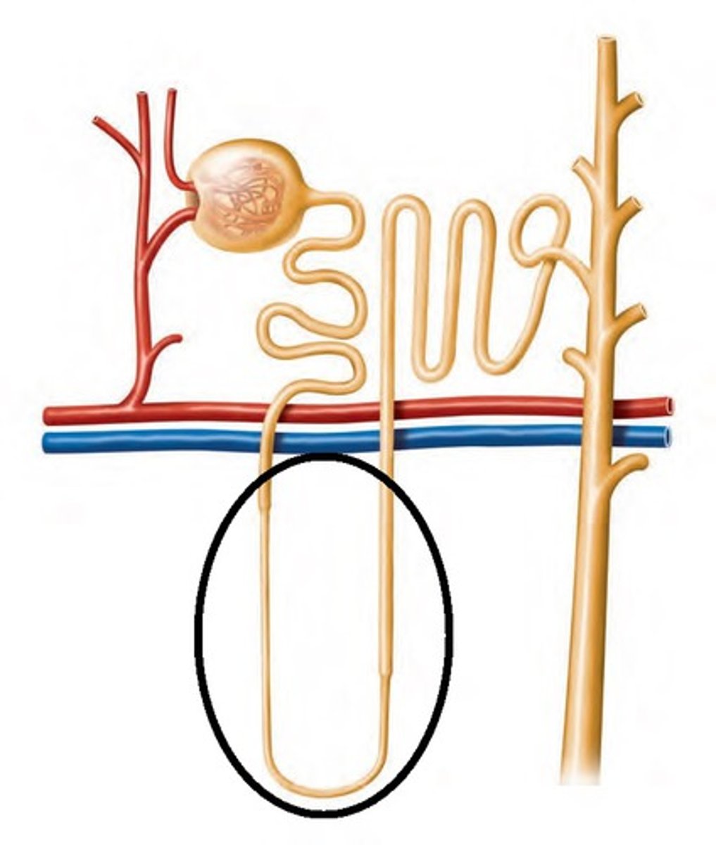
Nephron Loop
second part of tubule that dips into the medulla; reabsorbs water and salt (sodium and chloride ions); aka, loop of Henle
distal convoluting tubule
The furtherest end of the tubule before the collecting duct
G in diagram
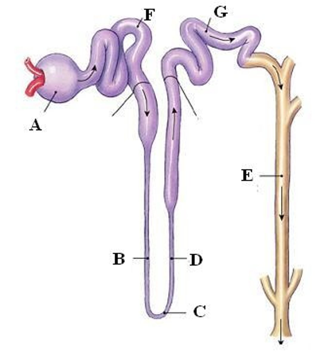
Collecting duct
A segment of the nephron that returns water form the filtrate to the bloodstream.
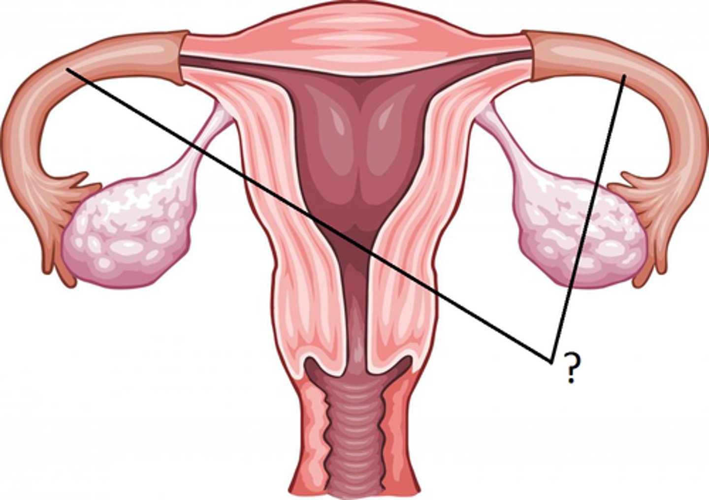
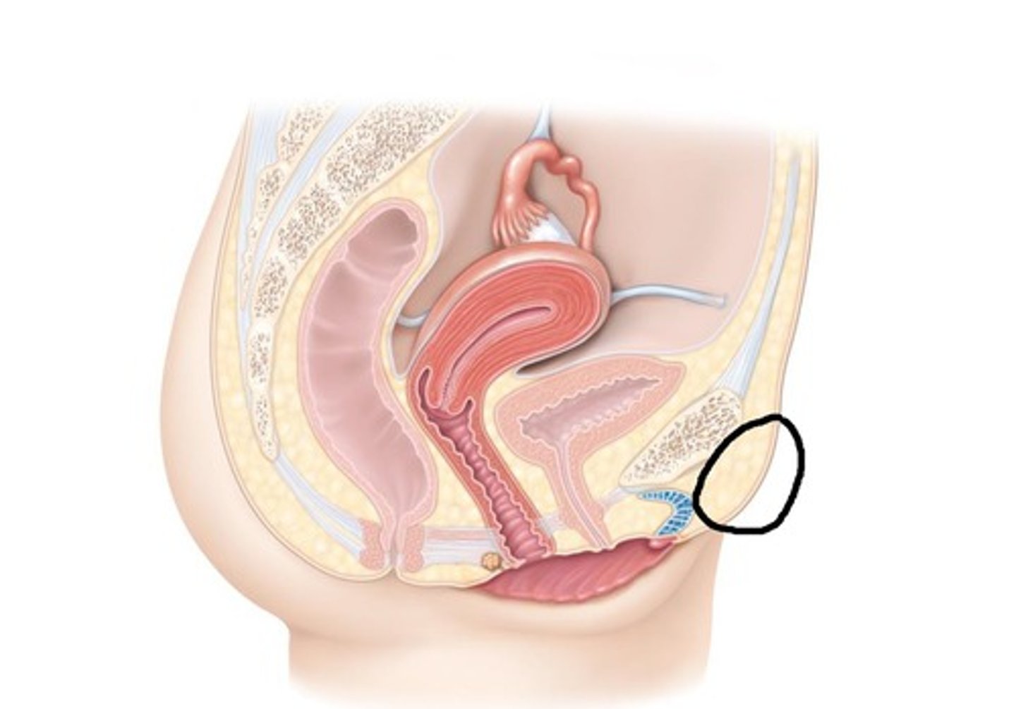
ovaries (right and left)
suprapubic
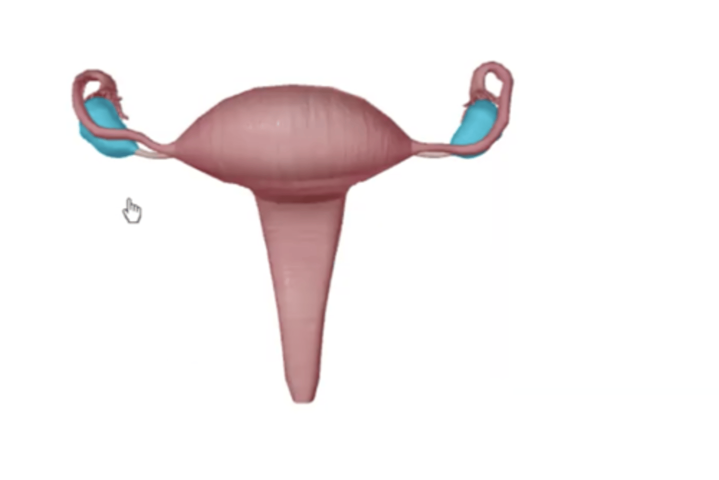
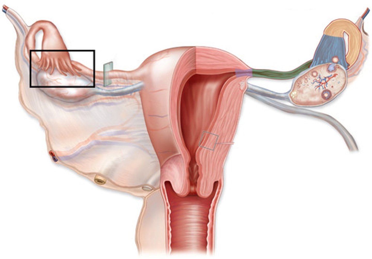
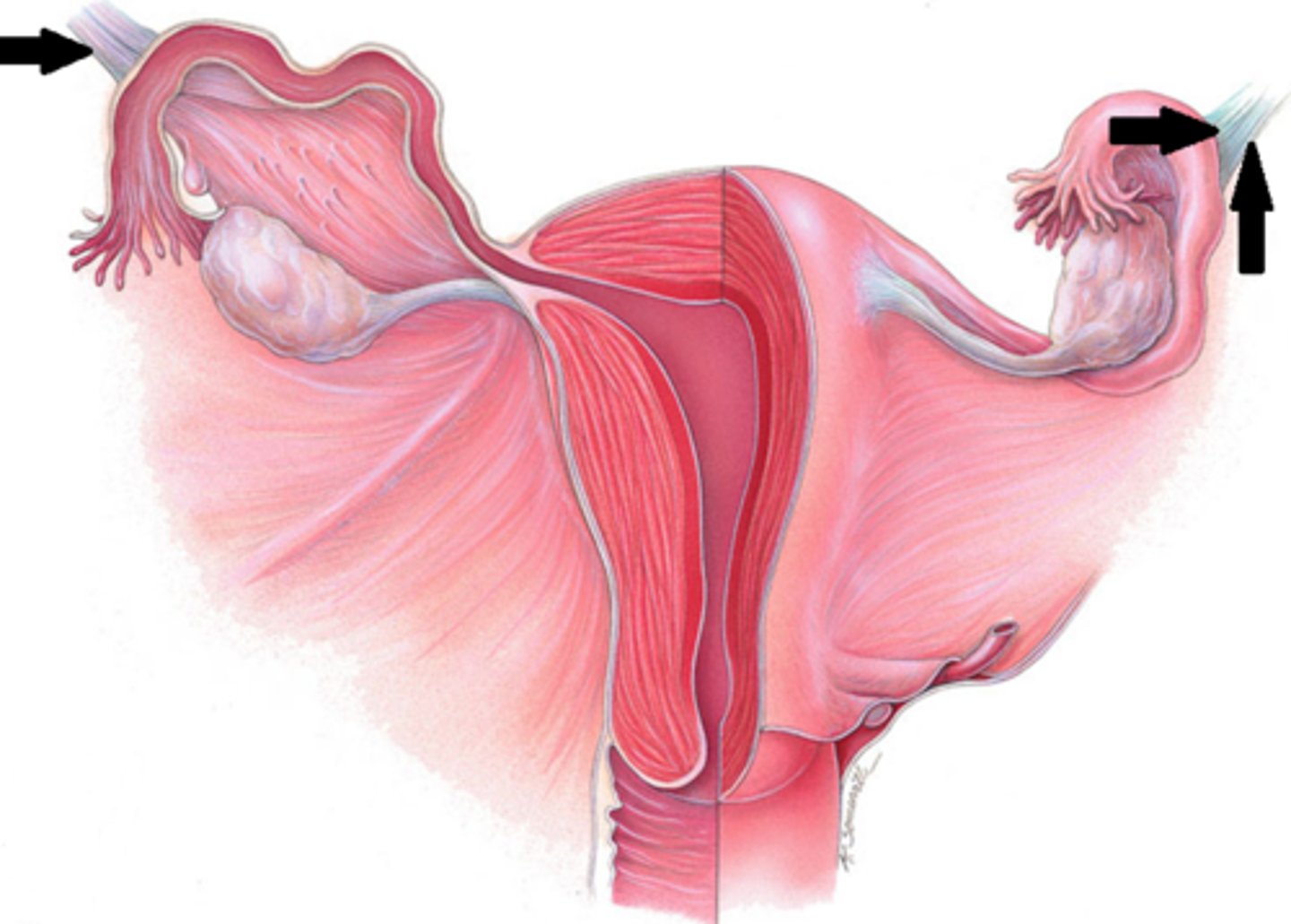
Uterine tubes (= oviduct, = Fallopian tube)
Tubes
infundibulum, ampulla, isthmus
3 parts of the fallopian tubes
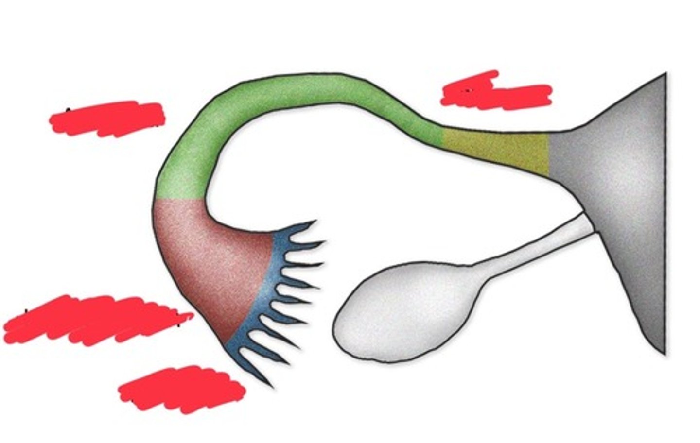
ostium (uterine tubes)
Follicle opening.
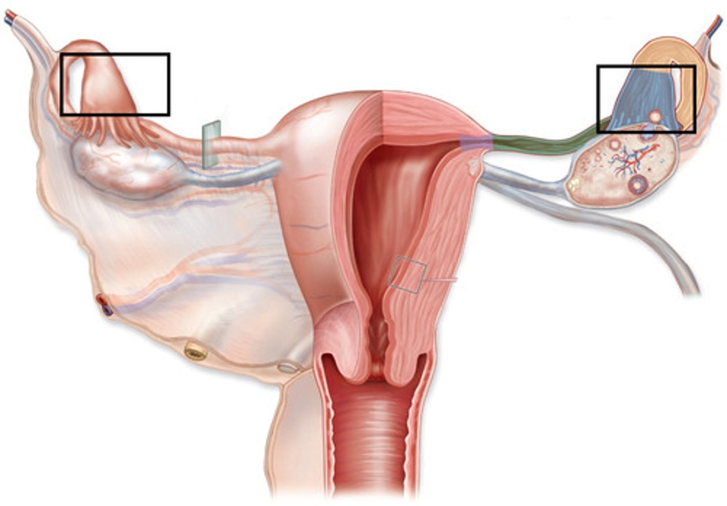
Fimbriae of infundibulum
fingerlike structures
Round ligament of uterus
restricts posterior movement of uterus
Broad ligament of uterus
runs parallel to the uterine horns and holds uterus in place
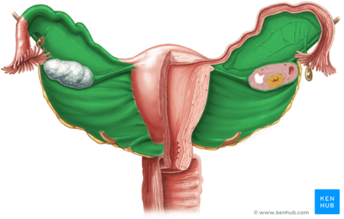
Suspensory ligament of ovary
Ligament that contains ovarian vessels
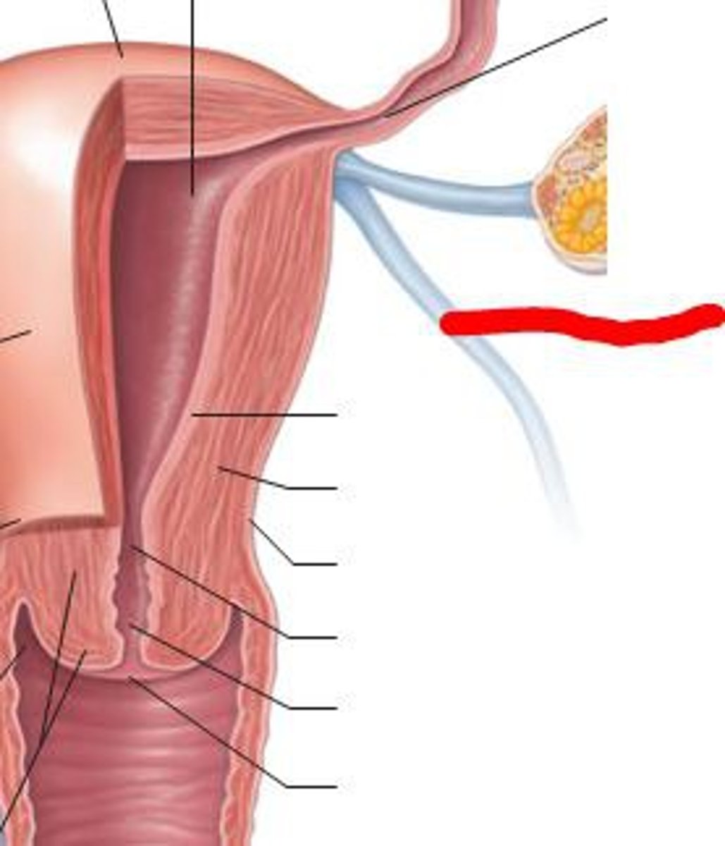
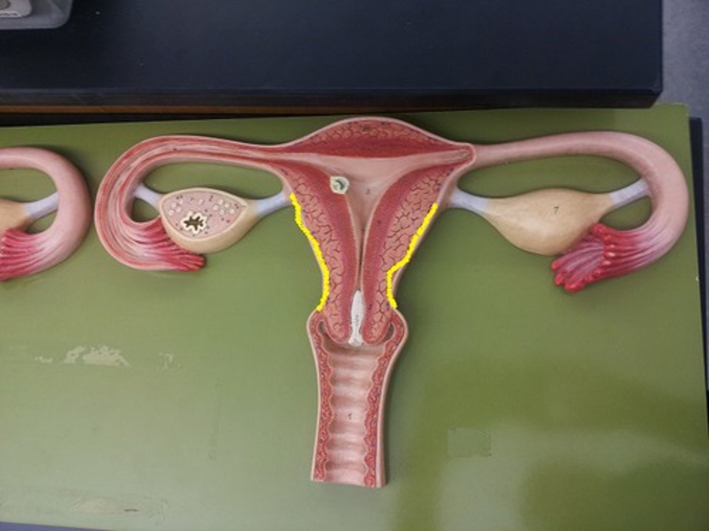
Uterus
Female organ of reproduction used to house the developing fetus.
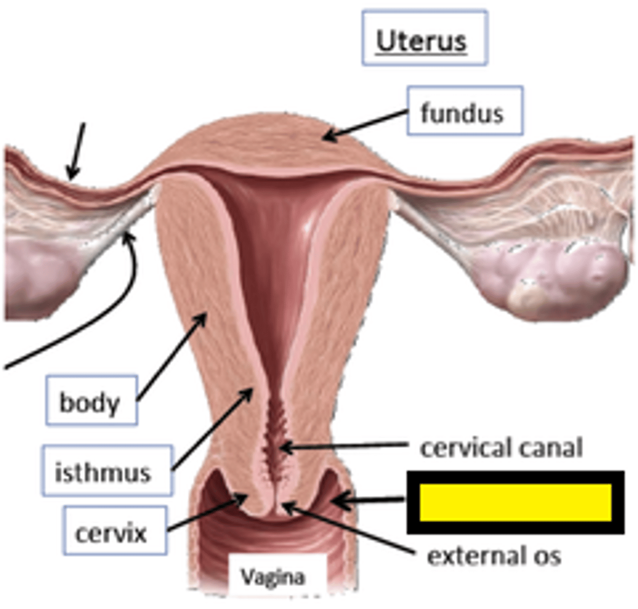
Fundus of uterus
Most superior & widest portion of uterus

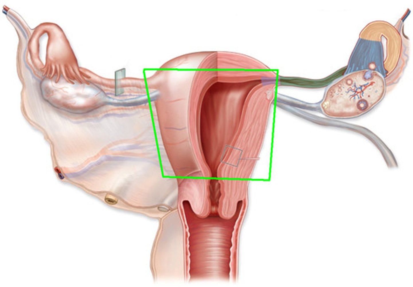
body of uterus
major portion of uterus
cervix of uterus
narrow neck which projects into the vagina inferiorly
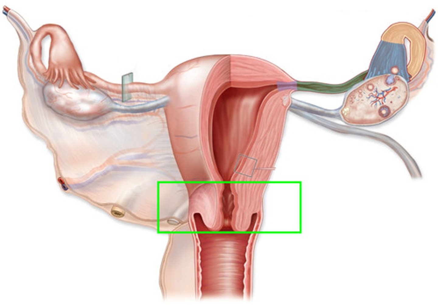
fornix vagina
The cranial portion of the vagina that forms a crypt extending cranially to the cervix.
Vaginal Rugae
ridges in vagina
(Lower portion in diagram)
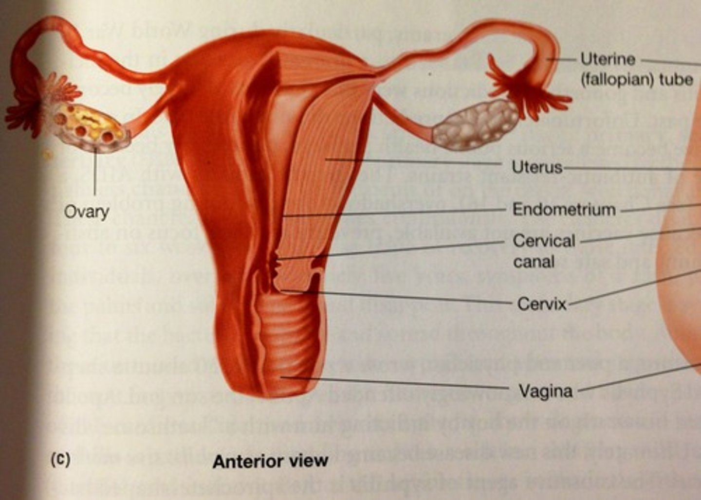
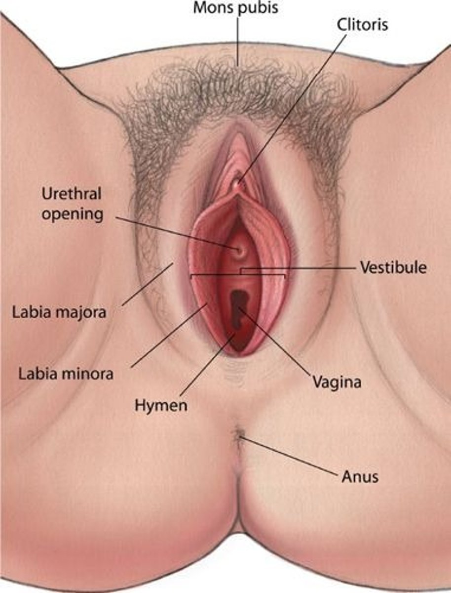
Vaginal Orifice
opening of the vagina
hymen
mucous membrane partially or completely covering the opening to the vagina
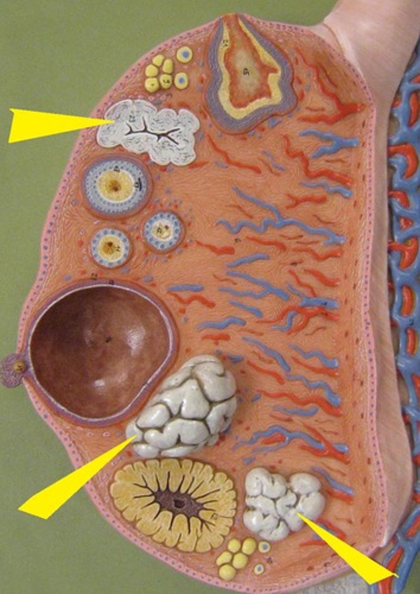
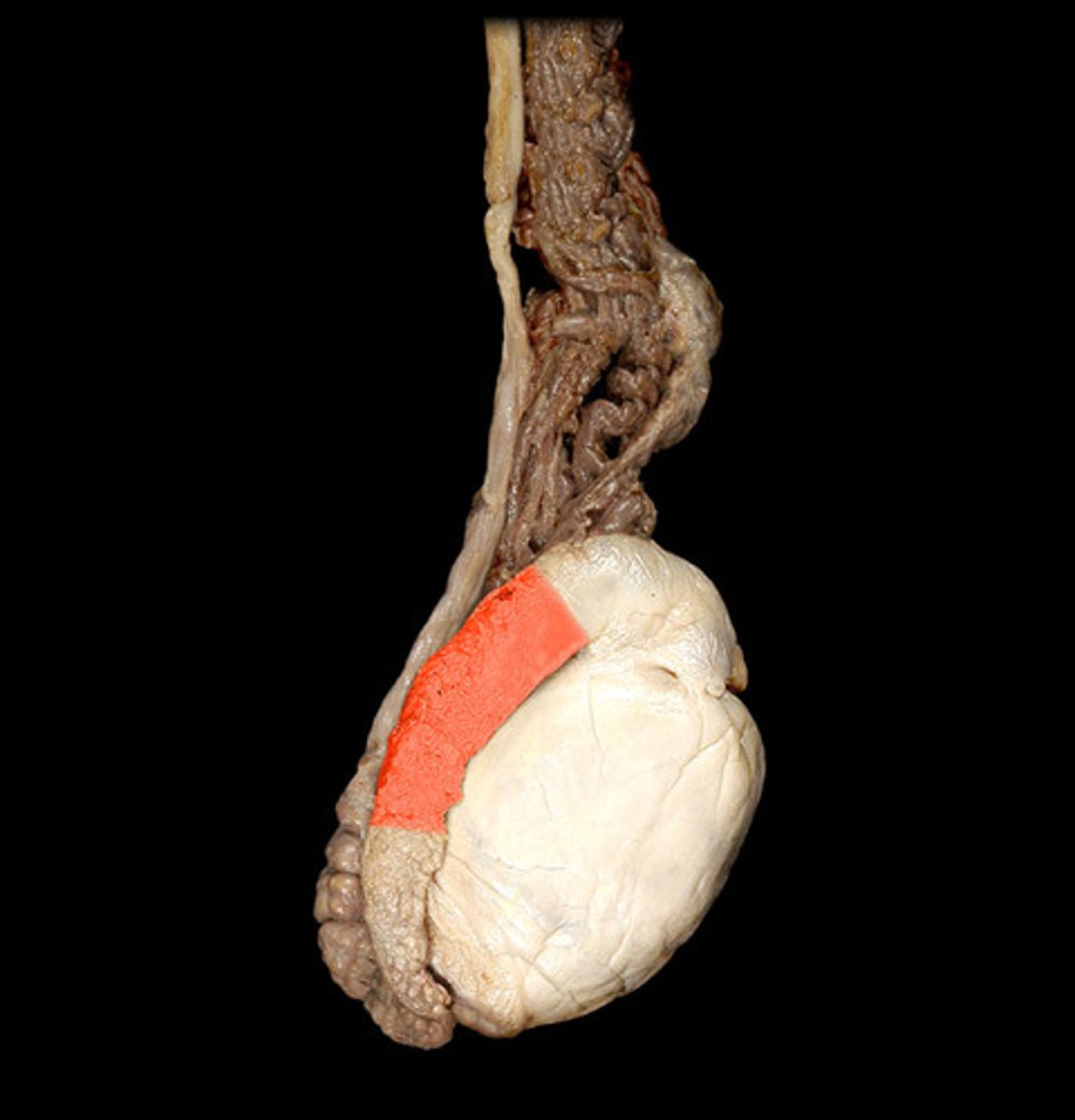
tunica albuginea
fibrous capsule of the testes
Corpus luteum
Endocrine tissue which produces hormones, estrogen, and progesterone which prepares the uterine lining for receiving an embryo
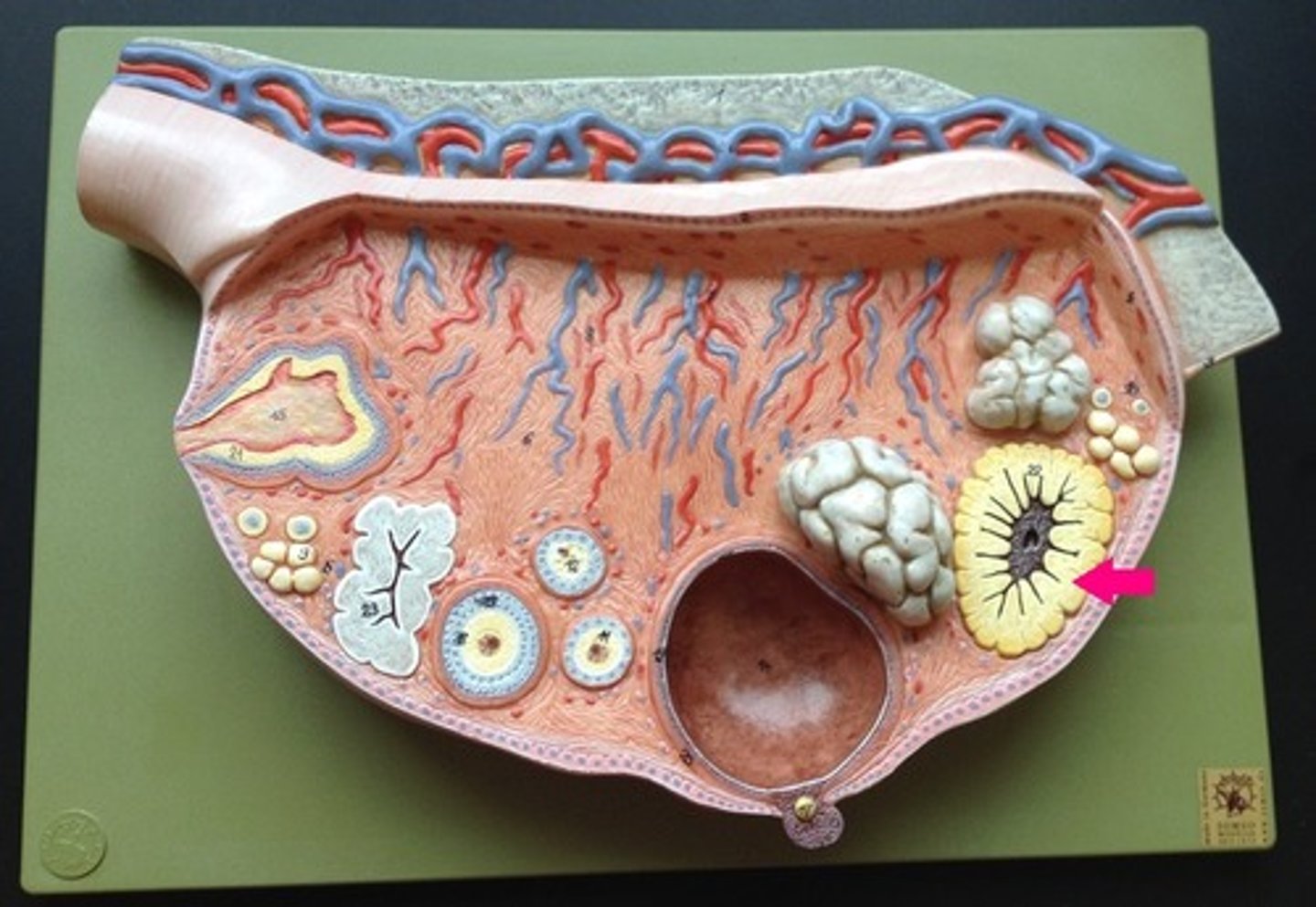
corpus albicans
degenerated corpus luteum
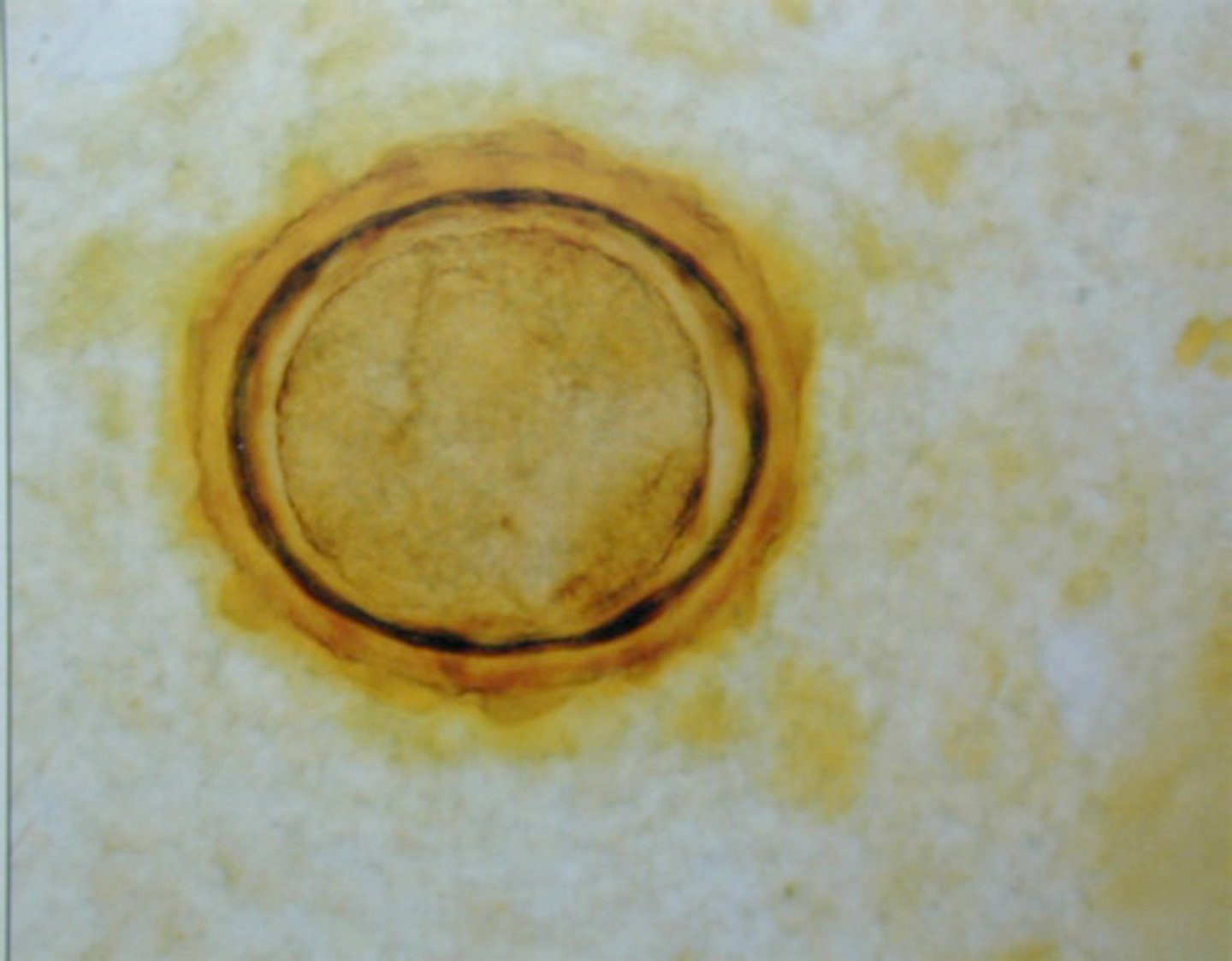
Ova
egg
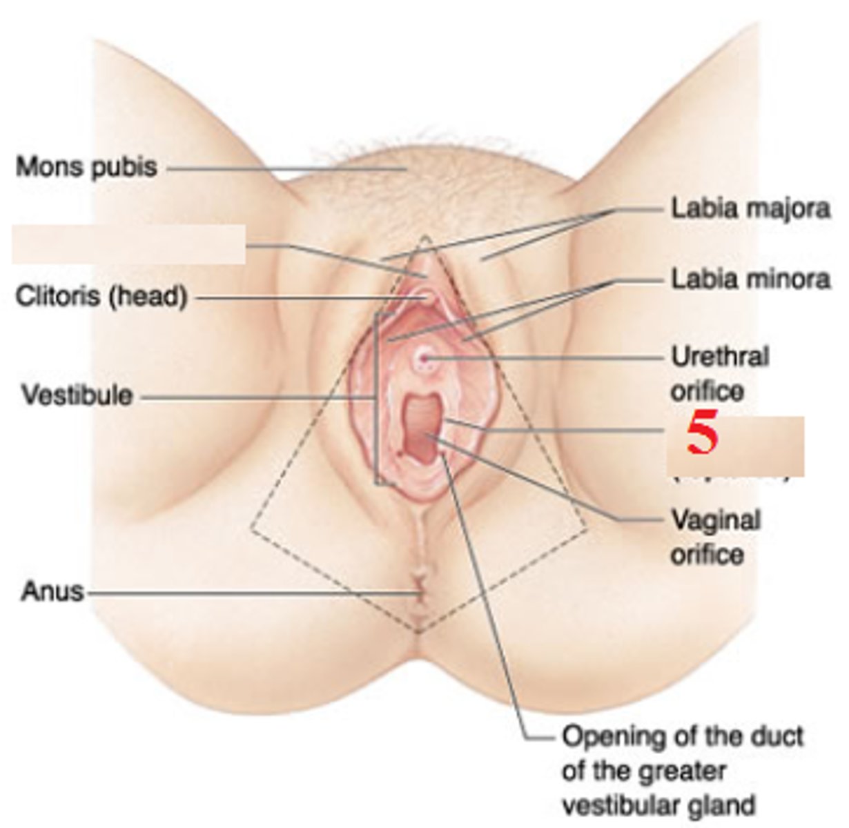
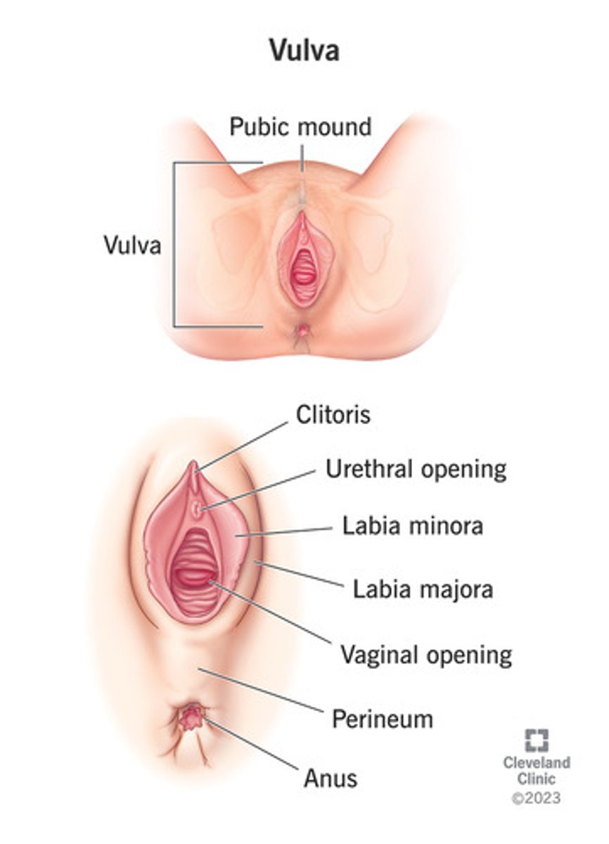
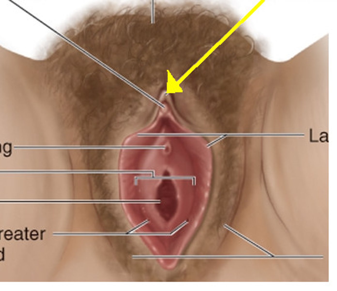
Vulva
external female genitalia
Labia majora
the larger outer folds of the vulva.

Labia minora
Smaller pair of skin folds that protect the vaginal opening
vestibule of vagina
structure located between the labia minora that houses the urethral orifice and the vaginal orifice
Prepuce
Vaginal hood over clitoris (foreskin in males)

Clitoris
organ of sensitive erectile tissue anterior to the opening of the female urethra
Mons pubis
a mound of fatty tissue covering the pubic area in women
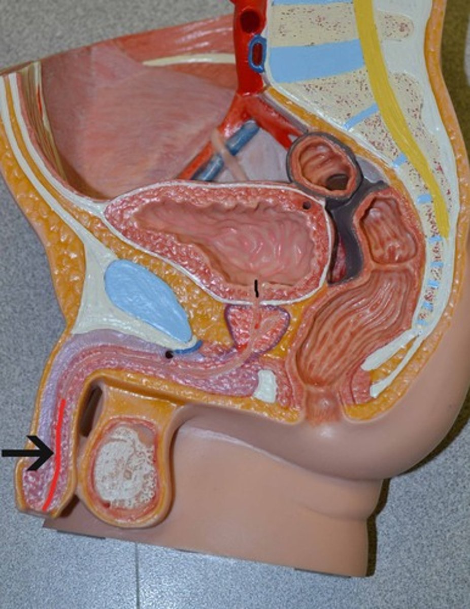
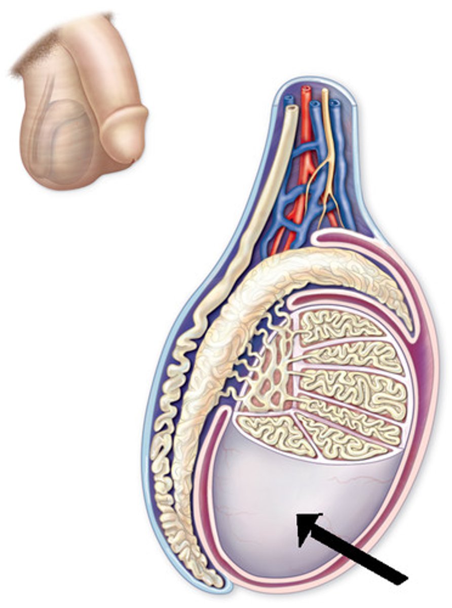
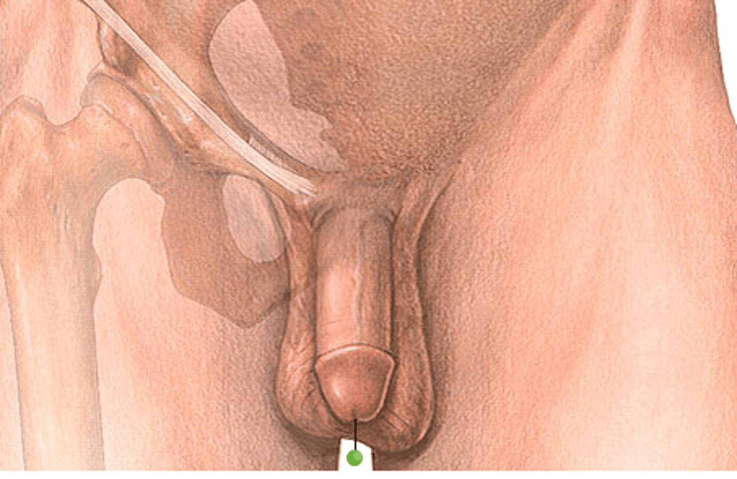
Glans (penis)
the conical mass of erectile tissue that forms the head of the penis
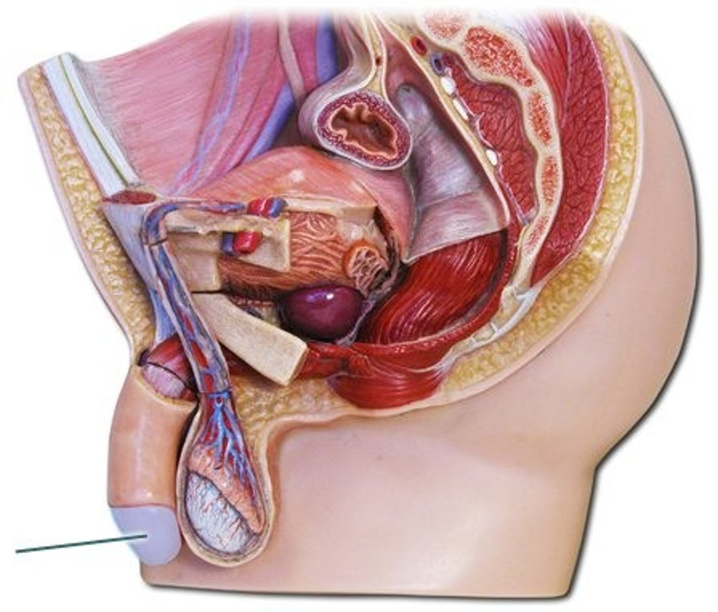
Shaft
Body of penis
Prepuce/foreskin
layer of skin that covers the glans penis in uncircumcised males
urethra meatus
external opening of the urethra
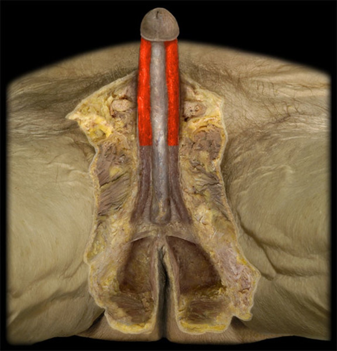
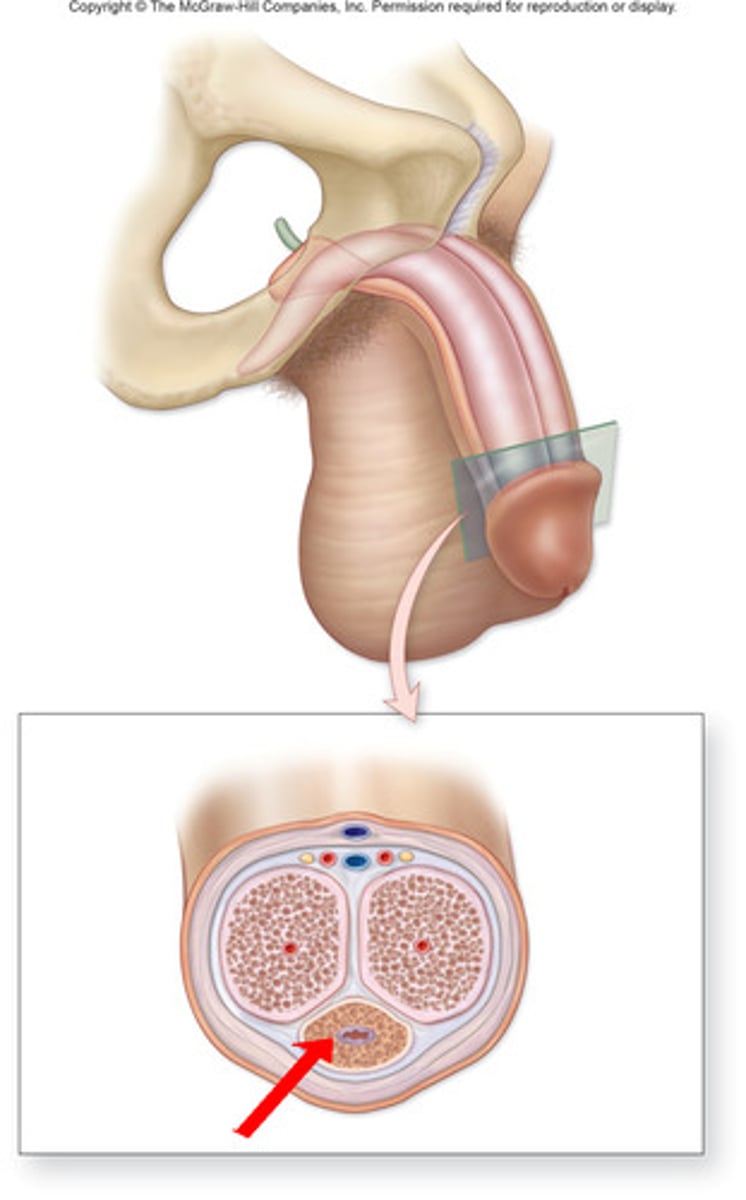
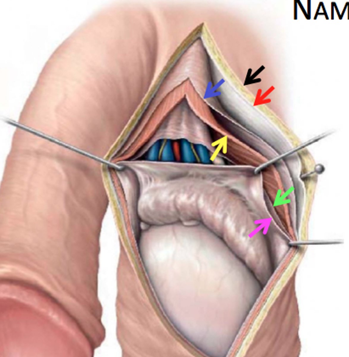
Corpus cavernosum
Pair of spongy tissue regions full of blood during erection
Corpus spongiosum
surrounds the urethra
Tunica dartos
the structure that separates the scrotum into two separate compartments internally
cremaster muscle
Muscle that pulls the scrotum closer to the body in cold temperatures and relaxes to let the testicles be farther away from the body in warmer weather
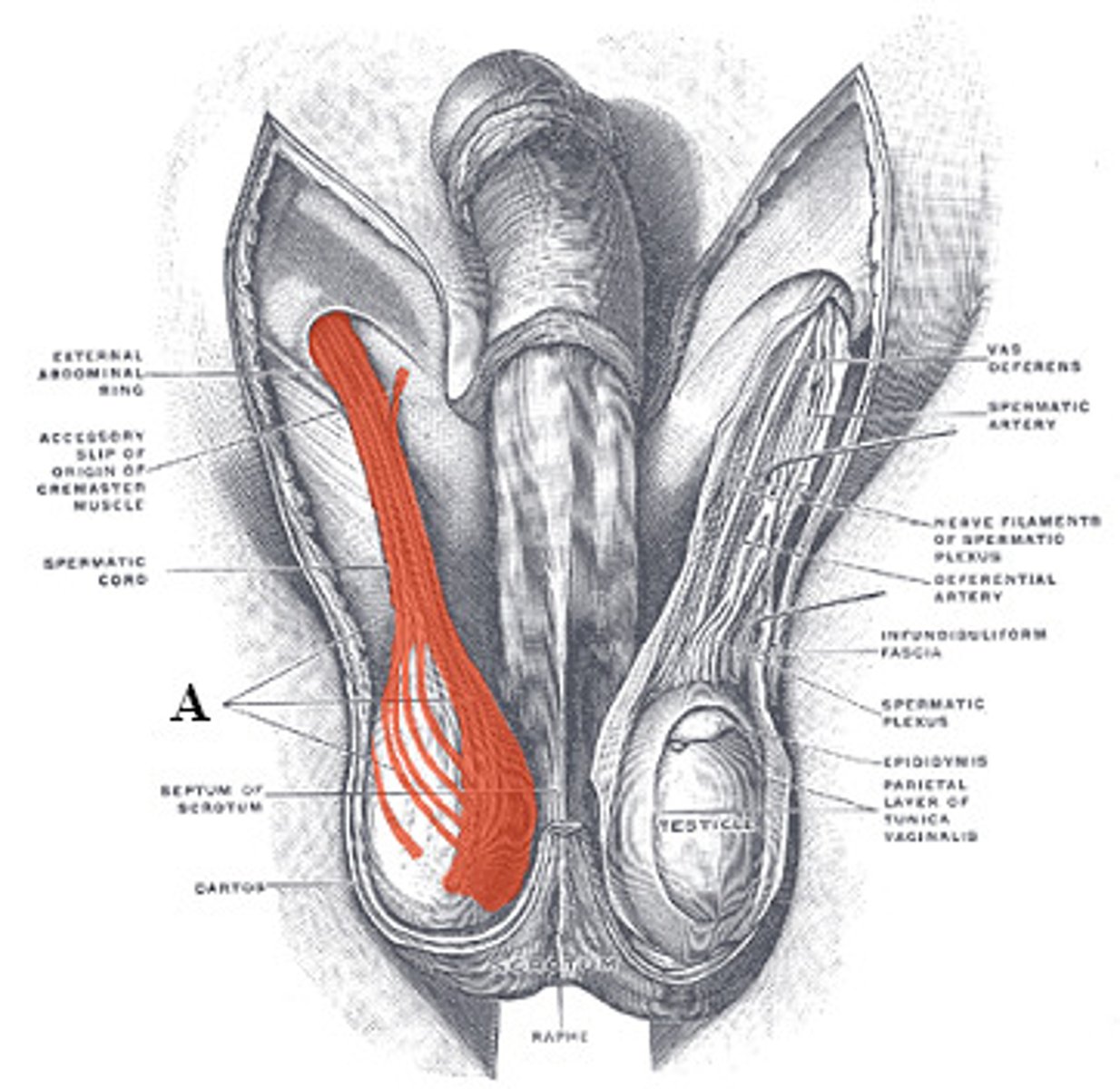
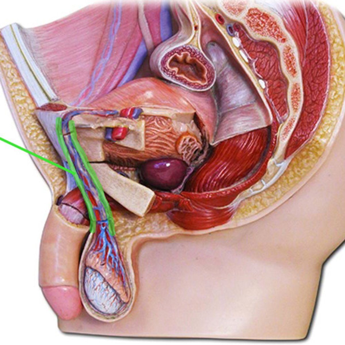
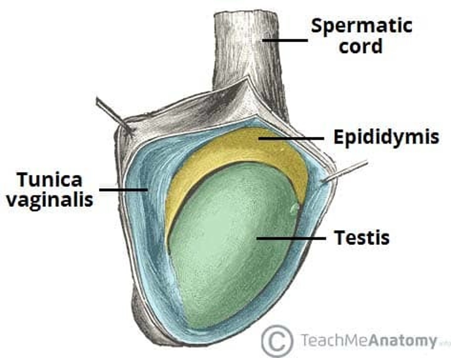
Spermatic cord
extends upward from the epididymis and is attached to each testicle
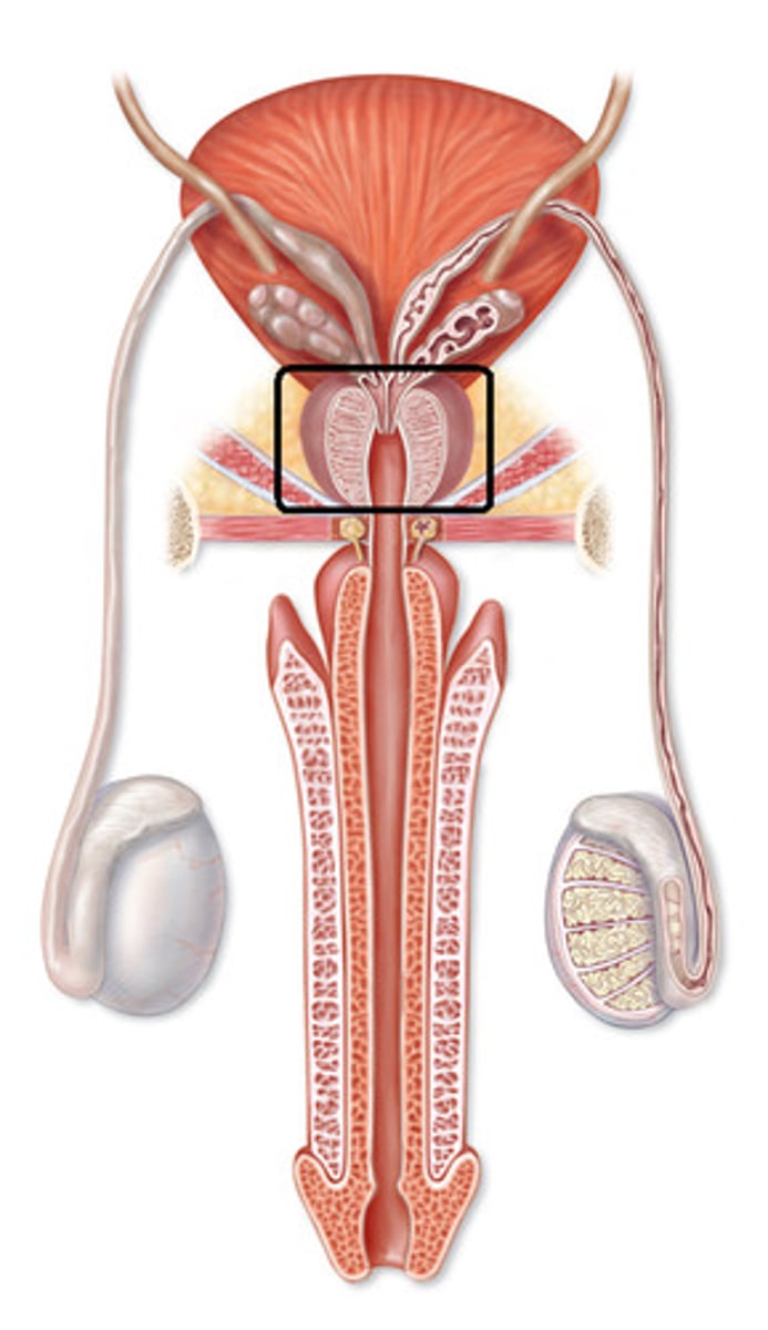
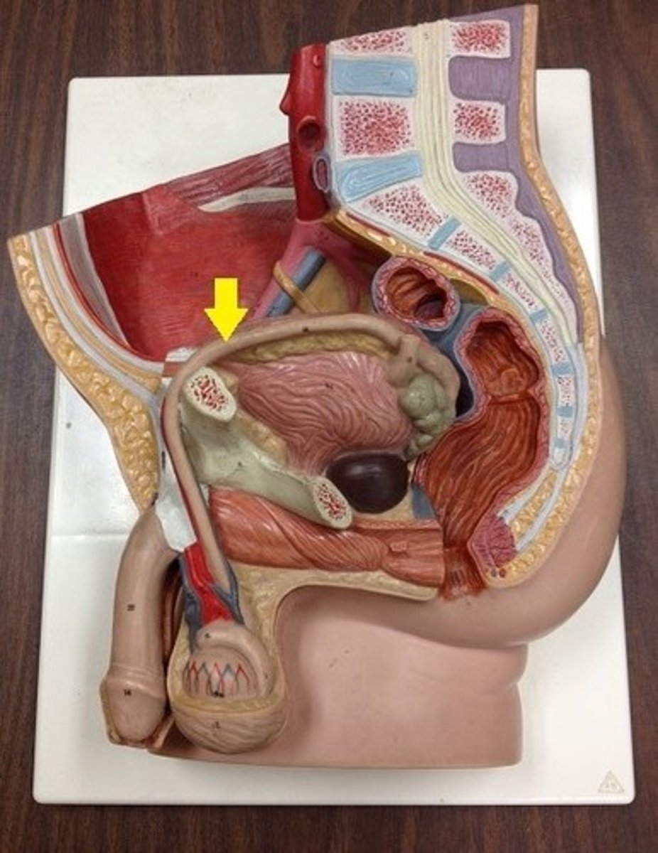
Prostate
a gland surrounding the neck of the bladder in male mammals and releasing prostatic fluid.
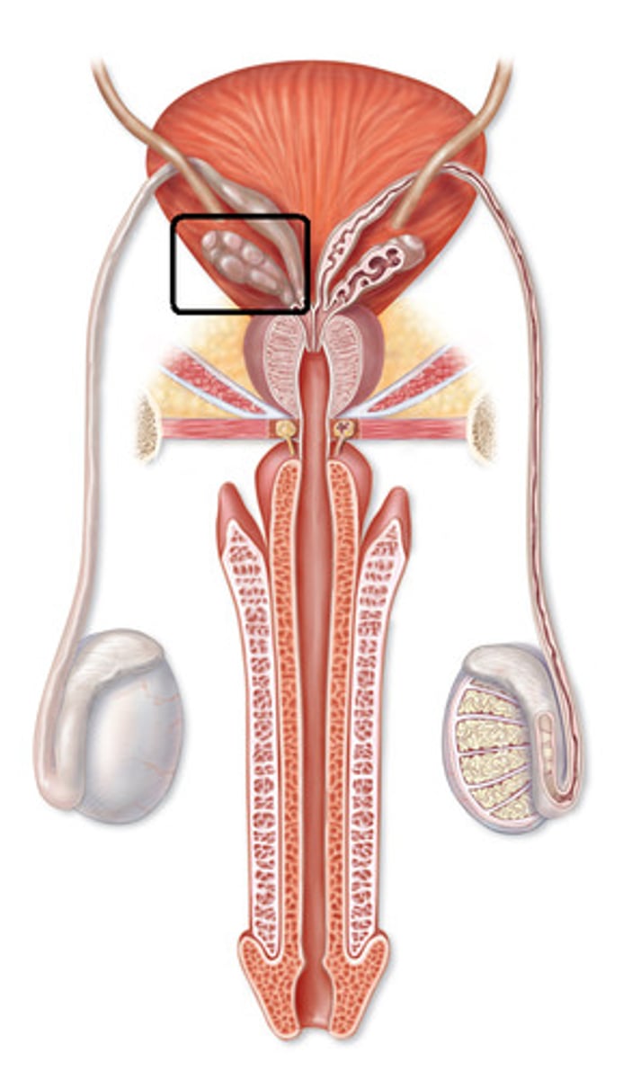
Bulburethral gland
one of a pair of exocrine glands near the male urethra
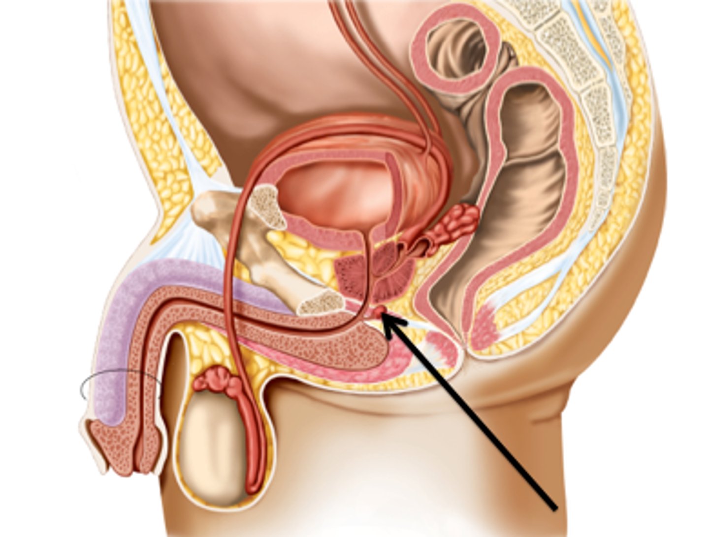
Seminal vesicles
two small glands that secrete a fluid rich in sugar that nourishes and helps sperm move
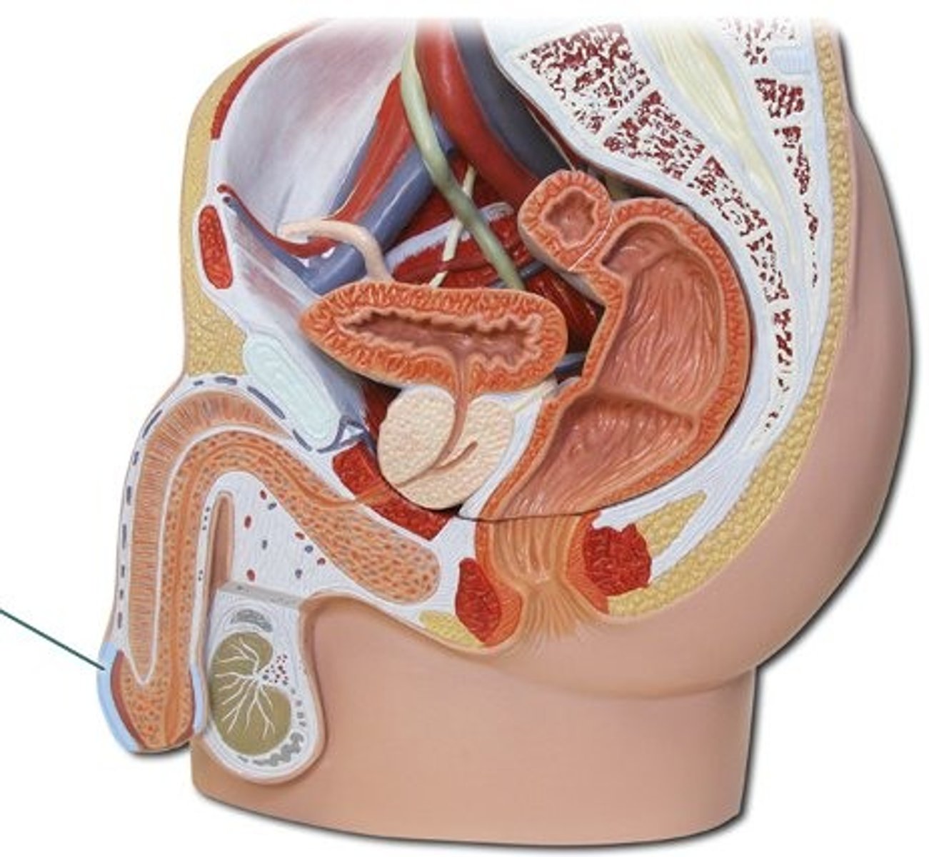
Tunica vaginalis
outer layer derived from peritoneum
Tunica albuginea
white fibrous capsule on testes

Septa of testis
multiple septa formed from the tunica albuginea that course toward the mediastinum testis and separate the testicle into lobules
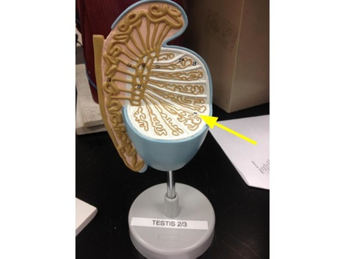
seminiferous tubules
Narrow, coiled tubules that produce sperm in the testes.
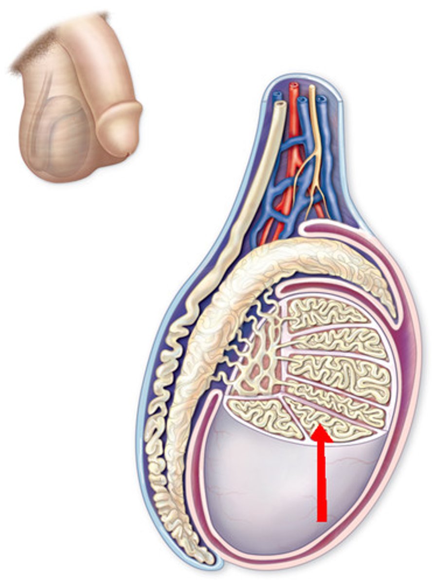
Rete testis
network of tubules between the seminiferous tubules and the epididymis
Vas deferens
tube that carries sperm from the epididymis to the urethra
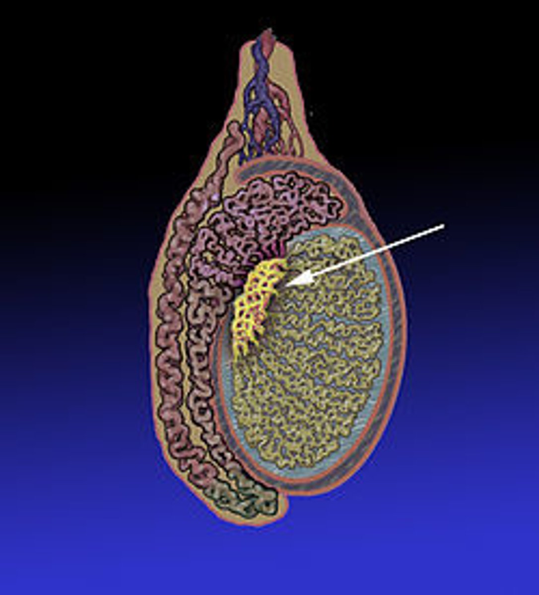
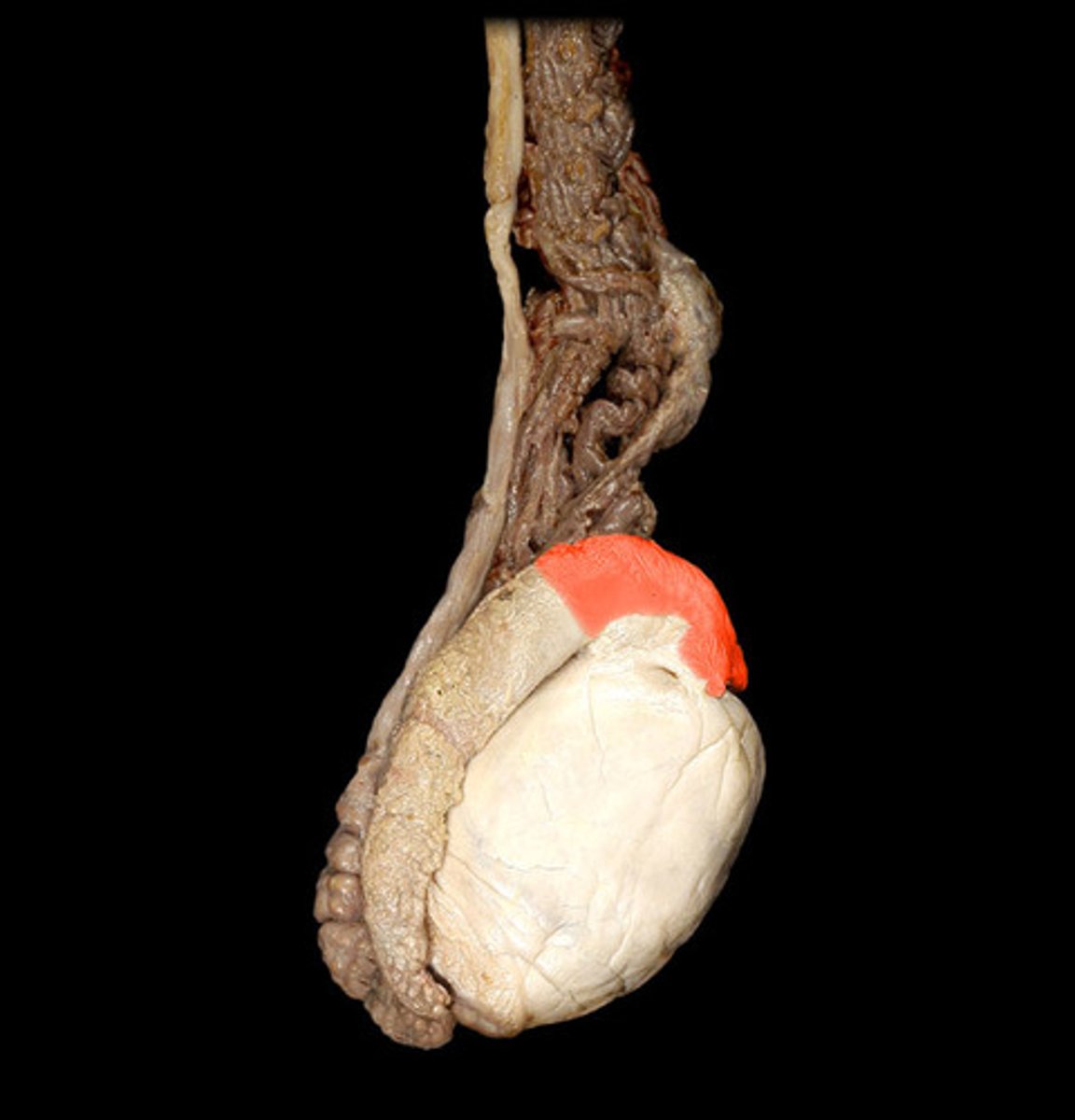
Epididymis
A long, coiled duct on the outside of the testis in which sperm mature.
epididymis head
contains efferent ductules and is located on superior aspect of testis
epididymis body
From last efferent ductule to posterior margin of testis
epididymis tail
-Begins near inferior border of testis where number of coils decreases
-Re-curves and ascends to connection with ductus deferens
-Primary storage location of spermatozoa