Week 2 - Special Senses - Hearing

0.0(0)
Studied by 0 people
call kaiCall Kai
learnLearn
examPractice Test
spaced repetitionSpaced Repetition
heart puzzleMatch
flashcardsFlashcards
GameKnowt Play
Card Sorting

1/17

encourage image

There's no tags or description

Looks like no tags are added yet.

Last updated 8:36 AM on 4/15/26
Name
Mastery
Learn
Test
Matching
Spaced
Call with Kai

No analytics yet

Send a link to your students to track their progress

18 Terms

1
New cards

Special Senses Breakdown

All special senses have corresponding ORGANS:

  • Organspecial sensestimulineural sensation

  • eyevisionlightcolour

  • Ear & cochleahearingsound wavespitch

  • Vestibular apparatusbalancehead movementmotion

  • Noes & olfactory epitheliumsmellairborne chemicalssmell

  • Tongue & taste budsgustationtastantsflavour

2
New cards

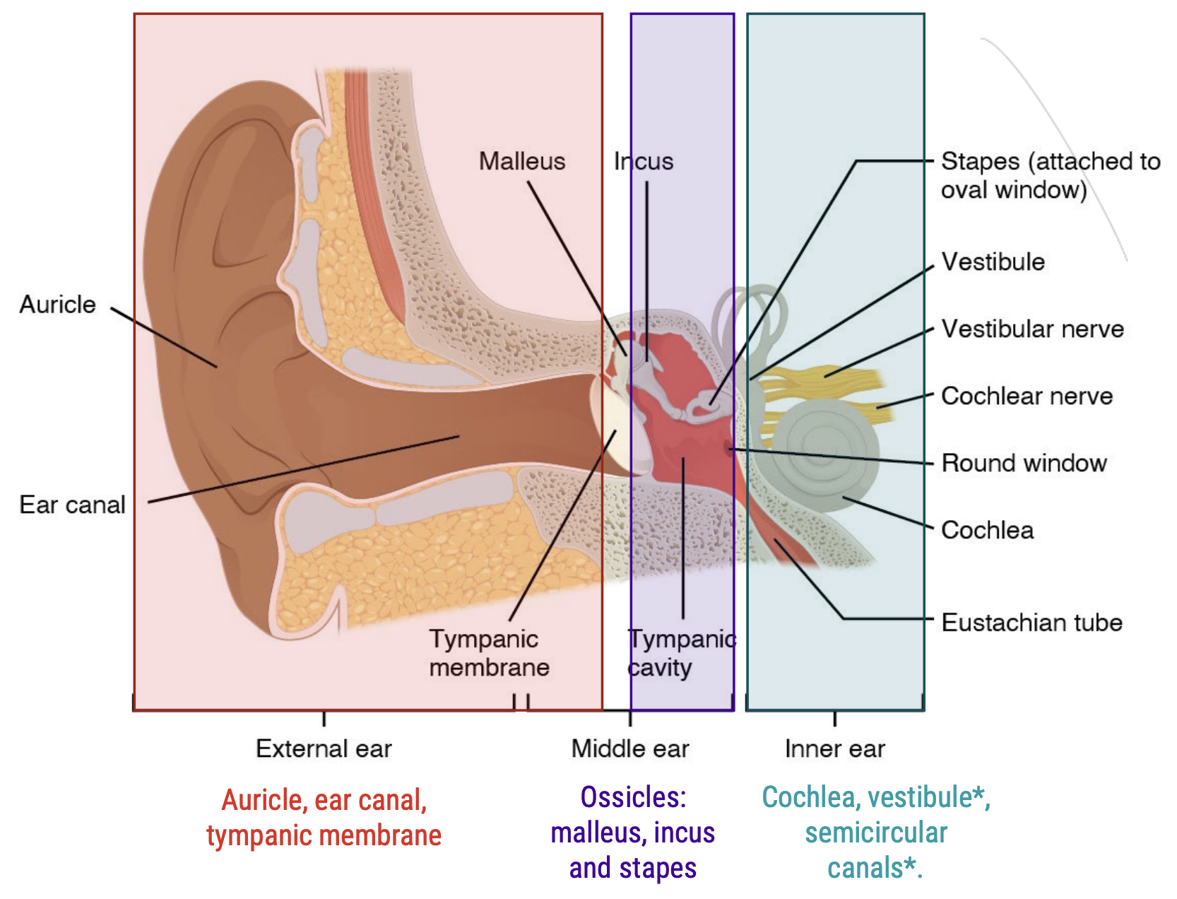
Hearing: Ear Anatomy

  • Large, fleshy external structure; auricle

  • Internal structures are protected by the skull (temporal bone)

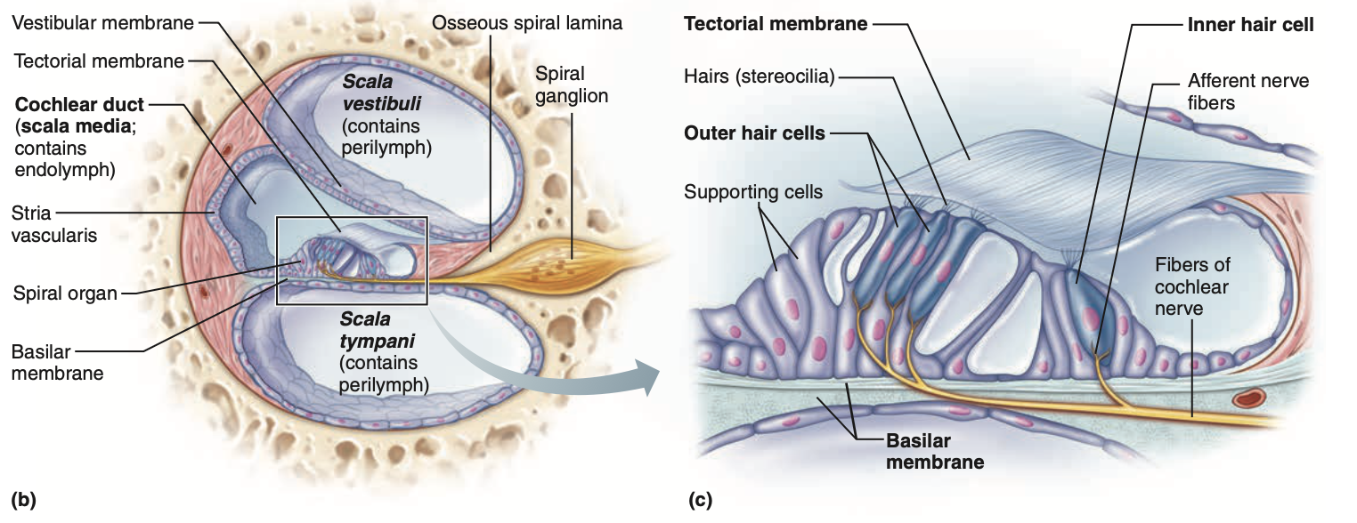
    • Cochlea is also a bony shell, called the osseous spiral lamina

      • Protects the scala vestibuli, scala media (cochlear duct) & scala tympani

3
New cards

Hearing: Ear Function

  1. Auricle catches soundwaves and funnels them into the ear canal

  2. Sound passes down ear canal.

  3. Soundwaves hit the eardrum/timpanic membrane, at the end of the ear canal.

  4. Eardrum separates outer & middle/inner ear

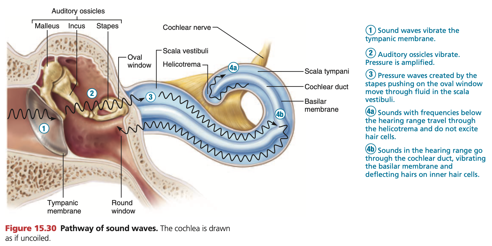
  5. Middle ear bones: Malleus, Incus & Stapes vibrate due the waves in that order, amplifying the soundwaves as they travel to the cochlea

<ol><li><p>Auricle catches soundwaves and funnels them into the ear canal</p></li><li><p>Sound passes down ear canal.</p></li><li><p>Soundwaves hit the eardrum/timpanic membrane, at the end of the ear canal.</p></li><li><p>Eardrum separates outer &amp; middle/inner ear</p></li><li><p>Middle ear bones: Malleus, Incus &amp; Stapes vibrate due the waves in that order, amplifying the soundwaves as they travel to the cochlea</p></li></ol><p></p>
4
New cards

Hearing: Sensation

  1. Middle ear bones: Malleus, Incus & Stapes vibrate due the waves, amplifying the soundwaves. The last bone, stapes hits the fleshy oval window, which propagates vibrations into the cochlea

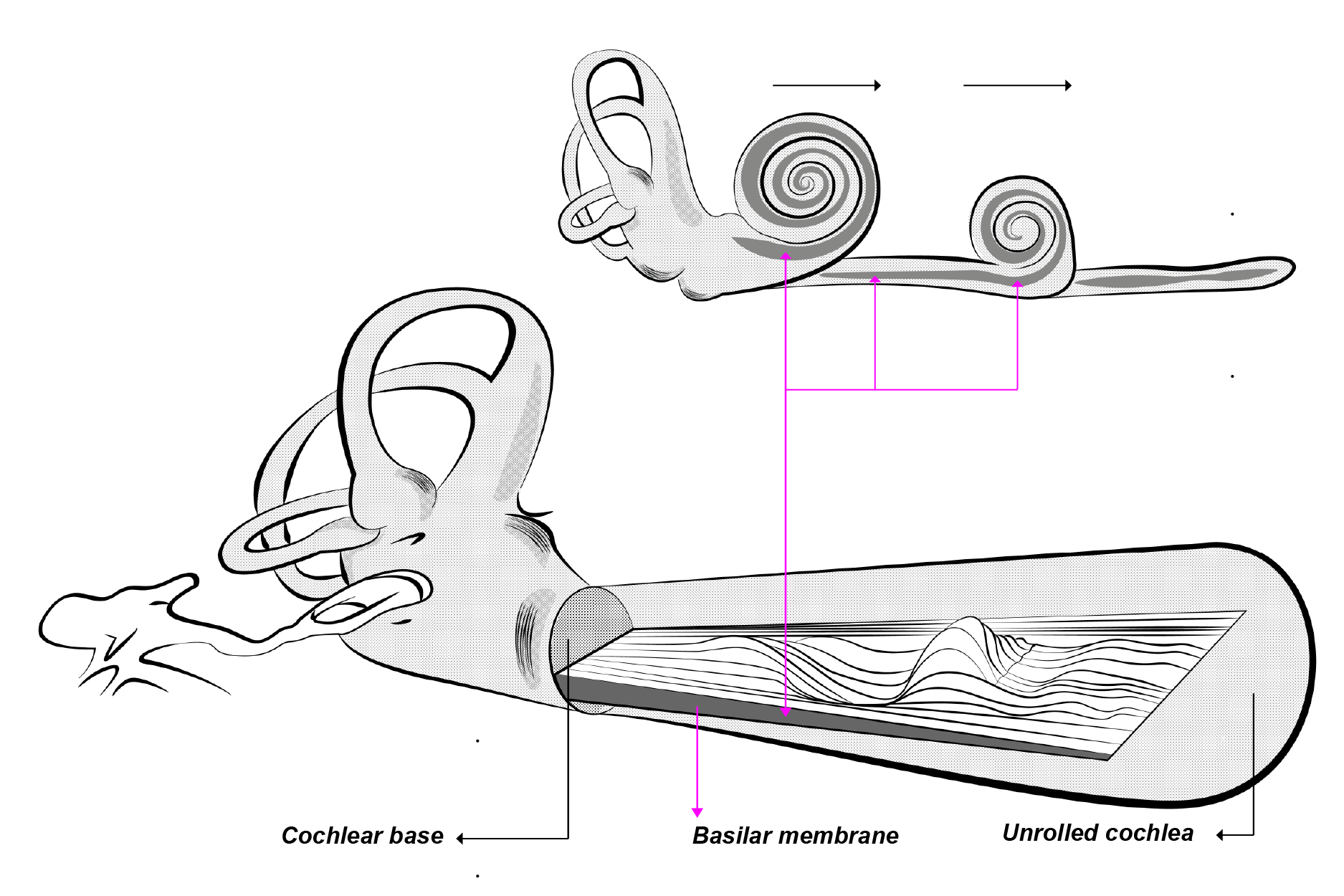
  2. Vibrations travel up the cochlea’s spiral through the scala vestibuli, towards the helicotrema (spiral apex), and back down the spiral through the scala tympani

  3. This order of movement causes the middle space, the cochlear duct (scala media), to move up and down, stimulating mechanoreceptors inside called ‘hair cells’.

  4. Hair cells have branches of the cochlear nerve attached to their bases (part of Vestibulocochlear nerve). Movement of hair cells stimulate this cochlear nerve.

<ol start="6"><li><p>Middle ear bones: <mark data-color="yellow" style="background-color: yellow; color: inherit;">Malleus, Incus &amp; Stapes</mark> vibrate due the waves, <strong>amplifying the soundwaves.</strong> The last bone, <mark data-color="yellow" style="background-color: yellow; color: inherit;">stapes</mark> hits the fleshy <strong><mark data-color="yellow" style="background-color: yellow; color: inherit;">oval window</mark></strong>, which propagates vibrations into the cochlea</p></li><li><p>Vibrations travel up the cochlea’s spiral through the <strong>scala vestibuli</strong>, towards the <strong>helicotrema</strong> (spiral apex), and back down the spiral through the <strong>scala tympani</strong></p></li><li><p>This order of movement causes the middle space, the <strong>cochlear duct (scala media)</strong>, to move up and down, stimulating <mark data-color="red" style="background-color: red; color: inherit;">mechanoreceptors</mark> inside called <mark data-color="red" style="background-color: red; color: inherit;">‘hair cells’</mark>.</p></li><li><p><mark data-color="red" style="background-color: red; color: inherit;">Hair cells</mark> have branches of the cochlear nerve attached to their bases (part of <mark data-color="blue" style="background-color: blue; color: inherit;">Vestibulo</mark><u><mark data-color="blue" style="background-color: blue; color: inherit;">cochlear</mark></u><mark data-color="blue" style="background-color: blue; color: inherit;"> nerve).</mark> Movement of hair cells stimulate this cochlear nerve.</p></li></ol><p></p>
5
New cards

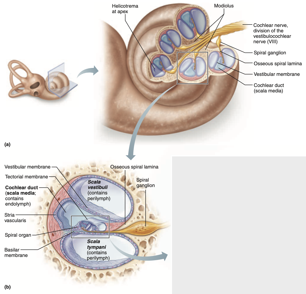
Hearing: Sensation: Cochlear duct (Scala media)

The cochlear duct is located as the middle of three spaces occupying the cochlea (view image), hence the alternate name scala media.

The basilar membrane is located as the border separating the lower scala tympani from the middle cochlear duct.

Within this cochlear duct, can the organ of corti be located (boxed in bottom image).

  • As the vibrations move the fluid within the cochlea, the basilar membrane in the cochlear duct moves, and the organ of corti is stimulated

<p>The <strong><mark data-color="yellow" style="background-color: yellow; color: inherit;">cochlear duct</mark></strong> is located as the middle of three spaces occupying the cochlea (view image), hence the alternate name <strong>scala media</strong>.</p><p>The <strong><mark data-color="yellow" style="background-color: yellow; color: inherit;">basilar membrane</mark></strong> is located as the border separating the lower <strong>scala tympani</strong> from the middle <strong>cochlear duct</strong>.</p><p>Within this cochlear duct, can the <strong><mark data-color="yellow" style="background-color: yellow; color: inherit;">organ of corti</mark></strong> be located (boxed in bottom image).</p><ul><li><p>As the vibrations move the fluid within the cochlea, the <strong>basilar membrane</strong> in the <strong>cochlear duct </strong>moves, and the<strong> organ of corti </strong>is stimulated</p></li></ul><p></p>
6
New cards

Hearing: Sensation: Organ of corti

The organ of corti is located at the bottom of the cochlear duct. With the basilar membrane bordering the bottom of it.

Connected above the basilar membrane are mehanoreceptors, called “hair cells”.

Connected to the base of these hair cells are fibres of the cochlear nerve running through the basilar membrane.

Directly above the hair cells is the tectorial membrane.

  • The ‘hairs’ of some hair cells can be connected to the tectorial membrane

  1. Vibrations travelling through the fluid in the scala vestibuli & scala tympani pushes and moves the basilar membrane up and down.

  2. Pushes hair cells up, and the hairs are smushed into the tectorial membrane

  3. As the hair cells are mechanoreceptors, the physical deformations in their cell membranes activate then, sending graded receptor potentials which can become action potentials down the cochlear nerve

  • Transduces physical sensation (movement of hair cells from vibrations) into electrical signals

<p>The <strong><mark data-color="yellow" style="background-color: yellow; color: inherit;">organ of corti</mark></strong> is located at the bottom of the <strong>cochlear duct</strong>. With the <strong><mark data-color="yellow" style="background-color: yellow; color: inherit;">basilar membrane</mark></strong> bordering the bottom of it.</p><p>Connected above the <strong>basilar membrane</strong> are <strong><mark data-color="purple" style="background-color: purple; color: inherit;">mehanoreceptors</mark></strong>, called “<strong><mark data-color="purple" style="background-color: purple; color: inherit;">hair cells</mark></strong>”.</p><p>Connected to the base of these <strong><mark data-color="purple" style="background-color: purple; color: inherit;">hair cells</mark></strong> are <strong><mark data-color="purple" style="background-color: purple; color: inherit;">fibres of the cochlear nerve</mark></strong> running through the basilar membrane.</p><p>Directly above the hair cells is the <strong><mark data-color="yellow" style="background-color: yellow; color: inherit;">tectorial membrane</mark>.</strong></p><ul><li><p>The ‘hairs’ of some hair cells can be connected to the <strong>tectorial membrane</strong></p></li></ul><p></p><ol><li><p>Vibrations travelling through the fluid in the <strong><mark data-color="yellow" style="background-color: yellow; color: inherit;">scala vestibuli</mark> </strong>&amp;<strong> <mark data-color="yellow" style="background-color: yellow; color: inherit;">scala tympani</mark></strong> pushes and moves the basilar membrane up and down.</p></li><li><p>Pushes <strong><mark data-color="purple" style="background-color: purple; color: inherit;">hair cells</mark></strong> up, and the hairs are smushed into the <strong><mark data-color="yellow" style="background-color: yellow; color: inherit;">tectorial membrane</mark></strong></p></li><li><p>As the hair cells are <strong><mark data-color="purple" style="background-color: purple; color: inherit;">mechanoreceptors</mark></strong>, the physical deformations in their cell membranes activate then, sending graded receptor potentials which can become action potentials down the <strong><mark data-color="purple" style="background-color: purple; color: inherit;">cochlear nerve</mark></strong></p></li></ol><ul><li><p>Transduces physical sensation (movement of hair cells from vibrations) into electrical signals</p></li></ul><p></p>
7
New cards

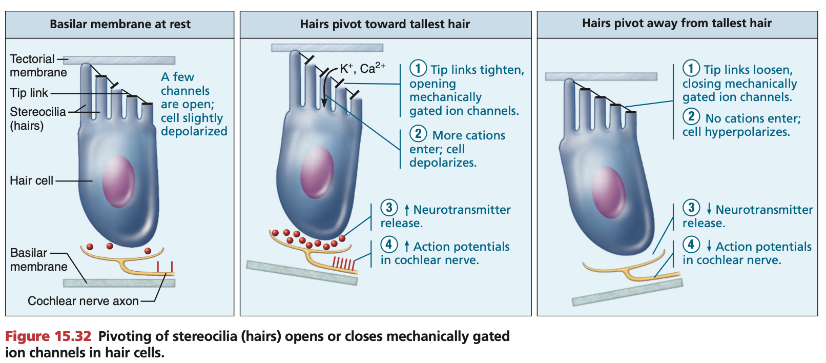
Hearing: Mechanoreceptors: Hair cells

Kinocilium: Tallest hair on each hair cell

Stereocilia: Remaining hairs on each hair cell (includes kinocilium)

Kinocilium and stereocilium are connected at their tips by fibres called “tip links”.

Activation of hair cells:

  1. Due to movement of the basilar membrane, hair cells are pushed up into the tectorial membrane.

  2. Due to the forces when the vibration waves arrive, stereocilia bend towards tallest hair, kinocilium.

  3. Tension forms at the tip links

  4. Tension physically opens K+ and Ca2+ ion-gated channels

  5. Causes rapid depolarisation of cell membranes in sensory neurons (mechanoreceptors; hair cells)

  6. Neurotransmitters released towards afferent cochlear fibres

  7. Action potentials propagate via afferent cochlear fibres towards the brain

    1. Special somatic afferent nerves of the Vestibulocochlear nerve (Cranial nerve 8)

Deactivation of hair cells:

  1. When vibration waves move away, due to the forces stereocilia bend away from the tallest hair, kinocilium

  2. Tip links relax and ion-gated channels remain closed.

  3. No neurotransmitters released.

Sound waves can increase or decrease cochlear nerve activity:

Tectorial membrane leaning towards kinocilium, more action potentials.

Tectorial membrane moving away from kinocilium, less action potentials.

  • Diagram: cochlear axon, NOT axon terminal

<p><strong><mark data-color="purple" style="background-color: purple; color: inherit;">Kinocilium</mark>:</strong> Tallest hair on each hair cell</p><p><strong><mark data-color="purple" style="background-color: purple; color: inherit;">Stereocilia</mark>:</strong> Remaining hairs on each hair cell (includes kinocilium)</p><p>Kinocilium and stereocilium are connected at their tips by fibres called <strong><mark data-color="purple" style="background-color: purple; color: inherit;">“tip links”</mark></strong>.</p><p><strong><em><u>Activation of hair cells:</u></em></strong></p><ol><li><p>Due to movement of the <strong><mark data-color="yellow" style="background-color: yellow; color: inherit;">basilar membrane</mark></strong>, <strong><mark data-color="purple" style="background-color: purple; color: inherit;">hair cells</mark> </strong>are pushed up into the <strong><mark data-color="yellow" style="background-color: yellow; color: inherit;">tectorial membrane</mark></strong>.</p></li><li><p>Due to the forces when the vibration waves arrive, <mark data-color="purple" style="background-color: purple; color: inherit;">stereocilia</mark> <em>bend towards</em> tallest hair, <strong><mark data-color="purple" style="background-color: purple; color: inherit;">kinocilium</mark></strong>.</p></li><li><p>Tension forms at the <strong><mark data-color="purple" style="background-color: purple; color: inherit;">tip links</mark></strong></p></li><li><p>Tension physically opens <strong><mark data-color="green" style="background-color: green; color: inherit;">K+</mark></strong> and <strong><mark data-color="green" style="background-color: green; color: inherit;">Ca2+ ion-gated channels</mark></strong></p></li><li><p>Causes rapid depolarisation of cell membranes in sensory neurons (mechanoreceptors; hair cells)</p></li><li><p>Neurotransmitters released towards <strong><mark data-color="blue" style="background-color: blue; color: inherit;">afferent cochlear fibres</mark></strong></p></li><li><p>Action potentials propagate via <strong><mark data-color="blue" style="background-color: blue; color: inherit;">afferent cochlear fibres</mark></strong> towards the brain</p><ol><li><p><strong><mark data-color="blue" style="background-color: blue; color: inherit;">Special </mark><mark data-color="purple" style="background-color: purple; color: inherit;">somatic</mark><mark data-color="blue" style="background-color: blue; color: inherit;"> afferent</mark></strong><mark data-color="blue" style="background-color: blue; color: inherit;"> nerves of the&nbsp;Vestibulo</mark><strong><u><mark data-color="blue" style="background-color: blue; color: inherit;">cochlear</mark></u><mark data-color="blue" style="background-color: blue; color: inherit;"> nerve (Cranial nerve 8)</mark></strong></p></li></ol></li></ol><p></p><p><strong><em><u>Deactivation of hair cells:</u></em></strong></p><ol><li><p>When vibration waves move away, due to the forces <mark data-color="purple" style="background-color: purple; color: inherit;">stereocilia</mark> <em>bend away from the</em> tallest hair, <strong><mark data-color="purple" style="background-color: purple; color: inherit;">kinocilium</mark></strong></p></li><li><p>Tip links relax and ion-gated channels remain closed.</p></li><li><p>No neurotransmitters released.</p></li></ol><p></p><p>Sound waves can increase or decrease cochlear nerve activity:</p><p>Tectorial membrane leaning <strong><em><u>towards</u></em></strong> kinocilium, <strong>more action potentials</strong>.</p><p>Tectorial membrane moving <strong><em><u>away</u></em></strong> from kinocilium, <strong>less action potentials</strong>.</p><ul><li><p><strong><em><u><mark data-color="blue" style="background-color: blue; color: inherit;">Diagram:</mark></u></em></strong><mark data-color="blue" style="background-color: blue; color: inherit;"> cochlear </mark><strong><mark data-color="blue" style="background-color: blue; color: inherit;">axon</mark></strong><mark data-color="blue" style="background-color: blue; color: inherit;">, NOT </mark><strong><mark data-color="blue" style="background-color: blue; color: inherit;">axon terminal</mark></strong></p></li></ul><p></p>
8
New cards

Hearing: Sound

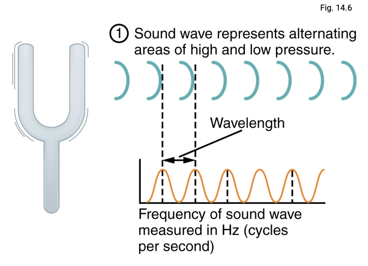
Sound is a pressure disturbance, alternating areas of high and low pressure of molecules travelling through air or water.

Peaks: high pressure (compressed areas)

Troughs: low pressure (rarified areas)

Wavelengths: Distance between wave peaks

<p>Sound is a <strong>pressure disturbance</strong>, alternating areas of high and low pressure of molecules travelling through air or water.</p><p><strong>Peaks:</strong> high pressure (compressed areas)</p><p><strong>Troughs:</strong> low pressure (rarified areas)</p><p><strong><em><u><mark data-color="red" style="background-color: red; color: inherit;">Wavelengths:</mark></u></em></strong> Distance between wave peaks</p>
9
New cards

Hearing: Sound Frequency: Sensitivity

Humans can sense from 2,000 to 20,000 frequencies (measured in hertz).

  • more sensitive to frequencies between 1,500 to 4,000 hertz

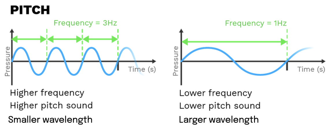
Frequency: Number of soundwaves/wavelengths propagated in a certain period of time

  • More frequency, lower wavelength, vice versa

  • Frequency determines pitch

  • Smaller wavelengths → Higher frequency → higher pitch

  • Larger wavelengths → Lower frequency → lower pitch

<p>Humans can sense from 2,000 to 20,000 <strong>frequencies</strong> (measured in <strong>hertz</strong>).</p><ul><li><p>more sensitive to frequencies between <strong><em><u>1,500 to 4,000 hertz</u></em></strong></p></li></ul><p></p><p><strong><em><u><mark data-color="red" style="background-color: red; color: inherit;">Frequency:</mark></u> </em></strong>Number of soundwaves/wavelengths propagated in a certain period of time</p><ul><li><p><mark data-color="red" style="background-color: red; color: inherit;">More frequency, lower wavelength, vice versa</mark></p></li><li><p><strong><mark data-color="red" style="background-color: red; color: inherit;">Frequency</mark></strong><mark data-color="red" style="background-color: red; color: inherit;"> determines </mark><strong><mark data-color="red" style="background-color: red; color: inherit;">pitch</mark></strong></p></li><li><p>Smaller wavelengths → Higher frequency → higher pitch </p></li><li><p>Larger wavelengths → Lower frequency → lower pitch</p></li></ul><p></p>
10
New cards

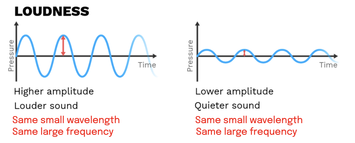
Hearing: Sound intensity/loudness

The loudness of a sound is proportional to its amplitude in soundwaves.

Higher amplitude: louder

Lower amplitude: quieter

  • Frequency/pitch/wavelengths are irrelevant

  • However, usually lower amplitudes (quieter) are associated with deeper sounds with low pitches (e.g tummy grumbling)

  • Contrarily, higher amplitudes (louder) are associated with sharper sounds with high pitches (e.g mouse squeak)

<p>The loudness of a sound is proportional to its <strong><mark data-color="red" style="background-color: red; color: inherit;">amplitude</mark></strong> in soundwaves.</p><p>Higher amplitude: louder</p><p>Lower amplitude: quieter</p><ul><li><p>Frequency/pitch/wavelengths are irrelevant</p></li><li><p>However, usually lower amplitudes (quieter) are associated with deeper sounds with low pitches (e.g tummy grumbling)</p></li><li><p>Contrarily, higher amplitudes (louder) are associated with sharper sounds with high pitches (e.g mouse squeak)</p></li></ul><p></p>
11
New cards
<p><mark data-color="yellow" style="background-color: yellow; color: inherit;">Hearing:</mark> Cochlea: Basillar membrane - <strong><u><mark data-color="purple" style="background-color: purple; color: inherit;">Tonotopically organised</mark></u></strong></p>

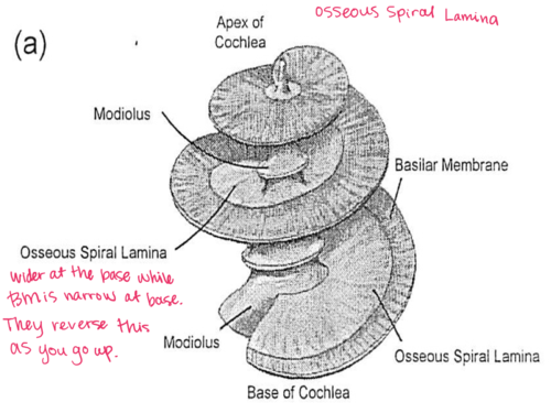
Hearing: Cochlea: Basillar membrane - Tonotopically organised

The basilar membrane is a horizontal lining of fibres extending down towards the cochlear apex. Its structure changes as it moves towards the cochlear apex and is called tonotopical organisation.

Base of cochlea:

  • Shorter fibres, more taut & rigid

  • Thicker fibres

  • Less flexible

    • Requires more energy and force to be moved by cochlear fluid

    • Vibrates at shorter wavelengths, high frequencies, higher pitch, higher amplitude

Apex of cochlea:

  • Longer fibres, more floppy & malleable

  • Thinner fibres

  • More flexible

    • Requires less energy and force to be moved by cochlear fluid

    • Vibrates at lower wavelengths, lower frequencies, lower pitch, lower amplitude

<p>The <strong>basilar membrane</strong> is a horizontal lining of fibres extending down towards the <strong>cochlear apex</strong>. Its <em>structure changes</em> as it moves towards the cochlear apex and is called <strong><mark data-color="purple" style="background-color: purple; color: inherit;">tonotopical organisation</mark></strong>.</p><p></p><p><strong><em><u>Base of cochlea:</u></em></strong></p><ul><li><p>Shorter fibres, more taut &amp; rigid</p></li><li><p>Thicker fibres</p></li><li><p><mark data-color="yellow" style="background-color: yellow; color: inherit;">Less flexible</mark></p><ul><li><p>Requires <strong><em><u>more energy and force</u></em></strong> to be moved by cochlear fluid</p></li><li><p><mark data-color="red" style="background-color: red; color: inherit;">Vibrates at </mark><strong><mark data-color="red" style="background-color: red; color: inherit;">shorter wavelengths, high frequencies, higher pitch, higher amplitude</mark></strong></p></li></ul></li></ul><p></p><p><strong><em><u>Apex of cochlea:</u></em></strong></p><ul><li><p>Longer fibres, more floppy &amp; malleable</p></li><li><p>Thinner fibres</p></li><li><p><mark data-color="yellow" style="background-color: yellow; color: inherit;">More flexible</mark></p><ul><li><p>Requires <strong><em><u>less energy and force</u></em></strong> to be moved by cochlear fluid</p></li><li><p><mark data-color="red" style="background-color: red; color: inherit;">Vibrates at </mark><strong><mark data-color="red" style="background-color: red; color: inherit;">lower wavelengths, lower frequencies, lower pitch, lower amplitude</mark></strong></p></li></ul></li></ul><p></p>
12
New cards

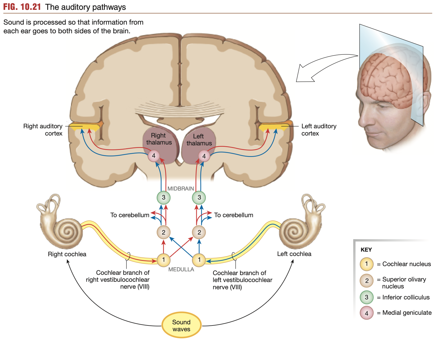
Hearing: Neural pathways

Special somatic afferent nerves of the Vestibulocochlear nerve (Cranial nerve 8)

Where?

  1. Action potentials propagate from hair cells

  2. Travel along the cochlear branch of the vestibulocochlear nerve

  3. Synapse with the cochlear nuclei within the brain stem’s medulla

  4. Information from both ears combines within the cochlear nuclei

  5. Fibres travel up the brain stem, some staying on their respective sides

  6. Fibres reach the temporal lobe, and respect left & right auditory cortices (cortexes)

  7. Hearing is perceived

  8. Auditory info is processed by other sections of the brain to enable interpretation & integration of the image

Beginning: In the inner ear, specifically base of hair cells lining the cochlea

Ending: Primary auditory cortex, temporal lobe

<p><strong><mark data-color="blue" style="background-color: blue; color: inherit;">Special </mark><mark data-color="purple" style="background-color: purple; color: inherit;">somatic</mark><mark data-color="blue" style="background-color: blue; color: inherit;"> afferent</mark></strong><mark data-color="blue" style="background-color: blue; color: inherit;"> nerves of the&nbsp;Vestibulo</mark><strong><u><mark data-color="blue" style="background-color: blue; color: inherit;">cochlear</mark></u><mark data-color="blue" style="background-color: blue; color: inherit;"> nerve (Cranial nerve 8)</mark></strong></p><p><strong><em><u>Where?</u></em></strong></p><ol><li><p>Action potentials propagate from hair cells</p></li><li><p>Travel along the <mark data-color="blue" style="background-color: blue; color: inherit;">cochlear branch</mark> of the vestibulo<strong><u>cochlear</u> </strong>nerve</p></li><li><p>Synapse with the <strong>cochlear nuclei</strong> within the <strong>brain stem’s medulla</strong></p></li><li><p>Information from both ears combines within the <strong>cochlear nuclei</strong></p></li><li><p>Fibres travel up the brain stem, some staying on their respective sides</p></li><li><p>Fibres reach the <strong>temporal lobe</strong>, and respect left &amp; right <strong>auditory cortices</strong> (cortexes)</p></li><li><p>Hearing is perceived</p></li><li><p>Auditory info is processed by other sections of the brain to enable <strong>interpretation</strong> &amp; <strong>integration</strong> of the image</p></li></ol><p><strong><em>Beginning:</em></strong> In the inner ear, specifically base of hair cells lining the cochlea</p><p><strong><em>Ending:</em></strong> <mark data-color="green" style="background-color: green; color: inherit;">Primary auditory cortex, temporal lobe</mark></p>
13
New cards

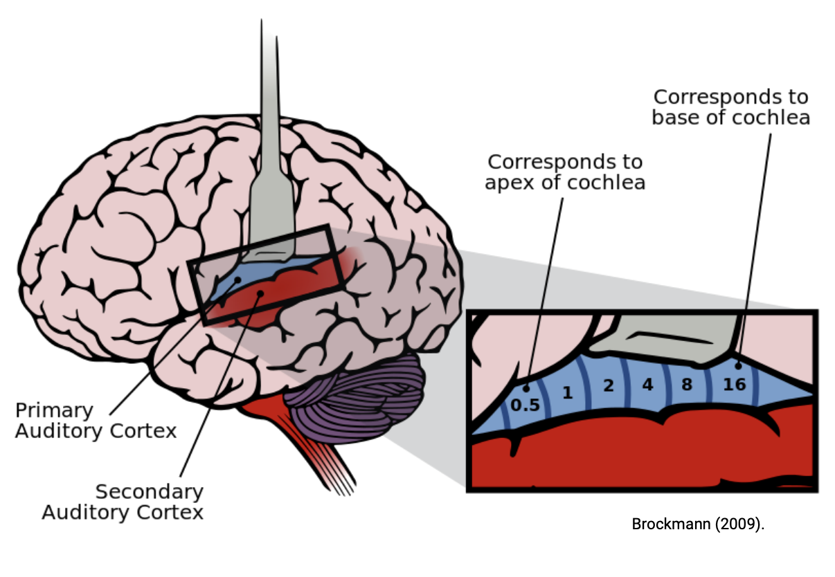
Hearing: Temporal lobe, Primary & secondary auditory cortex map

  • Each segment of the primary auditory cortex responds to impulses arriving from specific nerve fibres. Each section responds to a certain cochlear nerve fibre that reacts to a certain pitch/frequency

  • From anterior to posterior, the primary auditory cortex transduces cochlear sensation from the apex, to the base.

  • Left & right sides respond to information received from both ears

    • Hence, damage to one side of the brain’s temporal lobe (e.g left temporal lobe), will cause hearing deficits in both ears

<ul><li><p>Each segment of the primary auditory cortex responds to impulses arriving from specific nerve fibres. Each section responds to a certain cochlear nerve fibre that reacts to a certain pitch/frequency</p></li><li><p>From anterior to posterior, the primary auditory cortex transduces cochlear sensation from the apex, to the base.</p></li><li><p><mark data-color="yellow" style="background-color: yellow; color: inherit;">Left &amp; right sides respond to information received from </mark><strong><mark data-color="yellow" style="background-color: yellow; color: inherit;">both ears</mark></strong></p><ul><li><p><mark data-color="yellow" style="background-color: yellow; color: inherit;">Hence, damage to one side of the brain’s temporal lobe (e.g left temporal lobe), will cause hearing deficits in </mark><strong><mark data-color="yellow" style="background-color: yellow; color: inherit;">both ears</mark></strong></p></li></ul></li></ul><p></p>
14
New cards

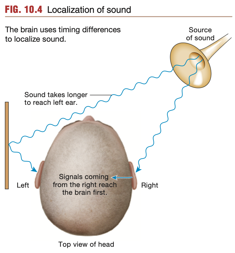
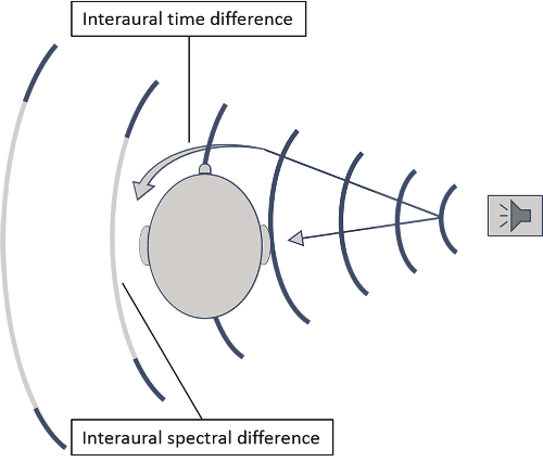
Hearing: Locating sounds: Interaural time difference (Spatial audio)

Unless positioned directly in front of a listener, sounds heard angled away from the listener will be heard microseconds apart between both ears.

  • This time difference is termed the Interaural time difference

  • Helps brain position sound sources

<p>Unless positioned directly in front of a listener, sounds heard <strong>angled</strong> away from the listener will be heard <strong>microseconds apart</strong> between both ears.</p><ul><li><p>This time difference is termed the <u><mark data-color="purple" style="background-color: purple; color: inherit;">Interaural </mark></u><strong><u><mark data-color="purple" style="background-color: purple; color: inherit;">time</mark></u></strong><u><mark data-color="purple" style="background-color: purple; color: inherit;"> difference</mark></u></p></li><li><p>Helps brain position sound sources</p></li></ul><p></p>
15
New cards

Hearing: Locating sounds: Interaural intensity difference (Spatial audio)

Unless positioned directly in front of a listener, sounds heard angled away from the listener will be heard a different intensities between ears, due to the head blocking some soundwaves.

  • This time difference is termed the Interaural intensity difference

  • Helps brain position sound sources

<p>Unless positioned directly in front of a listener, sounds heard <strong>angled</strong> away from the listener will be heard a <strong>different intensities</strong> between ears, due to the <strong>head blocking some soundwaves</strong>.</p><ul><li><p>This time difference is termed the <u><mark data-color="purple" style="background-color: purple; color: inherit;">Interaural </mark></u><strong><u><mark data-color="purple" style="background-color: purple; color: inherit;">intensity</mark></u></strong><u><mark data-color="purple" style="background-color: purple; color: inherit;"> difference</mark></u></p></li><li><p>Helps brain position sound sources</p></li></ul><p></p>
16
New cards

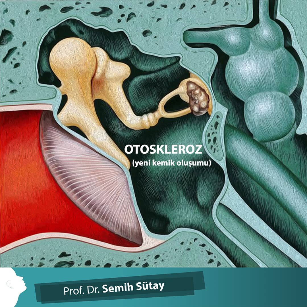
Hearing Disorders: 1 of many: Conductive hearing loss

  • Soundwaves entering the ear canal cannot be transmitted across the eardrum into the inner ear.

Temporary causes: Ear wax buildup, ruptured eardrum

Permanent causes: Otosclerosis - inner ear bones (Maleus, incus & stapes) become malformed and cannot vibrate

  • Stapes fuses to oval window

  • Maleus, incus and stapes become fused in some form, preventing vibration

Conductive hearing loss: Soundwaves cannot be “conducted” to the cochlea

<ul><li><p>Soundwaves entering the ear canal cannot be transmitted across the eardrum into the inner ear.</p></li></ul><p><strong>Temporary causes</strong>: Ear wax buildup, ruptured eardrum</p><p><strong>Permanent causes</strong>: Otosclerosis - inner ear bones (Maleus, incus &amp; stapes) become malformed and cannot vibrate</p><ul><li><p>Stapes fuses to oval window</p></li><li><p>Maleus, incus and stapes become fused in some form, preventing vibration</p></li></ul><p></p><p><strong>Conductive hearing loss: </strong>Soundwaves cannot be “conducted” to the cochlea</p>
17
New cards

Hearing Disorders: 1 of many: Sensorineural hearing loss

Sensorineural hearing loss: Issues with the sensory neurons or neural pathways that cause hearing loss.

  • Damage to the cochlea or neural pathways towards the brain

  • Often permenant

18
New cards

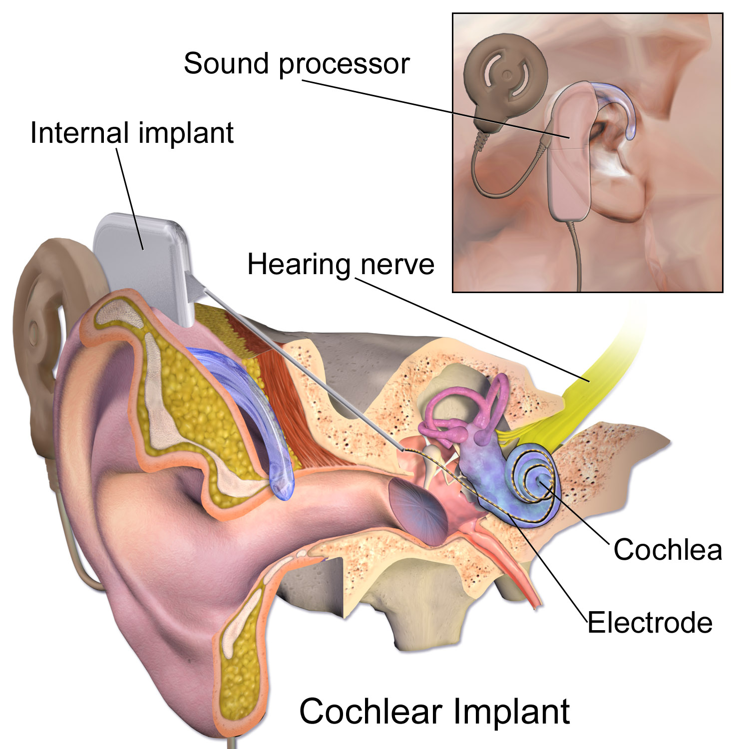
Hearing Disorders: Technology

Hearing aids: Amplify incoming sounds, making soundwaves more conductive and able to pass the ear canal towards the cochlea.

Cochlear implants: Directly stimulate the cochlear nerve via electrical impulses, allowing soundwaves to bypass the cochlea.

  • Used when substantial hearing damage is present, and hearing aids provide no effect.

<p><strong>Hearing aids:</strong> Amplify incoming sounds, making soundwaves more conductive and able to pass the ear canal towards the cochlea.</p><p><strong>Cochlear implants:</strong> Directly stimulate the <em><u>cochlear nerve</u></em> via electrical impulses, allowing soundwaves to <em><u>bypass the cochlea</u></em>. </p><ul><li><p>Used when substantial hearing damage is present, and hearing aids provide no effect.</p></li></ul><p></p>