Test 3
1/220
There's no tags or description
Looks like no tags are added yet.
Name | Mastery | Learn | Test | Matching | Spaced | Call with Kai |
|---|
No analytics yet
Send a link to your students to track their progress
221 Terms
optic nerve
neurons/nerve bundles closest to retina
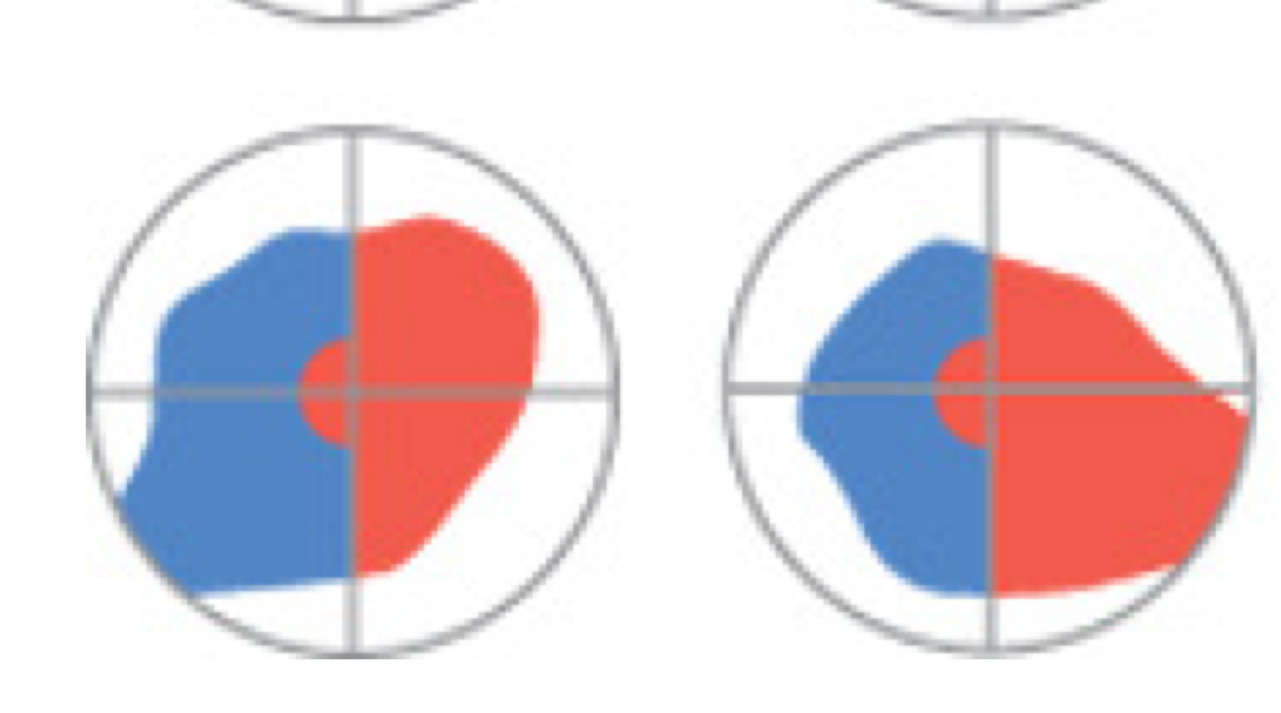
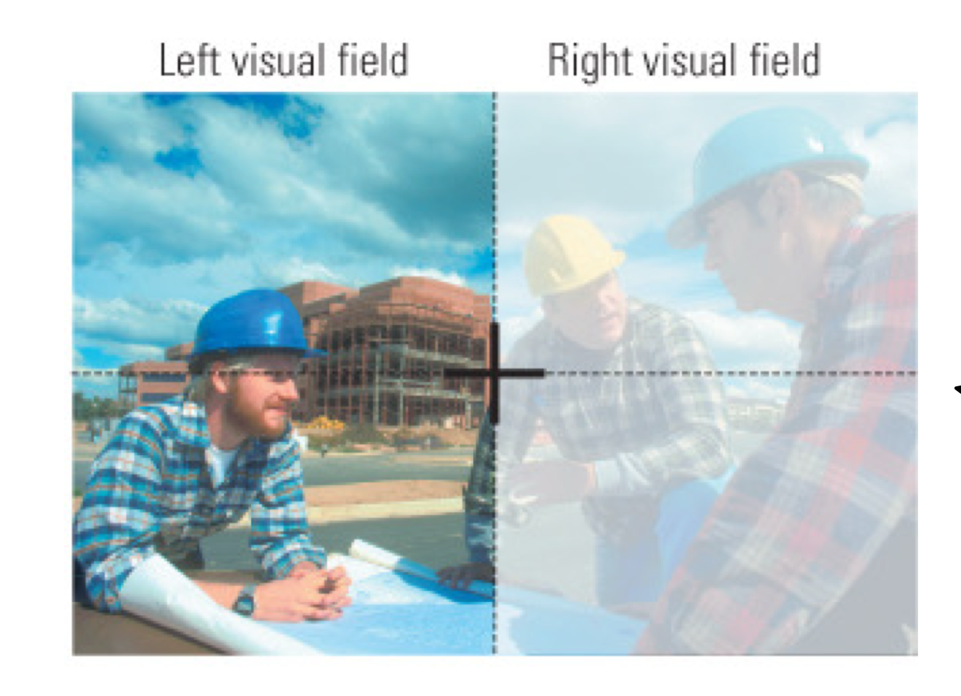
both sides of both eyes collect info from both sides of the visual field
before optic chiasm
optic chiasm
where criss crossing of visual information occurs
info from left visual field (collected from both eyes) gets processed in the right hemisphere
optic tract
visual information makes its way to the thalamus
after the optic chiasm
Lateral Geniculate Nucleus (LGN)
nucleus in the thalamus
APs make their way to primary visual cortex (V1)
optic radiation
tells us we’ve crossed the thalamus and going to occipital lobe
primary visual cortex
information that is first landing in the occipital lobe (V1)
first place info lands where processing occurs
moves information toward higher order processing (visual processing areas 2, 3, 4, 5) → more complexity in processing (higher area, more complex - colour, motion, orientation)
Calcarine sulcus
location of area V1; divides the upper and lower halves of the world (what we’re looking at)
Lingual gyrus
visual cortical regions V2 and VP (ventral posterior area)
VP is at the posterior area of the temporal lobe, closest to occipital
Fusiform gyrus
area V4; more anterior in the temporal
Occipital cortex
could have more than 6 layers in the occipital cortex as opposed to everywhere else
possibly because V1 does a lot of work
first cortical area involved in visual processing
decides where information should be going
sends visual info received from LGN to extrastriate cortex areas for higher order processing (colour, motion, orientation)
receives visual input from the LGN of the thalamus
Area V1
laminar organization: most distinct of all cortical layers (in layer 4)
distinct layers, not uniform across the cortex
layer 1: axons, dendrites → layer 3: lots of pyramidal cells
layer 1 looks different from layer 3 = laminar organization
in this case, layer 4 is the most distinct
heterogenous
has more than one distinct function (since there are different layers)
preserved in V2 (and V1)
Striate cortex
another name for visual cortex, due to its striped appearance in layer 4
layer 4 also has a striated appearance because different cell types
staining - cytoarchitecture (V1 & V2)
tells us the unique characteristics of the different cells in a particular layer
Area V1
blobs = sensitive to colour
more metabolically active than other cells
interblobs = sensitive to orientation (found between the cells/blobs)
Area V2
thin stripes = colour perception
thick stripes = form (shape) and motion perception
which area has the primary job of colour vision?
Area V4
but distributed throughout the occipital cortex
also plays a role in detection of movement, depth, and position (colour can change)
connections of the visual cortex - V1 (primary visual cortex)
input from LGN
output to all other levels
connections of the visual cortex - V2 (secondary visual cortex)
works closely w/ V1 to move info to other areas of the brain
output to all other levels
connections of the visual cortex - after V2
streams of processing
output to parietal lobe: dorsal stream
output to temporal lobe: ventral stream
output to superior temporal sulcus (STS): STS stream
streams of processing
dorsal stream: visual guidance of movements (where)
ventral stream: object perception (what)
STS stream: visuospatial functions
collecting info from both where and what pathways (it lies between them)
has access to info from both parietal and inferior temporal
Case study: organization of social perception & cognition within the STS
STS is involved in:
language (stories vs nonsense speech)
voices (voices vs environmental sounds)
faces (moving faces vs moving objects)
biological motion (point light humans vs point light objects)
theory of mind (what’s happening in someone else’s mind)
Wernicke’s area = posterior section of the superior temporal gyrus and middle temporal gyrus, and by extension, the cortex in between them in the posterior STS.
BOLD fMRI studies linked other cognitive functions (e.g., social cognition and perception to STS)
what was the first cognitive function ascribed to STS?
language comprehension
Hierarchical organization of the occipital lobe
Vision begins in V1 is heterogenous, then moves to more specialized cortical zones
blobs (V1): Area V4 → colour
interblobs (V1): Area MT/V5 → motion
V1 & V2: Area V3 → (dynamic form: shape of objects in different orientations, like during motion)
selective lesions up the hierarchy produce specific visual deficits
lesions to V1 are not aware of seeing
patients report not seeing anything, but will catch ball thrown at them
vision beyond the occipital lobe
vision-related areas in the brain make up about 55% of the total cortex
vision is not just in the occipital lobe
multiple visual regions in temporal, parietal, and frontal lobes
5 categories for vision
vision for action
action for vision
recognition
space
visual attention/how to not get overwhelmed
vision for action
parietal visual areas in the dorsal stream
reflex-based (not using your attention)
bottom-up
ducking, catching
action for vision
attention-based (using your attention to visually scan an environment)
visual scanning → top-down
eye movements and selective attention
normal subject: eye movements concentrate on facial features and directed more to left side of photograph (eye movements track shape of stimuli)
agnosic subject: eye tracking is disorganized, not connected to shape of stimulus
visual recognition
temporal lobes
recognizing object, face, person → temporal lobe processing
object recognition
what pathway (ventral)
visual space
parietal lobes
where something is in the environment
where pathway (dorsal stream)
spatial location
egocentric space: location of object relative to person
allocentric space: location of object relative to another
visual attention
paying attention to important characteristic of an image
aka: how to not get overwhelmed
binding problem
brain doesn’t encode everything, lots of things we ignore in our visual system
Milner-Goodale Model
Ventral and Dorsal streams
there is hierarchical processing in vision
Ventral stream:
V3: dynamic form (perception of objects/shapes that change over time)
V4: colour
Dorsal stream:
V3A: form (shape)
V4: motion (speed)
what’s the difference between areas V3 and V3A?
V3: part of ventral stream; detects dynamic form
V3A: part of dorsal stream; detects form/shape
monocular blindness
loss of sight in one eye
due to damage to retina or optic nerve

bitemporal hemianopia
loss of vision in both temporal fields
due to tumour to pituitary gland that puts pressure on the optic chiasm
preserved nasal vision

right nasal hemianopia
lesion in the lateral chiasm leading to loss of vision in one nasal field

homonymous hemianopia
blindness of one visual field due to damage in either: optic tract, LGN, V1

quadrant-anopia
loss of vision in one-quarter of the visual field
due to visual cortex lesions, particularly near calcarine sulcus

macular sparing
differentiates lesions of the optic tract or thalamus from cortical lesions
lesions of occipital lobe will often spare the macula
macular vision is preserved (spared from damage)
macula = small specialized area of high visual acuity, near retina

why/when does macular sparring occur?
likely because the macular part of area V1 might receive double vascular supply from medial and cerebral artery
lesions of occipital lobe often spare the macular region of the visual field (as opposed to lesions of the optic tract or thalamus)
scotoma
small visual cortex lesions, particularly near the calcarine sulcus
produces blindspots (scotomas)

monocular blindness

bitemporal hemianopia

right nasal hemianopia

homonymous hemianopia

quadrant-anopia
macular sparing
hemianopia (loss of vision in one visual field)

quadrant-anopia

scotoma
Visual agnosia (definition)
neurological condition that affects person’s ability to recognize or interpret visual information, despite having normal vision
broad term, need to specify what type of agnosia
types of visual agnosia
Object agnosia
Apperceptive agnosia
Simultagnosia
Associative agnosia
Other types of agnosia
Prosopagnosia (facial agnosia)
Alexia (dyslexia)
Visuospatial
apperceptive agnosia
difficulty with tasks like matching or identifying objects by shape, size, colour
due to: gross bilateral damage to occipital lobe
simultagnosia: inability to perceive more than one object at a time
difficulty with tasks that require processing multiple objects/scenes at once
e.g., reading, finding objects in cluttered space
associative agnosia
difficulty in recognizing objects due to a problem with connecting visual information with knowledge about the object
can perceive objects, but cannot identify them
due to: lesions of anterior temporal lobe
e.g., able to copy drawing, but unable to name what they drew
semantic problem (intact perception, but cannot understand its meaning)
prosopagnosia
facial agnosia → loss of knowledge associated with faces
cannot recognize previously known faces
due to: bilateral damage in temporal cortex
alexia
dyslexia → inability to read
due to: damage in left fusiform and lingual areas
visuospatial agnosia
topographic disorganization
inability to find one’s way
due to: damage to occipitotemporal regions and medial fusiform and lingual areas
symptom of dimentias
other visual deficits can accompany it (facial recognition)
Case study: V1 damage and a scotoma
MRI with lesion in occipital lobe + showing area of reduced visual acuity
right infarct (dead tissue) in occipital lobe
quadrantanopia (evidence from visual acuity)
experienced blindsight: could perceive a prior location once the light moved into a “visual” quadrant
vision without consciousness → occurs if there’s damage to V1
blindsight
vision without consciousness
dedicated V1 function
COME BACK TO THIS TO WATCH THE VIDEO
Case study: symptoms of hemianopia and cortical blindness (blindsight)
MRI shows an angioma in right calcarine fissure
symptoms include hemianopia and blindsight
probably due to damage in V1
Case study: vascular abnormality resulting in damage to V5
damage to medial temporal area (area MT/V5)
symptoms include inability to intercept moving objects by using their hand
therefore, there is loss of movement vision
Case study: bilateral hemorrhages in the occipitoparietal regions
symptoms include optic ataxia → a deficit in visually guided hand movements
visually guiding hand to move in a direction — where pathway (where something is in space)
optic ataxia
impairments in using visually guided hand movements
Case study: right occipitotemporal lesion
what pathway
symptoms include prosopagnosia
deficit in facial recognition
unable to give an identity to someone
Case study: left occipitotemporal lesion
Left → L → language
symptoms include alexia
inability to read
mental rotation
the cognitive ability to imaginatively turn 2D or 3D objects in one’s mind to determine if they match another object, regardless of orientation
although V1 appears to be anatomically homogenous…
staining it with cytochrome oxidase (enzyme for making energy available to cells), shows it to be heterogenous
what differentiates cat/dog vision from humans?
colour-related info processing enriches our capacity to detect motion, depth, position
in the absence of colour analysis, dogs/cats have overall reduced visual capacity compared to humans
single-celled organism Euglena
alters its swim pattern as a function of ambient light in different parts of the pond
since sunlight helps food production, it follows it to feed
example of how vision for motion evolved before vision for recogntion
Milner & Goodale
distinguished between ventral and dorsal streams
blind patient, but still had unconscious vision (dorsal stream intact)
vs patients with damaged dorsal stream who can’t reach accurately
proposed that dorsal stream = set of systems for visual control of action, based on:
visual neurons in posterior parietal regions are unique in that they are only active when the brain acts on visual information
visual posterior parietal neurons act as an interface between analysis of the visual world and motor action taken on it
most visual impairments associated with lesions to parietal cortex can be characterized as visuomotor or visuospatial
Limitation to the Milner—Goodale model
it mentions 2 distinct visual streams
dorsal: guiding movements
ventral: identifying objects
a third stream of visual processing: STS stream
associated with both the parietal and temporal pathways
the STS is part of the multimodal cortex characterized by…
polysensory neurons: neurons responsive to both visual and auditory or both visual and somatosensory input
parietal lobe intro slide
spatial awareness
body awareness: where you are located in the room
proprioception
attention/sensory attentional control
mathematical cognition (+, -, x, /)
numerical cognition: understanding that there are multiple vs one
2 main functions of the parietal lobes
process and integrate somatosensory information
process and integrate visual information
somatosensory system
comprises the receptors and processing centres to produce the experience of:
touch
temperature
proprioception
nociception
anterior border of parietal lobe
marked by central fissure (border between frontal and parietal lobe)
separates precentral and postcentral gyri
ventral border of parietal lobe
marked by Sylvian/lateral fissure
parietal lobe is dorsal to the…
cingulate gyrus
connects occipital, temporal, parietal lobes to frontal
mediates attention (prayer, meditation)
posterior border
marked by the parieto-occipital sulcus
postcentral gyrus
main sensory receptive area for the sense of touch
very specific, could think of it as unimodal (as in only sense of touch)
inferior parietal lobe (what does it contain?)
contains a multimodal associative area that receives auditory, visual, and somatosensory inputs
where pathway processing
includes 2 gyri:
supramarginal gyrus
angular gyrus
one of the last structures to mature
superior temporal gyrus
includes Wernicke’s area
as opposed to inferior temporal gyrus, which is the what pathway (ventral)
inferior parietal lobe is involved in…
comprehension of written language
it’s one of the last structures to mature, which may explain why children typically do not begin to read/write till they’re 5-6
angular gyrus (of the inferior parietal lobe)
involved in language processing/reading
converts written words into meaningful information by integrating visual information from eyes w/ language-related processing areas
reading comprehension, word recognition, semantic processing
word recognition in terms of parietal cortex
organizing words/letters (spatial organization)
word recognition in terms of temporal cortex
giving an identity to the words being read
Vogt and Vogt + Forster
architectonic mapping of the brain
electrophysiological mapping
forsees development of modern brain mapping
brodmann subdivided cerebral cortex into numerous areas based on cytoarchitecture
anterior zone: areas 1, 2, 3, 43
closest to frontal cortex
posterior zone: all other areas of parietal cortex
closest to occipital cortex
Von Economo’s cytoarchitectonic regions
for the labelling of the posterior zone
PE, PF, PG
functional zones of the parietal lobe
anterior zone: somatosensory cortex
posterior zone: where/how pathway
posterior parietal areas
PE, PF, PG
somatotopic organization
somatosensory cortex (postcentral gyrus) → anterior zone
contains the sensory homunculus
assists with somatosensation:
intensity
timing
location
temperature
pressure
pain
where does somatotopic organization also exist (other than somatosensory cortex)?
in the cerebellum
Area PE
Connections:
somatosensory cortex (postcentral gyrus)
motor cortex (precentral gyrus)
PF
Function:
somatosensory
role in guiding movement by providing info about limb position
if someone has difficulty with a sport they have played for years/dancer, and have trouble moving their limbs in that way, what area is most likely damaged?
area PE
Area PF
Connections:
Somatosensory cortex
Motor cortex
Premotor cortex
PG
Function:
part of the mirror neuron system
theory of mind: facilitated by mirror neurons
Area PG
Connections are multimodal:
receives complex connections (visual, somesthetic, proprioceptive, auditory, vestibular, oculomotor, cingulate)
Function:
dorsal stream
parieto-temporo-occipital crossroads
posterior parietal cortex is also closely related to…
the prefrontal cortex and limbic system
memory
spatially guided behaviour
spatial navigation
pathways from posterior parietal regions
posterio-premotor: “where”/”how” pathway
posterio-prefrontal: working memory
posterio-medial temporal: spatial navigation
Theory of parietal frontal lobe function
anterior zones process somatic sensations and perceptions
posterior zones integrate vision with multimodal info
viewer-centered object identification
parietal processing → where pathway (orientation, location in space)
works with temporal processing which helps figure out what we’re looking at (object identity)
binding problem
3 somatosensory symptoms of parietal lobe lesions
afferent paresis
astereognosis
abnormally high sensory threshold
afferent paresis
clumsy finger movement due to lack of feedback about finger position