Block 8: Push me pull you
1/13
Earn XP
Description and Tags
Week 5
Name | Mastery | Learn | Test | Matching | Spaced | Call with Kai |
|---|
No analytics yet
Send a link to your students to track their progress
14 Terms
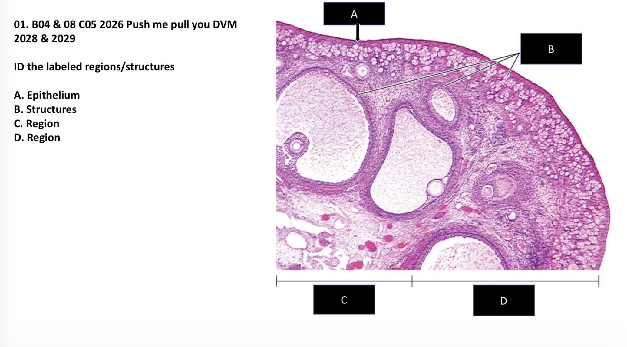
ID the labeled regions/structures.
A. Epithelium
B. Structures
C. Region
D. Region
A. Germinal/Simple cuboidal
B. Follicles
C. Medulla
D. Cortex


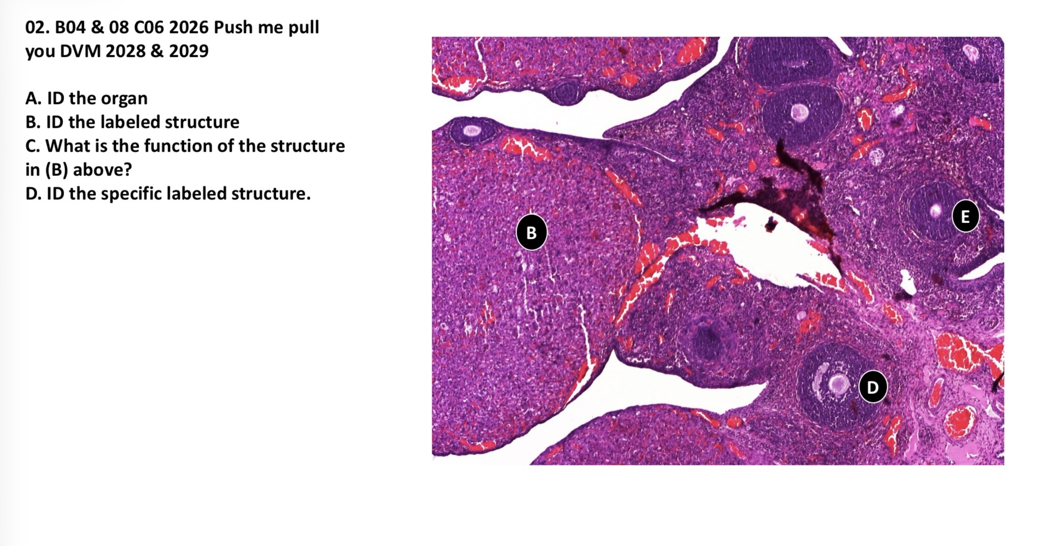
A. ID the organ
B. ID the labeled structure
C. What is the function of the structure in (B) above?
D. ID the specific labeled structure
E. ID the specific labeled structure
A. Ovary
B. Corpus luteum
C. Produce progesterone
D. Secondary follicle
E. Multi-laminar primary follicle

A. ID the labeled structure
B. What is the function of the inner layer?
C. Name the labeled structure
A. Theca layer
B. Secrete androgen hormone —androstenedione
C. Cumulus oophorus
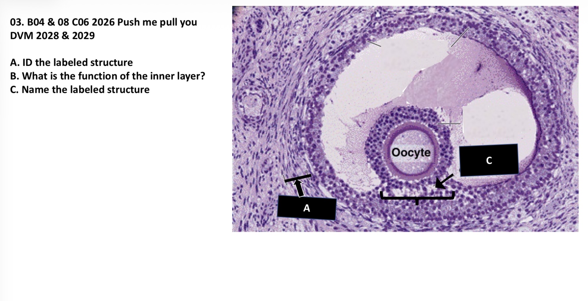
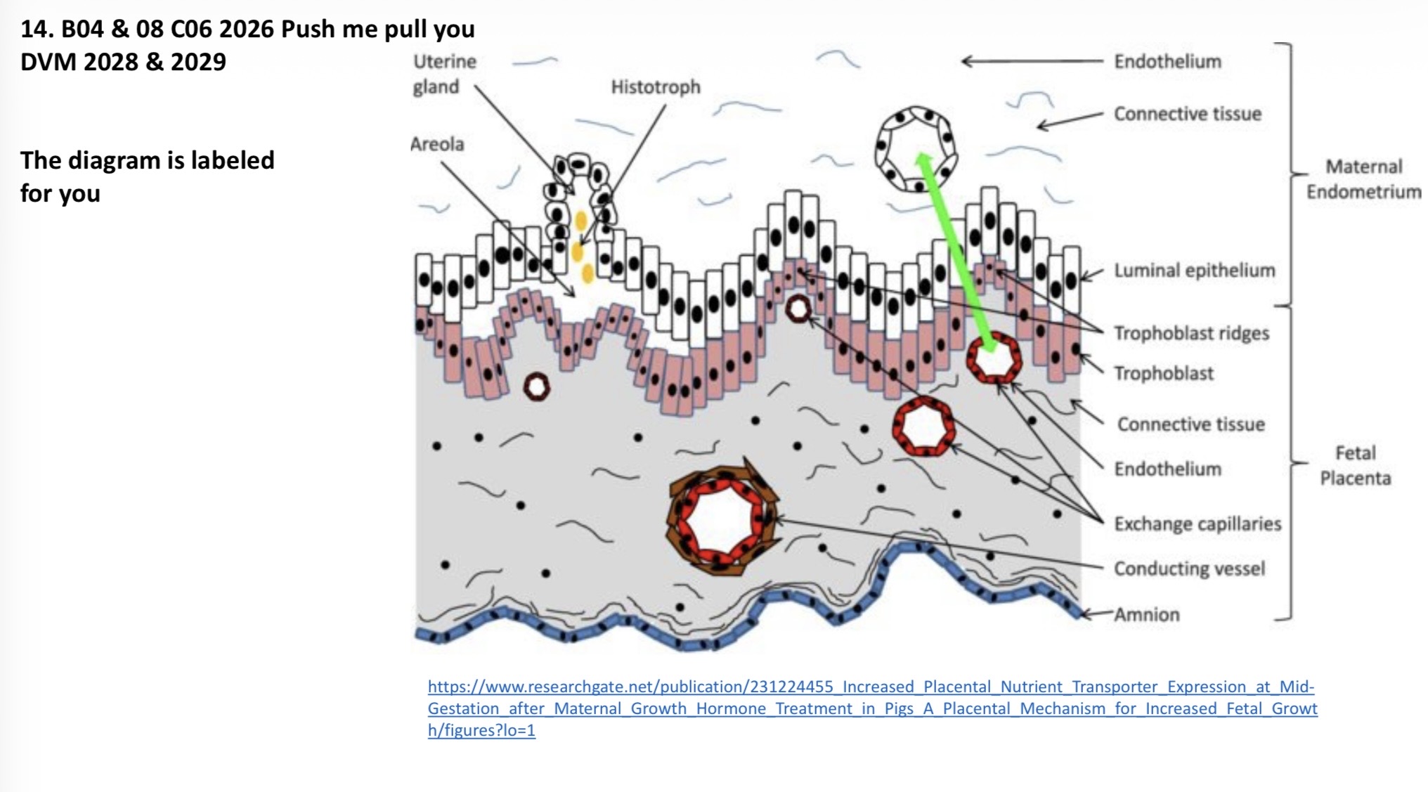
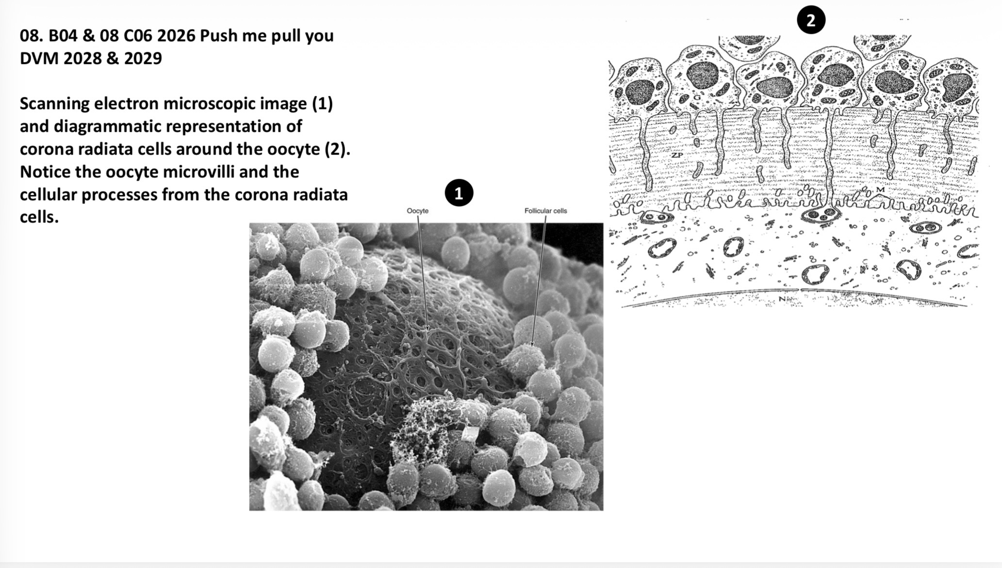
Image is labeled.


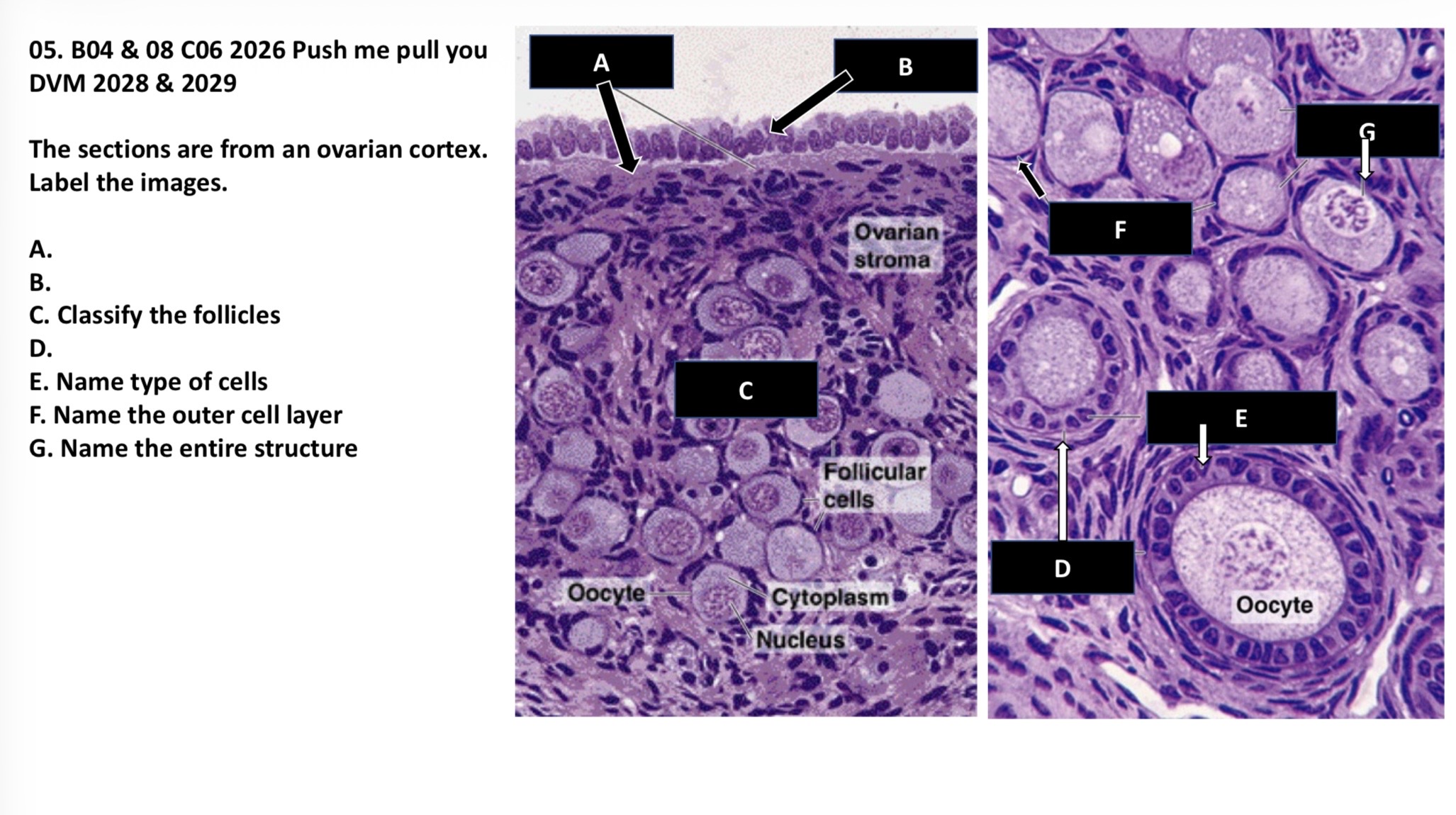
The sections are from an ovarian cortex. Label the images.
A.
B.
C. Classify the follicles
D.
E. Name the type of cells
F. Name the outer cell layer
G. Name the entire structure
A. Tunica albuginea
B. Germinal epithelium (simple cuboidal)
C. Primordial follicles
D. Primary follicle
E. Granulosa cells
F. Stromal (follicular) cells
G. Primordial follicle
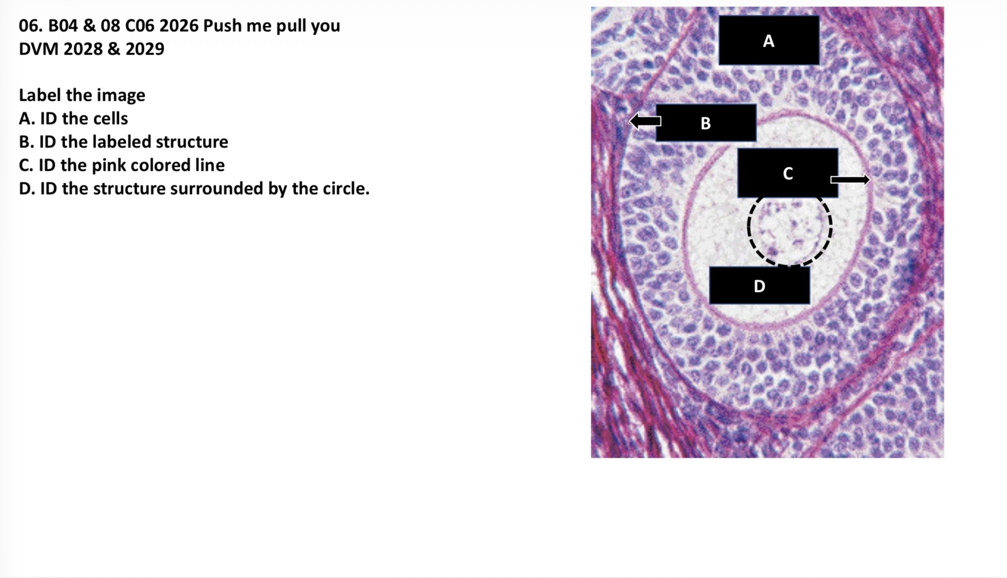
Label the image.
A. ID the cells
B. ID the labeled structure
C. ID the pink colored line
D. ID the structure surrounded by the circle
A. Granulosa cells
B. Basement membrane
C. Zona pellucida
D. Oocyte nucleus


This section is taken from the outer wall of a mature follicle. Label A-D.
A. Granulosa cells
B. Basement membrane
C. Theca interna
D. Theca externa
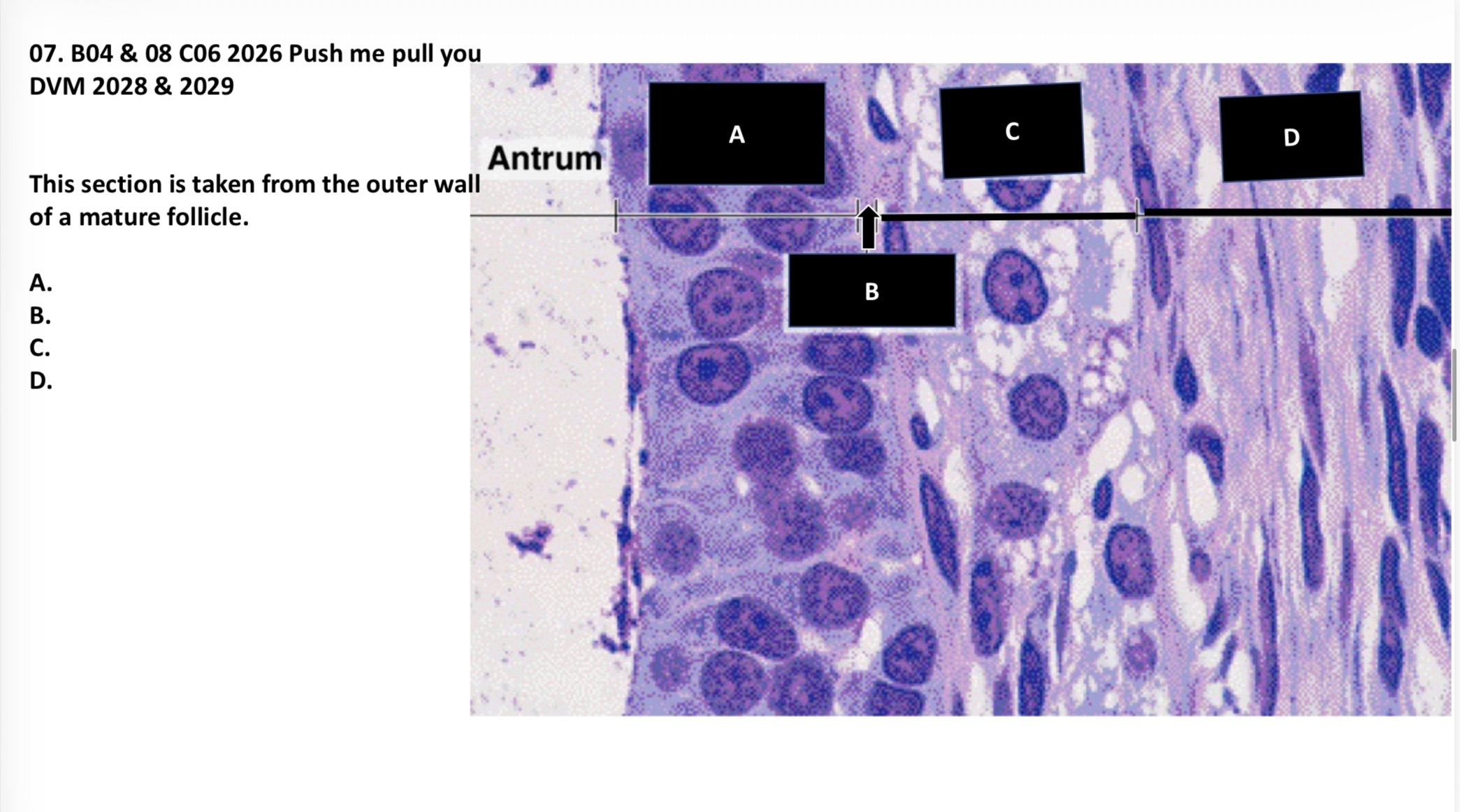

Image is labeled.

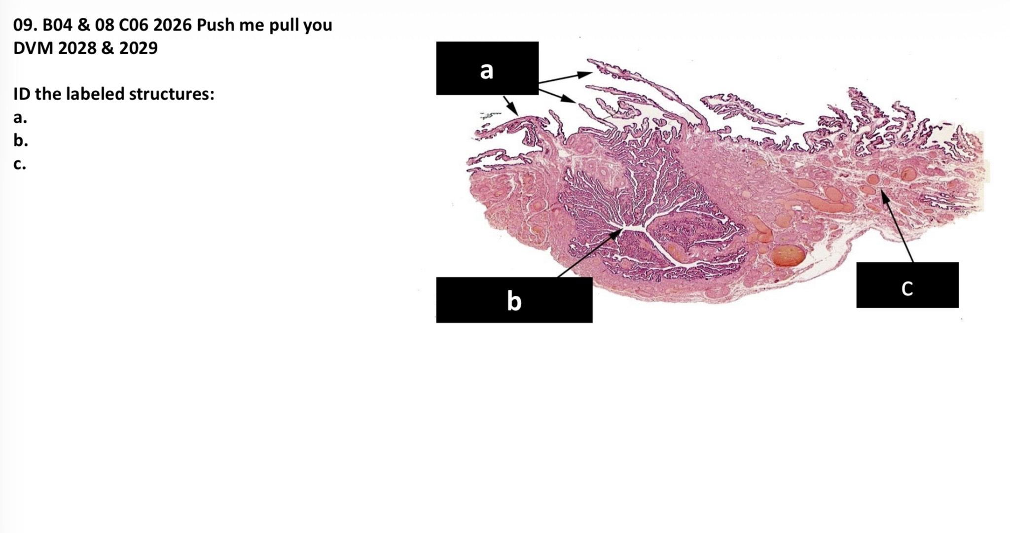
ID the labeled structures A-C
A. Fimbria of the uterine tube
B. Infundibulum of the uterine tube
C. Mesoalpinx
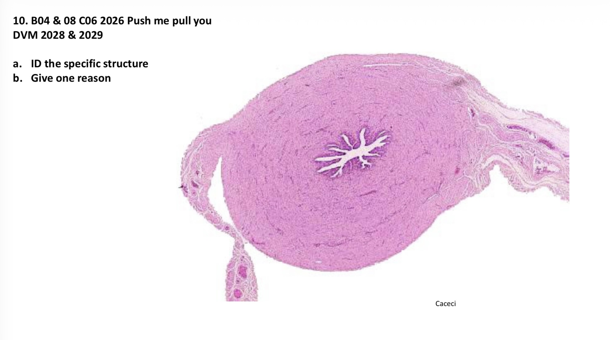
A. ID the specific structure
B. Give one reason
A. Isthmus of the uterine tube
B. Thick muscular layer, low number of folds

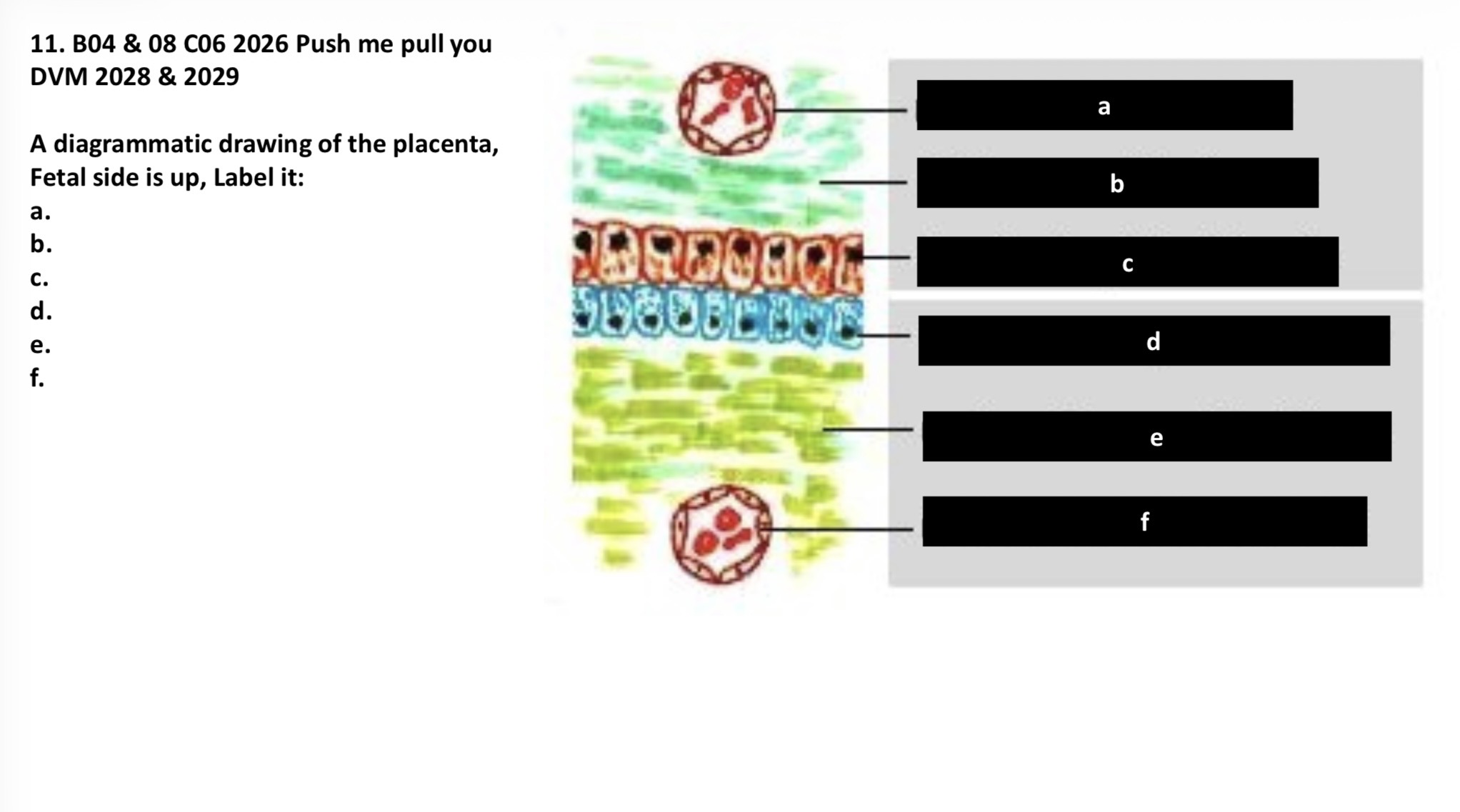
A diagrammatic drawing of the placenta, fetal side is up, label A-F.
A. Fetal capillary endothelium
B. Fetal connective tissue
C. Chorionic epithelium
D. Maternal (uterine) epithelium
E. Maternal connective tissue
F. Maternal capillary endothelium

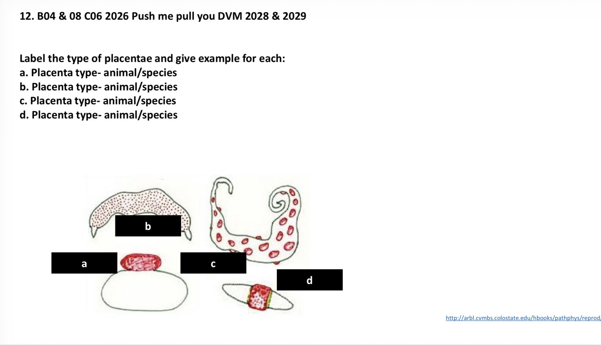
Label the type of placentae and give one example for each:
A-D: placenta type - animal/species
A. Discoidal - villi localized in one area
B. Diffuse - villi distributed across all of the chorion
C. Cotyledonary - villi gathered into distinct clusters
D. Zonary - villi gathered in a specific place on chorion

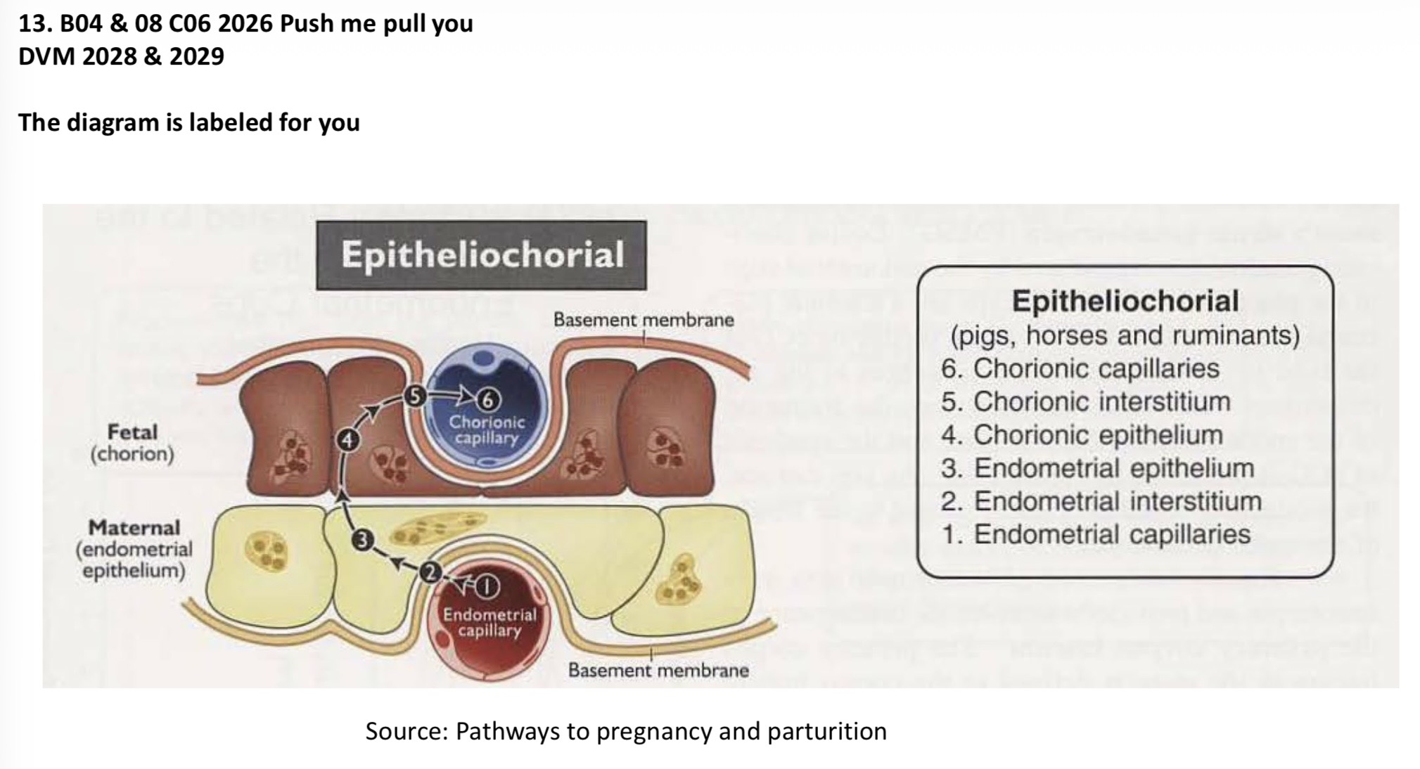
Image is labeled.


Image is labeled.