Chapter 11: Cranium, Facial Bones, Paranasal Sinuses
1/185
There's no tags or description
Looks like no tags are added yet.
Name | Mastery | Learn | Test | Matching | Spaced | Call with Kai |
|---|
No analytics yet
Send a link to your students to track their progress
186 Terms
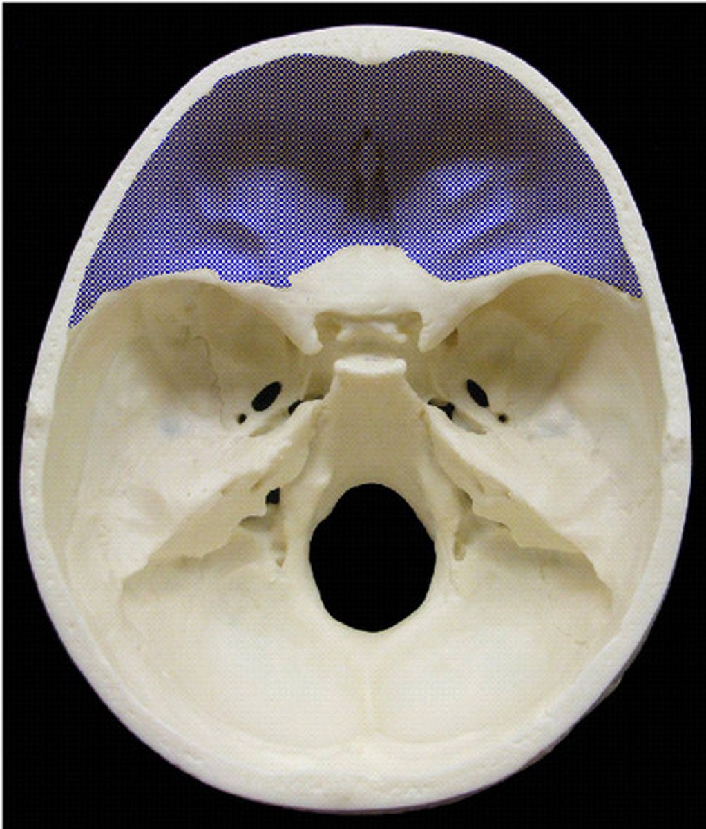
Which of the following bones is part of the floor of the cranium?
A) Temporal
B) Occipital
C) Frontal
D) Parietal
A) Temporal
pg. 377: Floor of the cranium bones
- Right temporal
- Left temporal
- Sphenoid
- Ethmoid
How many bones make up the facial bone region?
A) 6
B) 8
C) 12
D) 14
D) 14
pg. 377: the skull, or bony skeleton of the head, rests on the superior end of the vertebral column and is divided into two main sets of bones - 8 cranial bones and 14 facial bones.
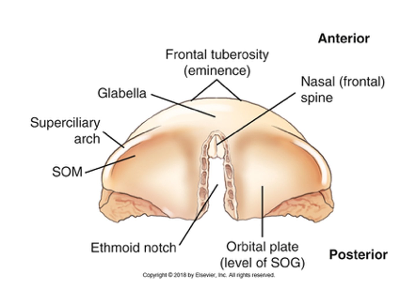
Which bony landmark represents the highest level of the facial bone mass?
A) Nasion
B) Acanthion
C) Orbital plates
D) Supraorbital notch
C) Orbital plates
pg. 378: The SOG becomes an important landmark because it corresponds to the floor of the anterior fossa of the cranial vault, which is also at the level of the orbital plate or at the highest level of the facial bone mass.
The orbital plate on each side forms the superior part of each orbit. Below the orbital plates lie the facial bones, and above the orbital plates is the anterior part of the floor of the brain case.
The widest portion of the cranium is found at the level of the:
A) parietal tubercles
B) right and left pterion
C) squamous portion of the temporal bone
D) external acoustic meatus (EAM).
A) parietal tubercles
pg. 379: the widest portion of the entire skull is located between parietal tubercles (eminences) of the two parietal bones.
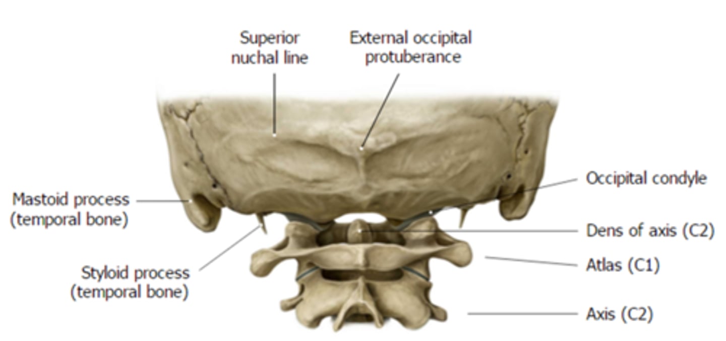
What is the name of the joint found between the lateral condylar processes of the skull and the atlas of C1?
A) Zygapophyseal joint
B) Intervertebral joint
C) Atlanto-occipital joint
D) Cervico-occipital joint
C) Atlanto-occipital joint
pg. 379: the two lateral condylar portions (occipital condyles) are oval processes with convex surfaces, with one on each side of the formen magnum. These articulate with depressions on the first cervical vertebrae, called the atlas. The two-part articulation between the skull and the cervical spine is called the atlanto-occipital joint.
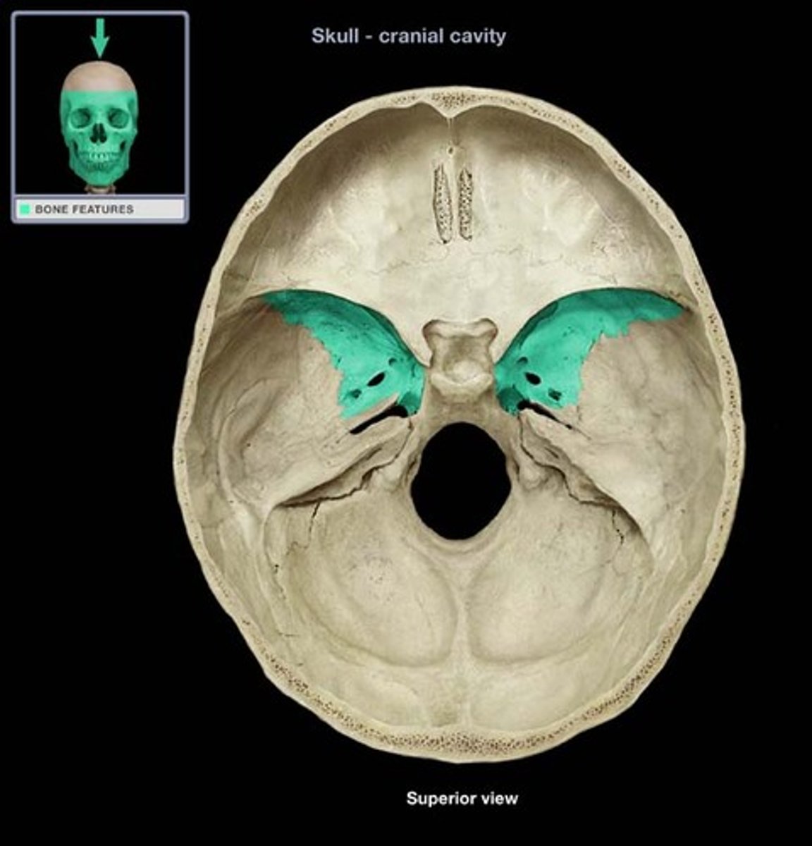
Which cranial bone articulates with all the other cranial bones?
A) Parietal
B) Ethmoid
C) Sphenoid
D) None of the above
C) Sphenoid
pg. 382: because of its central location, the sphenoid articulates with all seven of the other cranial bones. The sphenoid also articulates with five facial bones.
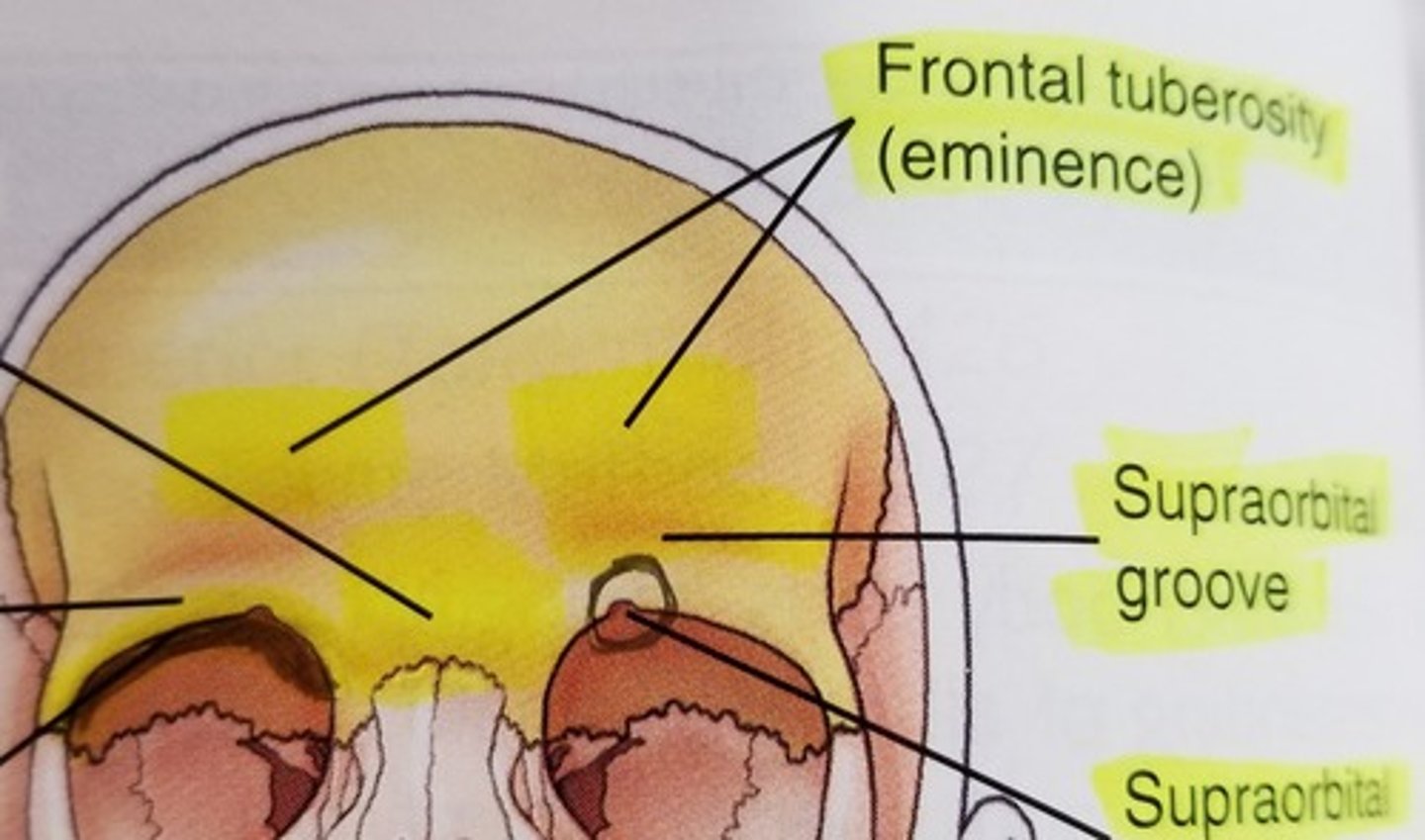
The slight depression above each eyebrow is termed the:
A) glabella
B) supraorbital foramina
C) supraorbital groove
D) frontal tuberosity.
C) supraorbital groove
pg. 378: the supraorbital groove is the slight depression above each eyebrow
Which of the following cranial bones does not articulate with the parietal bone?
A) Frontal
B) Sphenoid
C) Occipital
D) All of the above articulate with the parietal bone.
D) All of the above articulate with the parietal bone
pg. 379: each parietal bone articulates with five cranial bones - frontal, occipital, temporal, sphenoid, and opposite parietal.
The left mastoid fontanel becomes the ____ in an adult.
A) left asterion
B) left pterion
C) left bregma
D) squamosal suture
A) left asterion
Pg. 383: Two smaller lateral fontanels that close soon after birth are the sphenoid (pterion in an adult) and mastoid (asterion in an adult) fontanels.
There are a total of ____ fontanels in an infant.
A) four
B) two
C) six
D) eight
C) six
Pg. 383: Six fontanels occur in an infant as follows:
1. anterior fontanel (bregma)
2. posterior fontanel (lambda)
3. R sphenoid fontanel (R pterion)
4. L sphenoid fontanel (L pterion)
5. R mastoid fontanel (R asterion)
6. L mastoid fontanel (L asterion)
The frontal bone articulates with ____ cranial bones.
A) four
B) six
C) two
D) five
A) four
pg. 378: the frontal bone articulates with four cranial bones: right & left parietals, sphenoid, and ethmoid.
Which of the following landmarks corresponds with the level of the petrous ridge?
A) EAM
B) Top of ear attachment (TEA)
C) Squamosal suture
D) Inion
B) Top of ear attachment (TEA)
pg. 380: the petrous ridge of each pyramid corresponds to the level of an important external land mark, the TEA (top of the ear attachment).
The pituitary gland (hypophysis cerebri) is associated with and protected by the ____ bone.
A) temporal
B) ethmoid
C) palatine
D) sphenoid
D) sphenoid
pg. 381: the central portion of the sphenoid is the body ... the central depression on the body is termed the sella turcica. The sella turcica partially surrounds and protects a major gland of the body, the hypophysis cerebri, or pituitary gland.
Which cranial bone contains the foramen ovale?
A) Sphenoid
B) Occipital
C) Ethmoid
D) Temporal
A) Sphenoid
pg. 381: Three pairs of small openings or foramina exist in the greater wings (of the sphenoid) for passage of certain cranial nerves...
1. formen rotundum
2. foramen ovale
3. foramen spinosum
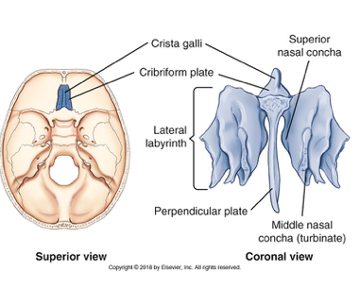
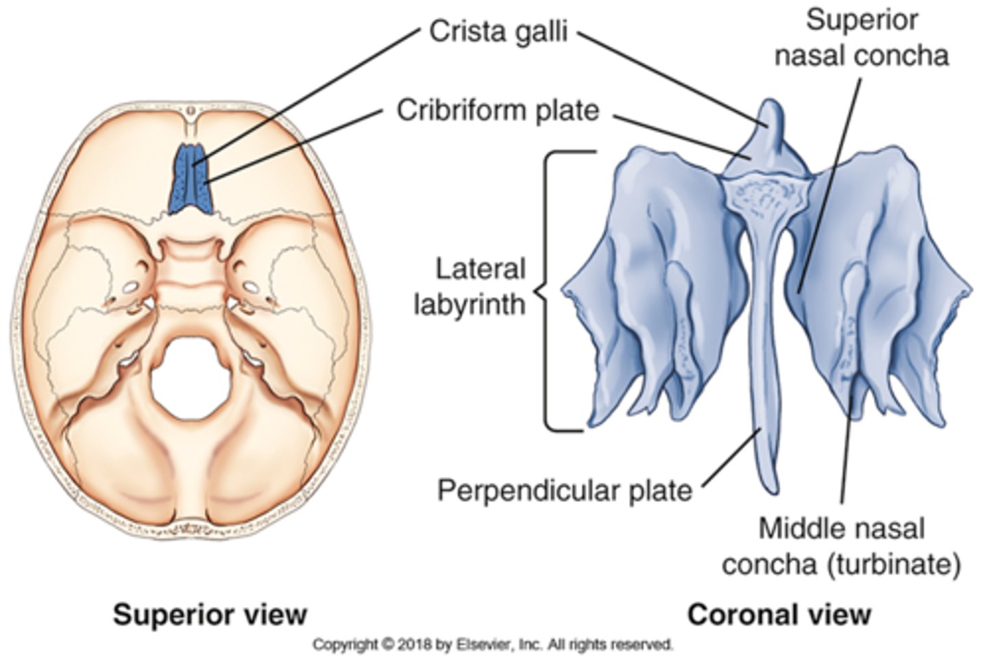
Which cranial bone contains the cribriform plate?
A) Sphenoid
B) Occipital
C) Temporal
D) Ethmoid
D) Ethmoid
pg. 382: the smaller upper horizontal portion of the ethmoid bone, termed the cribiform plate, contains many small openings or foramina through which segmental branches of the olfactory nerves pass.
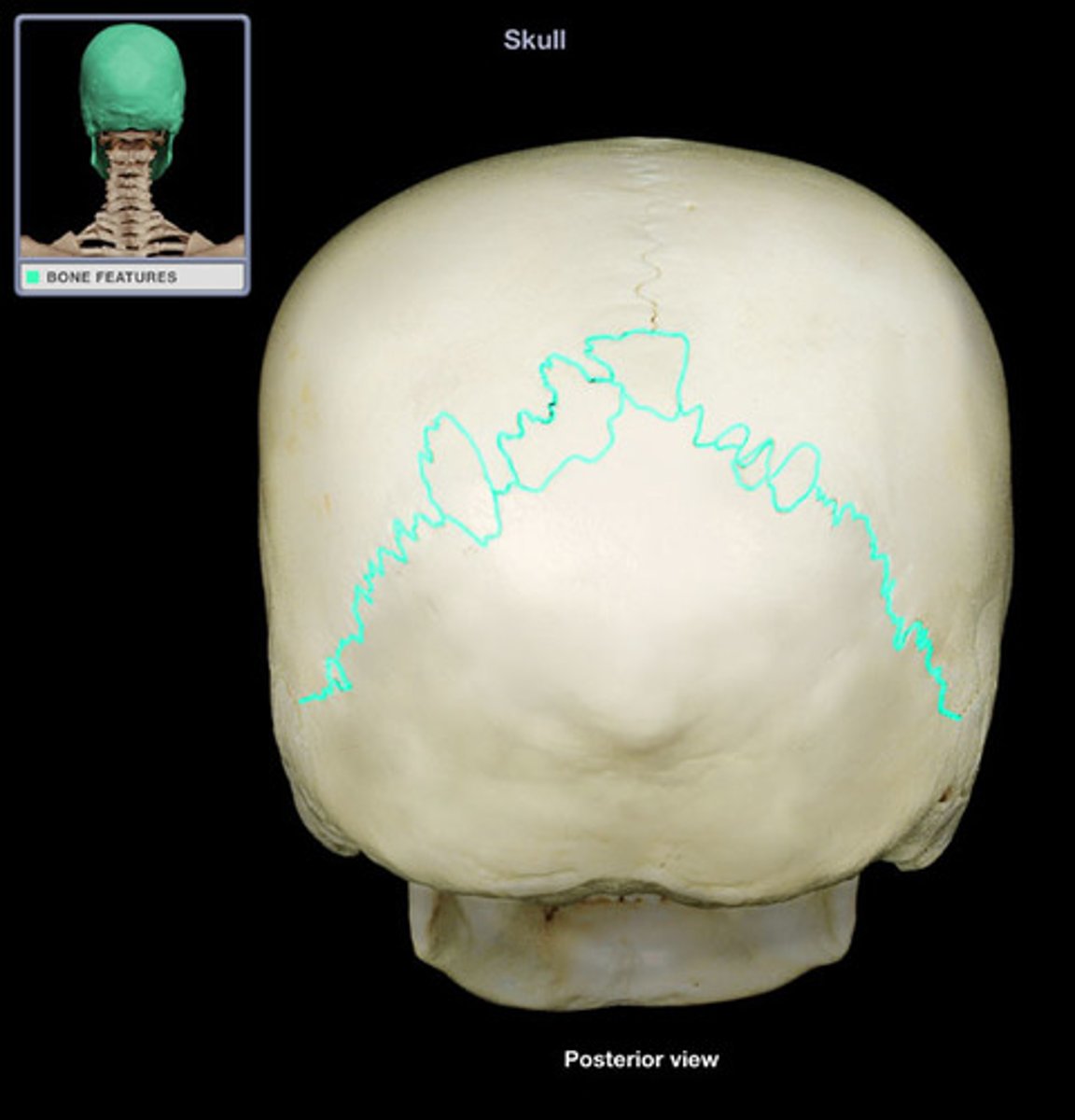
Which of the following sutures separates the parietal from the occipital bone?
A) Squamosal
B) Sagittal
C) Coronal
D) Lambdoidal
D) Lambdoidal
pg. 383: posteriorly, the lambdoidal suture separates the two parietal bones from the occipital bone.
Which of the following terms describes the anterior fontanel found in the adult skull?
A) Bregma
B) Pterion
C) Asterion
D) Lambda
A) Bregma
pg. 383: each end of the sagittal suture is identified as a point or area with a specific name as labeled. The anterior end of the sagittal suture is termed bregma.
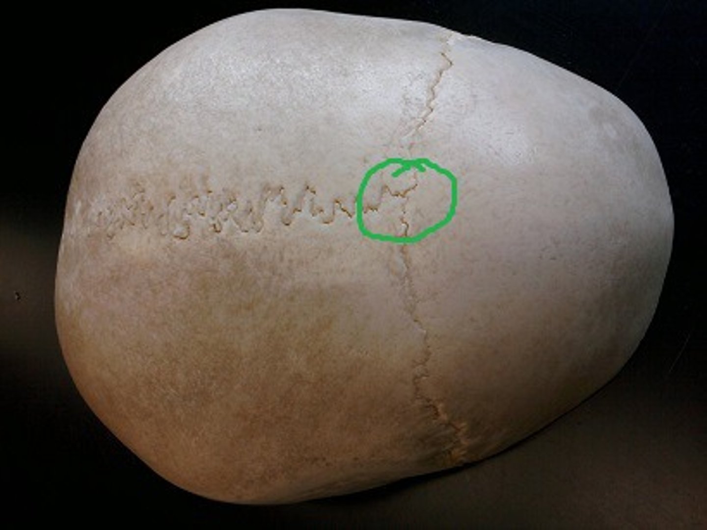
Which of the following terms describes the small irregular bones occasionally found in the sutures?
A) Asterion
B) Wormian
C) Sesamoid
D) Squamosal
B) Wormian
pg. 383: Certain small, irregular bones called sutural, or wormian, bones sometimes develop in adult skull sutures.
The ethmoid notch is part of which cranial bone?
A) Temporal
B) Ethmoid
C) Sphenoid
D) Frontal
D) Frontal
pg. 378: each orbital plate is separated from the body by the ethmoidal notch. The ethmoid bone, one of the bones of the floor of the cranium, fits into this notch.
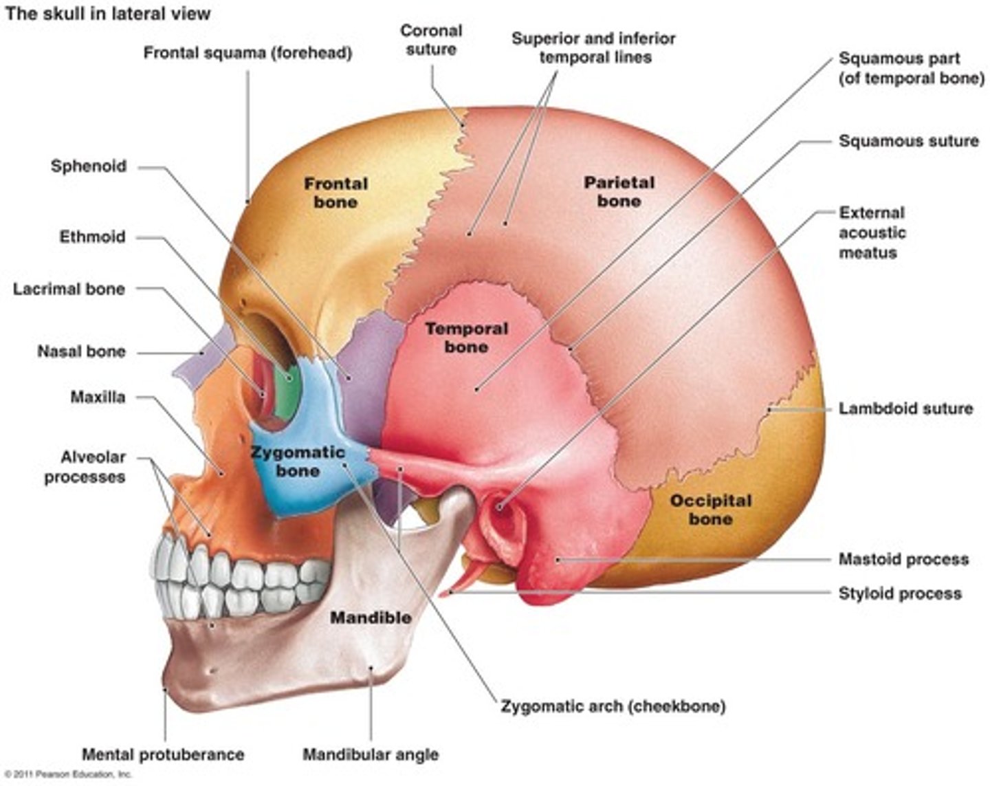
Which cranial bone is labeled vi in this figure? (Anatomic structures are labeled i through vi and sutures a through h.)
A) Temporal
B) Occipital
C) Greater wing of sphenoid
D) Parietal
C) Greater wing of sphenoid

Which bony structure is labeled v? (Anatomic structures are labeled i through vi and sutures a through h.)
A) EAM
B) Mastoid process
C) Styloid process
D) Zygomatic process
B) Mastoid process

The cranial bone labeled i is the: (Anatomic structures are labeled i through vi and sutures a through h.)
A) sphenoid.
B) lacrimal.
C) ethmoid.
D) palatine.
C) ethmoid.
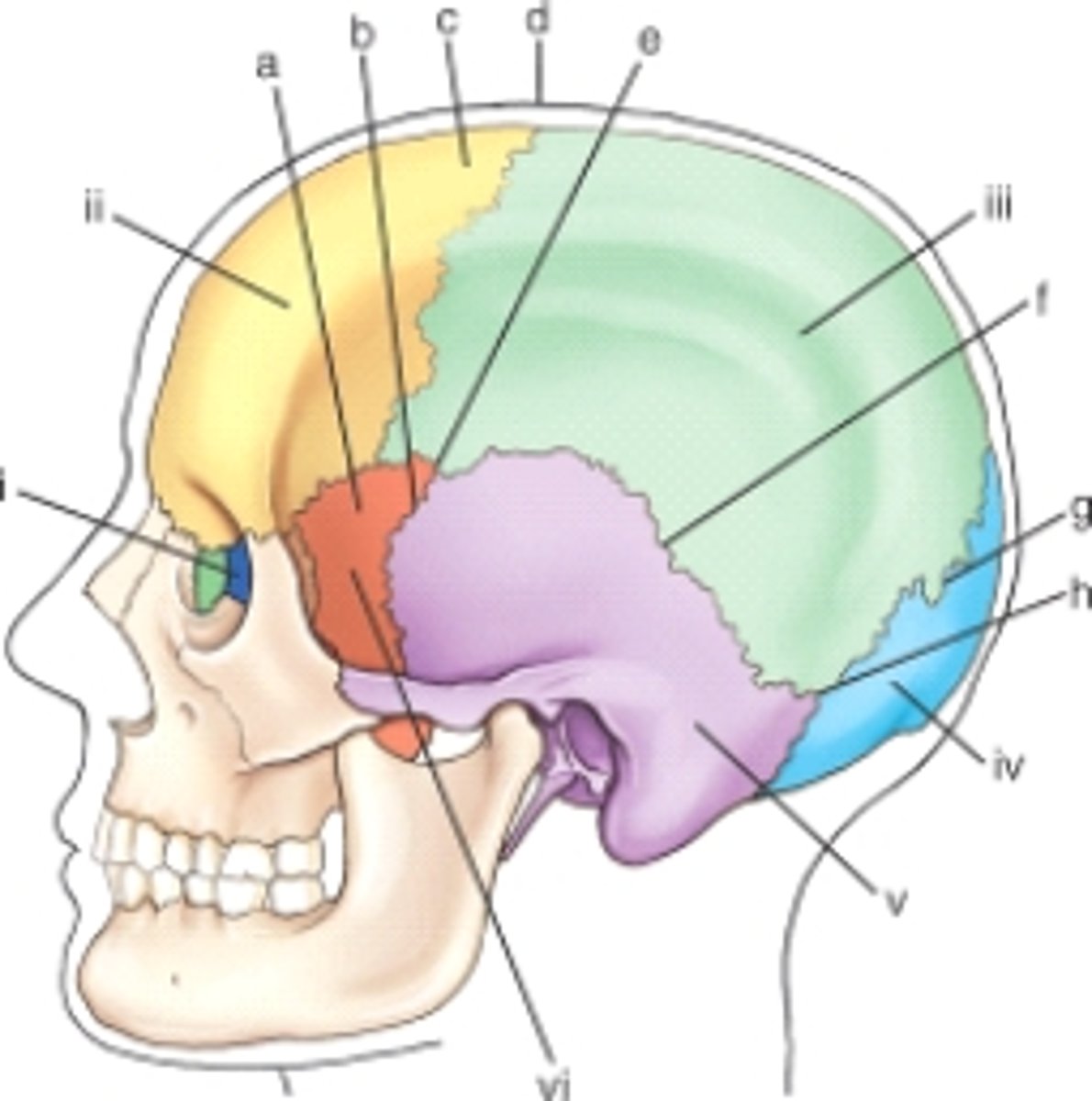
Which cranial suture is labeled g? (Anatomic structures are labeled i through vi and sutures a through h.)
A) Coronal
B) Squamosal
C) Lambdoidal
D) Sagittal
C) Lambdoidal

The sutural point or area labeled e is the: (Anatomic structures are labeled i through vi and sutures a through h.)
A) lambda.
B) asterion.
C) bregma.
D) pterion.
D) pterion

The sutural point or area labeled d is termed: (Anatomic structures are labeled i through vi and sutures a through h.)
A) bregma.
B) lambda.
C) asterion.
D) pterion.
A) bregma.

The suture labeled f is termed the: (Anatomic structures are labeled i through vi and sutures a through h.)
A) asterion.
B) lambdoidal.
C) coronal.
D) squamosal.
D) squamosal

Which of the fontanels is the last to close at about 18 months of age?
A) Sphenoid
B) Mastoid
C) Anterior
D) Posterior
C) Anterior
pg. 383: the anterior fontanel is the largest and at birth measures about 2.5 cm wide and 4 cm long. It does not completely close until about 18 months of age.
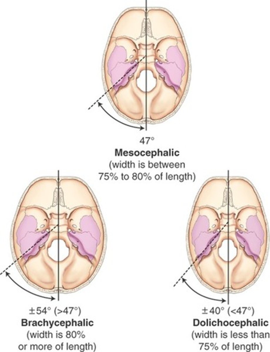
An average-shaped skull with a 47° angle between the petrous pyramids and the midsagittal plane is classified as:
A) mesocephalic
B) brachycephalic
C) dolichocephalic
D) morphocephalic.
A) mesocephalic
pg. 405: the shape of the average head is termed mesocephalic. In the mesocephalic head, the petrous pyramids form an angle of 47°.
For a mesocephalic skull, the width is __________ of the length
A) 75% to 80%
B) none of the above
C) less than 75%
D) 80% or more
A) 75% to 80%
pg. 405
An axiolateral oblique projection (Law method) for the temporomandibular joints on a brachycephalic type of skull would require ____ rotation as compared with an average-shaped skull.
A) more
B) less
C) the same
D) Rotation depends on the patient's age.
B) less
pg. 405: The positioning descriptions, including CR angles and head rotations, as described in this text are based on the average-shaped mesocephalic skull.
For example, the axiolateral oblique projection (Law method) for TMJs requires 15° of head rotation. A long, narrow, dolichocephalic head requires slightly more than 15° of rotation, and a short, broad, brachycephalic type requires less than 15°.
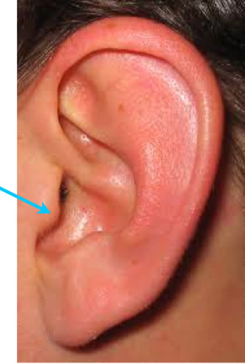
Which term describes the small flap of cartilage covering the opening to the ear?
A) Tragus
B) Pinna
C) Acanthion
D) EAM
A) Tragus
pg. 406: tragus, the small cartilaginous flap that covers the opening of the ear.
What is the difference, in degrees, between the infraorbitomeatal and orbitomeatal lines?
A) 10°
B) 15° to 2°
C) 7° to 8°
D) 20° to 25°
C) 7° to 8°
pg. 407: an average difference of 7° to 8° exists between the angles of the orbitomeatal line (OML) and the infraorbitalmeatal line (IOML).
Which one of the following technical considerations is most critical for demonstrating air and/or fluid levels within the cranium?
A) Medium kV range
B) Detail image receptor (IR)
C) Short exposure time
D) Horizontal x-ray beam
D) Horizontal x-ray beam
pg. 408: air-fluid levels in the sinuses or other cranial cavities may indicate certain pathologic conditions that are visible only in the erect position or with the use of a horizontal beam radiography.
A radiograph of an anteroposterior (AP) axial projection of the cranium reveals that the dorsum sellae is projected below the foramen magnum, but the posterior arch of C1 is visible within the foramen. Which of the following positioning errors led to this radiographic outcome?
A) Excessive central ray (CR) angulation
B) Insufficient CR angulation
C) Insufficient flexion of the head and neck
D) Tilt of the skull
A) Excessive central ray (CR) angulation
pg. 413: Overangulation of CR or excessive neck flexion superimposes the posterior arch of C1 over the dorsum sellae within the foramen magnum and produces foreshortening of the dorsum sellae.
Underangulation of CR or insufficient neck flexion projects the dorsum sellae superior to the foreman magnum.
A radiograph of a lateral projection of the facial bones reveals that the sella turcica is not a clear saddle. What specific positioning error is present on the radiograph?
A) Rotation
B) Excessive extension
C) Tilt
D) Excessive flexion
A) Rotation
Which one of the following positioning errors most often results in a repeat exposure of a cranial position?
A) Rotation
B) Incorrect central ray placement
C) Slight flexion
D) Slight extension
A) Rotation
pg. 409: five common positioning errors
1. Rotation
2. Tilt
3. Excessive neck flexion
4. Excessive neck extension
5. Incorrect CR angle
A radiograph of a posteroanterior (PA) axial projection (Caldwell method) of the cranium reveals that the petrous ridges are located at the level of the lower one third of the orbits. The technologist performed this projection with the CR angled 15° caudal to the orbitomeatal line (OML). How must positioning be altered if a repeat exposure is performed?
A) Increase the extension of the skull
B) Increase the flexion of the skull
C) Increase the CR angulation
D) None of the above; positioning was correct.
D) None of the above; positioning was correct
pg. 415: PA Axial 15° caudal angle - petrous pyramids are projected into the lower one-third of the orbits. Supraorbital margin is visualized without superimposition.
A radiograph of a submentovertex projection of the cranium reveals that the mandibular condyles are projected into the petrous pyramids. What must be altered during the repeat exposure to produce a more diagnostic radiograph?
A) Increase the extension of the skull.
B) Increase the flexion of the skull.
C) Decrease the CR angulation.
D) None of the above; it is an acceptable image.
A) Increase the extension of the skull.
pg. 432: correct extension of the neck puts mandibular condyles anterior to the petrous pyramids.
A radiograph of a lateral projection of the cranium reveals that the orbital roofs (plates) are not superimposed, one is slightly superior to the other. Which of the following positioning errors led to this radiographic outcome?
A) Rotation
B) Tilt
C) Excessive flexion
D) Excessive extension
B) Tilt
pg. 414: tilt is evident by superior and inferior separation of symmetric horizontal structures such as the orbital roofs (plates), and greater wings of the sphenoid.
A radiograph of a lateral cranium reveals that the mentum was cut off from the bottom of the radiograph. A 24 x 30-cm (10 x 12-inch) IR was used, and it was placed crosswise. What must be altered if a repeat exposure is performed?
A) Center the CR at the EAM.
B) Increase SID to reduce magnification.
C) Place the 24 30-cm (10 12-inch) IR lengthwise.
D) None of the above; all of the structures were demonstrated.
D) None of the above; all of the structures were demonstrated.
pg. 414: anatomy demonstrated = entire cranium, superimposed parietal bones, entire sellae turcica, anterior & posterior clinoid processes, dorsum sellae, and clivus.
IR size = 10" x 12", CW
A patient comes to radiology for a routine study of the cranium. He is unable to flex his head and neck sufficiently to place the OML perpendicular to the IR for the AP axial projection. What should the technologist do to compensate for this problem without creating excessive magnification of the occipital bone?
A) Use the inferior OML and increase the CR angulation by 7°.
B) Perform the Haas method.
C) Perform a submentovertex projection in place of the AP axial projection.
D) Use the AML and increase the CR angulation by 10°.
A) Use the inferior OML and increase the CR angulation by 7°.
pg. 413: if patient is unable to depress the chin sufficiently to bring OML perpendicular to IR even with a small spong under the head, IOML can be placed perpendicular instead and the CR angle increased to 37° caudad. This maintains the 30° angle between the OML and CR and demonstrates the same anatomic relationships (a 7° to 8° difference exists between the IMOL and OML).
A patient comes to radiology with a history of a possible erosion of the superior orbital fissures. Which of the following projections would best demonstrate this structure?
A) PA axial with a 15° caudal angle to OML
B) Submentovertex
C) PA axial with a 25° to 30° caudal angle to OML
D) AP axial with a 37° caudal angle to OML
C) PA axial with a 25° to 30° caudal angle to OML
pg. 415: Alternative 25° to 30° projection allows better visualization of the superior orbital fissures, foreman rotundum, and the inferior orbital rim.
A patient enters the emergency department (ED) with a possible basilar skull fracture. Which of the following skull projections would best demonstrate any blood present in the sphenoid sinus?
A) AP with a 15° cephalic angle
B) Haas method
C) Submentovertex
D) Horizontal beam lateral projection
D) Horizontal beam lateral projection
pg. 402: Basal skull fractures - if bleeding occurs, plain radiographic images may reveal an air-fluid level in the sphenoid sinus if a horizontal ray is used for the lateral view.
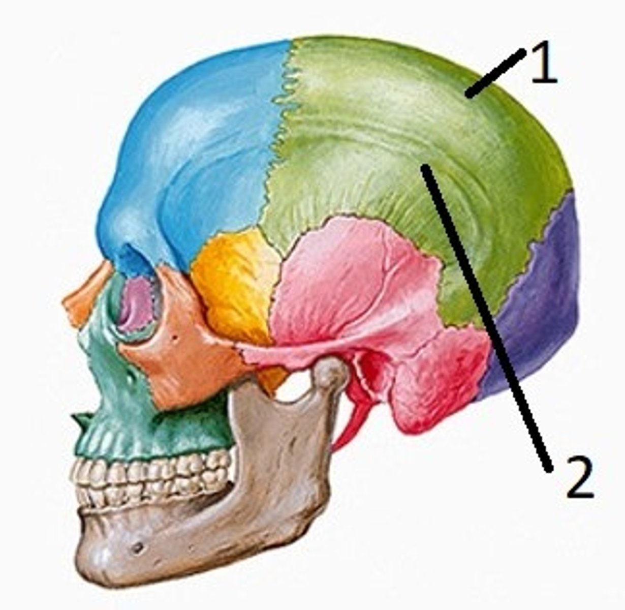
A patient comes to radiology with a possible bone cyst within the squamous portion of the frontal bone. Which of the following projections would best demonstrate this region with a minimal amount of distortion of the frontal bone?
A) AP axial with a 30° caudal angle to OML
B) PA axial with a 30° caudal angle to OML
C) PA axial with a 15° caudal angle to OML
D) PA with no CR angulation to OML
D) PA with no CR angulation to OML
pg. 416: PA projection, CR 0° - this projection is intended to demonstrate the frontal bone with minimal distortion.
A PA axial projection with a 25° caudad angle of the cranium reveals that the petrous ridges are at the level of the superior orbital margins. Which of the following modifications are required to correct this error?
A) Decrease CR angle.
B) Increase extension of cranium.
C) Increase flexion of cranium.
D) No corrections are required; this is an acceptable position.
B) Increase extension of cranium.
pg. 415: the patients neck is flexed too much, which puts the petrous ridges at the level of the SOM.
PA axial with a 25° to 30° caudal angle to OML - with proper flexion of the neck this projection should demonstrate the petrous pyramids projected at or just below the IOM to allow visualization of the entire orbital margin.
Which one of the following cranial projections will best demonstrate a possible basilar fracture?
A) PA axial-Caldwell method
B) Lateral recumbent
C) Horizontal beam lateral
D) AP axial (Townes method)
C) Horizontal beam lateral
pg. 402: Basal skull fractures - if bleeding occurs, plain radiographic images may reveal an air-fluid level in the sphenoid sinus if a horizontal ray is used for the lateral view.
Which positioning line should be perpendicular to the plane of the IR for the AP axial (Towne) projection with a 37° caudad CR angle?
A) OML
B) IOML
C) AML
D) LML
B) IOML
pg. 413: Anlge CR 30° caudad to OML, or 37° caudad to IOML
Which division of the temporal bone contains the organs of hearing and equilibrium?
A) Petrous
B) Mastoid
C) Squamous
D) Antrum
A) Petrous
pg. 385: The organs of hearing and equilibrium are the main structures found within the petrous portion of the temporal bones.
Which one of the following structures is part of the middle ear?
A) Tragus
B) Cochlea
C) Tympanic cavity
D) Eustachian tube
C) Tympanic cavity
pg. 385: three main parts of the middle ear = tympanic membrain, auditory ossicles, tympanic cavity (tympanic cavity proper and attic/epitympanic recess).
To which aspect of the ear does the eustachian tube attach?
A) External ear
B) Middle ear
C) Inner ear
D) Cochlea
B) Middle ear
pg. 385: three main parts of the middle ear = tympanic membrain, auditory ossicles, tympanic cavity (tympanic cavity proper and attic/epitympanic recess).
The tympanic cavity communicates anteriorly with the nasopharynx by way of the eustachian tube.
The aditus is defined as a(n):
A) large chamber containing the mastoid air cells.
B) thin plate of bone separating the mastoid air cells from the brain.
C) passageway for the auditory nerve.
D) opening between the epitympanic recess and the mastoid air cells.
D) opening between the epitympanic recess and the mastoid air cells.
pg. 386: The aditus is the opening between the epitympanic recess and the mastoid portion of the temporal bone
The incus auditory ossicle is labeled:
A) iii.
B) i.
C) iv.
D) ii.
E) vii.
A) iii.
pg. 387

The oval window is labeled:
A) vi.
B) ix.
C) vii.
D) i.
E) v.
A) vi.
pg. 387

The cochlea is labeled:
A) v.
B) vii.
C) viii.
D) ix.
C) viii.
pg. 387
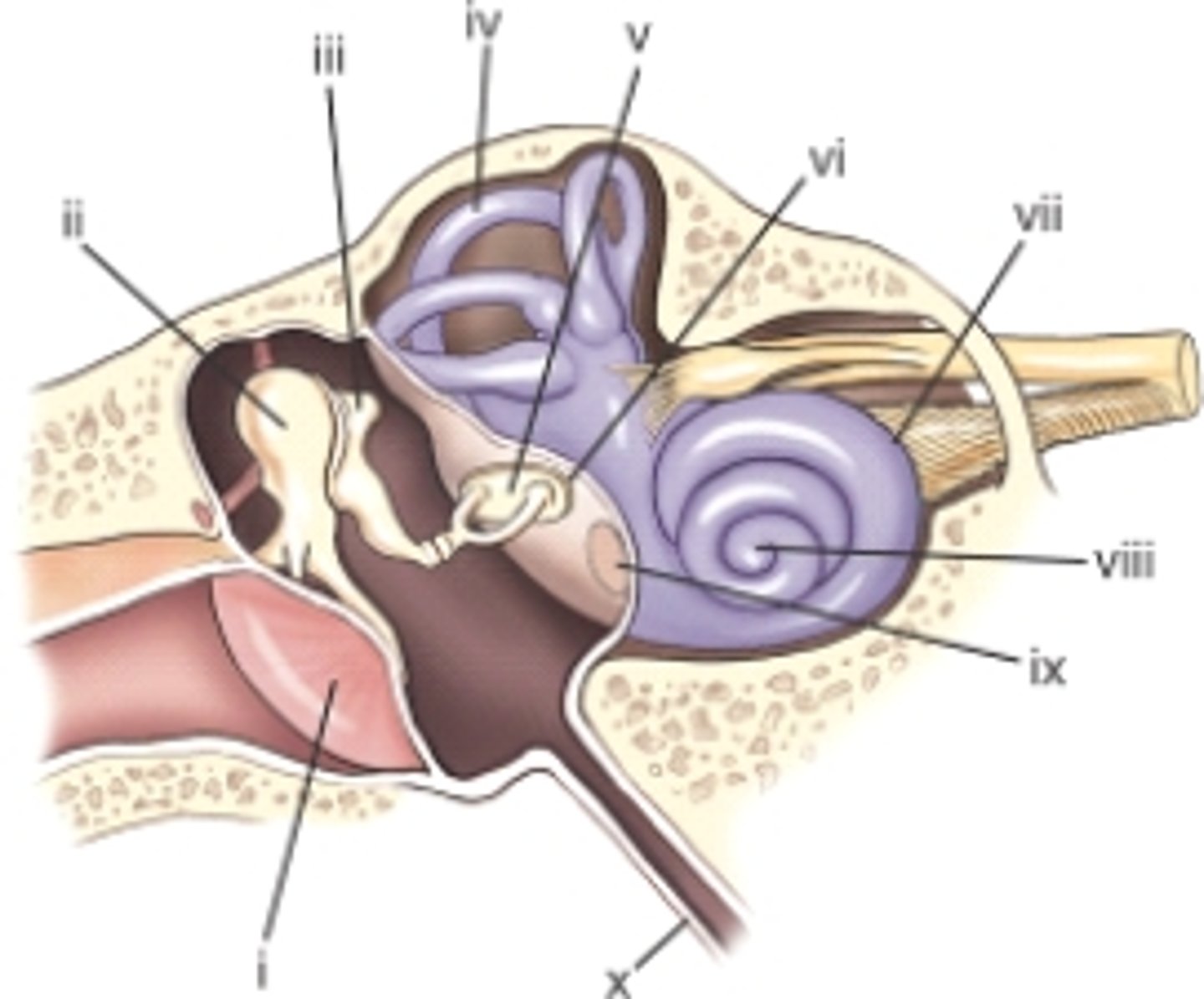
The part labeled ii is the:
A) stapes.
B) incus.
C) malleus.
D) cochlea.
C) malleus
pg. 387

The part labeled i is the:
A) external acoustic meatus.
B) tegmen tympani.
C) auditory tube.
D) tympanic membrane.
D) tympanic membrane.
pg. 387

The part labeled x is the:
A) eustachian tube.
B) internal auditory canal.
C) tympanic tube.
D) internal auditory meatus.
A) eustachian tube.
pg. 387

The mastoid air cells communicate with the:
A) inner ear.
B) middle ear.
C) external ear.
D) base of the brain.
B) middle ear.
pg. 386: A second direct communication into the middle ear occurs posteriorly to the mastoid air cells.
Which of the following structures of the inner ear is responsible for hearing?
A) Vestibule
B) Semicircular canals
C) Cochlea
D) Round window
C) Cochlea
pg. 387: the cochlea relates to the sense of hearing because of its connection to the stapes through the oval window.
Where is the CR centered for a lateral projection of the cranium?
A) EAM
B) Three-fourths inch (2 cm) anterior and 3/4 inch (2 cm) superior
C) Two inches (5 cm) superior to EAM
D) Midway between EAM and nasion
C) Two inches (5 cm) superior to EAM
pg. 414: CR is perpendicular, centered 2" superior to EAM or halfway between the glabella and the inion for other types of skull morphologies.
Which cranial bone possesses the sella turcica?
A) Temporal
B) Sphenoid
C) Ethmoid
D) Occipital
B) Sphenoid
pg. 381: the central portion of the sphenoid is the body. The central depression on the body is termed the sella turcica.
Which cranial bone possesses the superior nasal conchae?
A) Ethmoid
B) Sphenoid
C) Frontal
D) Temporal
A) Ethmoid
pg. 382: contains the superior and middle nasal conchae
Which cranial bone possesses the zygomatic process?
A) Frontal
B) Sphenoid
C) Temporal
D) Ethmoid
C) Temporal
pg. 380: extending anteriorly from the squamous portion of the temporal bone is an arch of bone termed the zygomatic process. This process meets the temporal process of the zygomatic bone (one of the facial bones) to form the zygomatic arch.
Which of the following modalities best demonstrates early signs of Paget's disease of the skull?
A) CT
B) Nuclear medicine
C) MRI
D) Sonography
B) Nuclear medicine
pg. 402: Paget disease - Nuclear medicine scans can demonstrate both regions of no (cold) and increased (hot) uptake of the radionuclide based on the stage of the disease.
A patient comes to radiology with a clinical history of an acoustic neuroma. Which of the following imaging modalities will provide the best assessment for this tumor?
A) Radiography
B) Ultrasound
C) Nuclear medicine
D) MRI
D) MRI
pg. 402: acoustic neuroma - it typically is diagnosed with the use of CT or MRI, but may be visualized on plain images in advanced cases.
A patient comes to radiology with severe mastoiditis. Which one of the following imaging modalities will best demonstrate possible bony destruction within the mastoid region?
A) CT
B) Nuclear medicine
C) Ultrasound
D) MRI
A) CT
pg. 402: mastoiditis - CT scan demonstrates a fluid-filled abscess that replaces air-filled mastoid air cells.
A patient comes in with a clinical history of a possible pituitary adenoma. Because this is a rural hospital, CT and MRI are not available. Which radiographic projection or position would best demonstrate signs of bony erosion of the sella turcica because of the tumor?
A) AP axial-Towne method
B) PA-Caldwell method
C) Lateral position
D) Both A and C
D) Both A and C
pg. 402: pituitary adenomas - investigated primarily by CT or MRI. Plain radiographic images may demonstrate enlargement of the sella turcica and erosion of the dorsum sellae (AP axial-Towne & lateral).
What is the largest immovable bone of the face?
A) Vomer
B) Mandible
C) Maxilla
D) Zygomatic
C) Maxilla
pg. 389: the two maxillae are the largest immovable bones of the face.
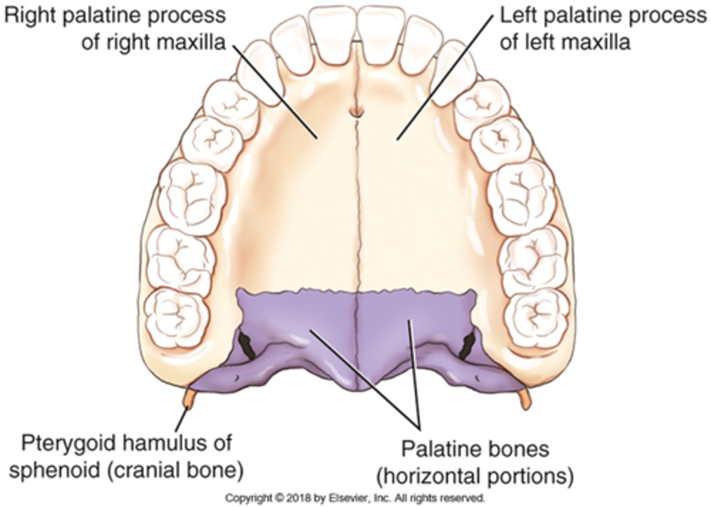
Which facial bone forms the majority of the hard palate?
A) Mandible
B) Palatine
C) Maxilla
D) Zygomatic
C) Maxilla
pg. 390: the fourth process of each maxillary bone is the palatine process, which can be demonstrated only on an inferior view of the two maxillae. The two palatine processes form the anterior portion of the roof of the mouth called the hard, or bony, palate.
Which three cranial bones articulate directly with the zygomatic bone?
A) Frontal, ethmoid, temporal
B) Frontal, sphenoid, temporal
C) Sphenoid, frontal, occipital
D) Ethmoid, parietal, frontal
B) Frontal, sphenoid, temporal
pg. 391: each zygoma articulates with three cranial bones (frontal, sphenoid, and temporal) and with one facial bone (maxilla).
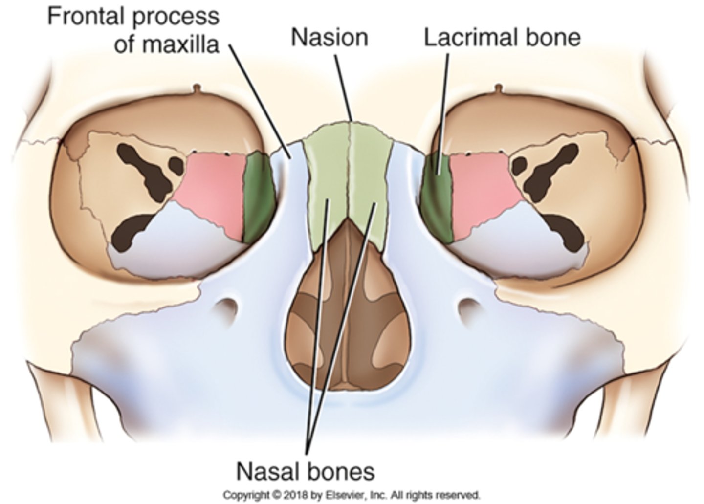
Which of the following terms describes the junction of the two nasal bones?
A) Nasion
B) Acanthion
C) Glabella
D) Supraorbital groove
A) Nasion
pg. 391: The point of junction of the two nasal bones with the frontal bone is a surface landmark called the nasion.
Which of the following structures are described as scroll-like projections found in the nasal cavity?
A) Perpendicular plate
B) Pterygoid processes
C) Septal cartilage
D) Conchae
D) Conchae
pg. 392: Within the nasal cavity are two plate-like, curved (or scroll-shaped) facial bones called the inferior nasal conchae.
There are three pairs of nasal conchae. The superior and middle pairs are parts of the ethmoid bone, and the inferior pair consists of separate facial bones.
Which two bones form the bony nasal septum?
A) Superior and inferior nasal conchae
B) Ethmoid and vomer
C) Vomer and maxilla
D) Sphenoid and ethmoid
B) Ethmoid and vomer
pg. 392: Two bones - the ethmoid and the vomer - form the bony nasal septum.
The upper and lower teeth are embedded in the:
A) symphysis menti.
B) condyloid processes.
C) palatine processes.
D) alveolar processes.
D) alveolar processes.
pg. 390: the alveolar process is the inferior aspect of the body of each maxilla. Eight upper teeth occur along the inferior margin of each alveolar process.
pg. 393: the lower teeth are rooted in the mandible. An alveolar process extends along the entire superior portion of the body of the mandible.
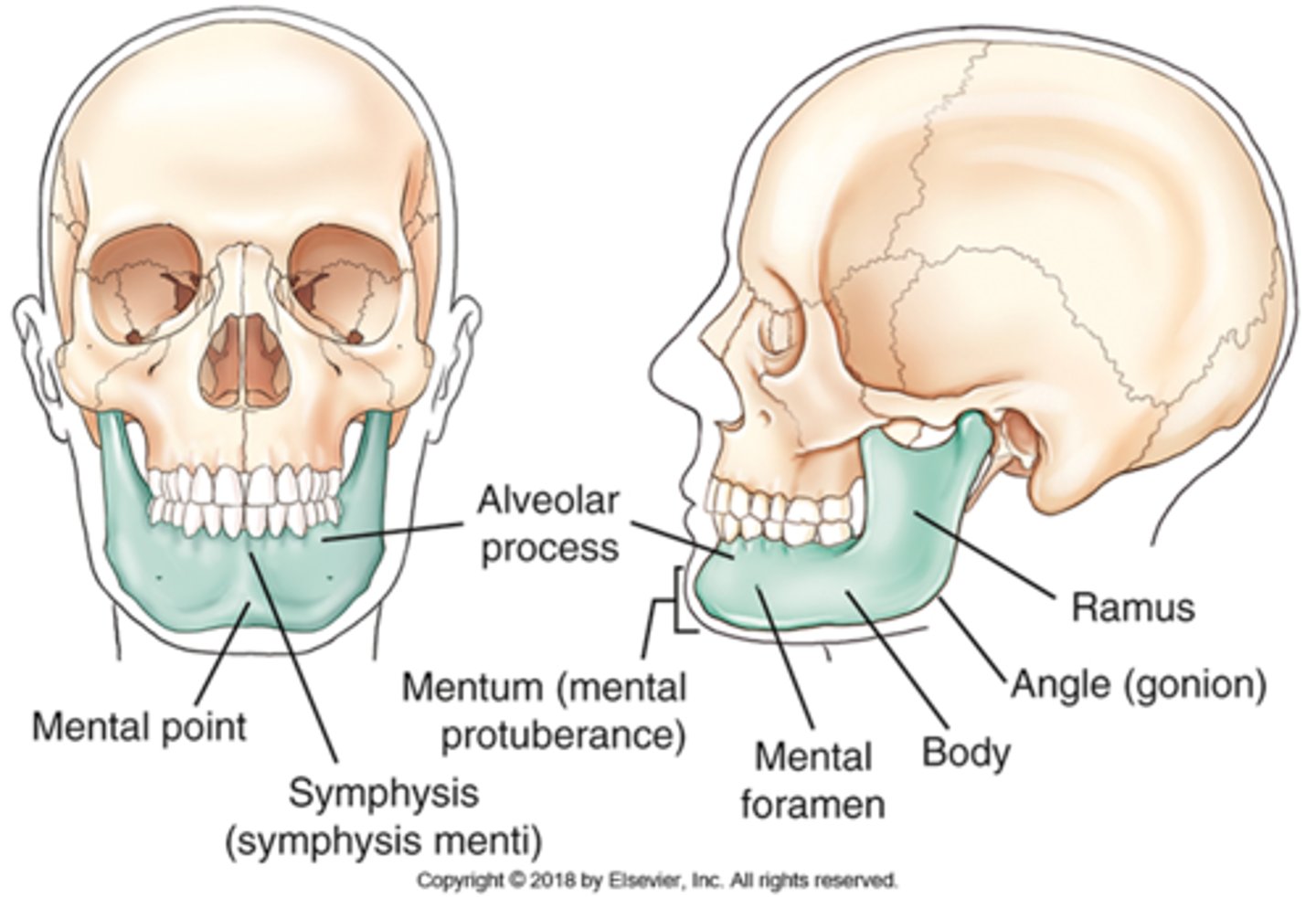
The point of union between both halves of the mandible is termed:
A) gonion.
B) ramus.
C) symphysis menti.
D) mental foramina.
C) symphysis menti.
pg. 393: the anterior aspect of the adult mandible is best seen on a frontal view. The single body forms each lateral half and unites at the anterior midline. This union is called the symphysis of the mandible (symphysis menti)
What primary type of joint movement occurs with the temporomandibular joint?
A) Plane
B) Gomphosis
C) Spheroidal
D) Bicondylar
D) Bicondylar
pg. 394: The complex TMJ is classified as a synovial type of joint ... the TMJ is classified as a bicondylar joint similar to the knee.
What is the classification of the joint found between the teeth and maxilla?
A) Synovial
B) Fibrous
C) Cartilaginous
D) Synarthrodial
B) Fibrous
pg. 394: two types of fibrous joints involve the skull, both of which are synarthrodial (immovable). First are the sutures between cranial bones. Second is a unique type of fibrous joint involving the teeth with the mandible and maxillae. This is a gomphosis subclass type of fibrous joint that is found between the roots of the teeth and the alveolar processes of both the maxillae and mandible.
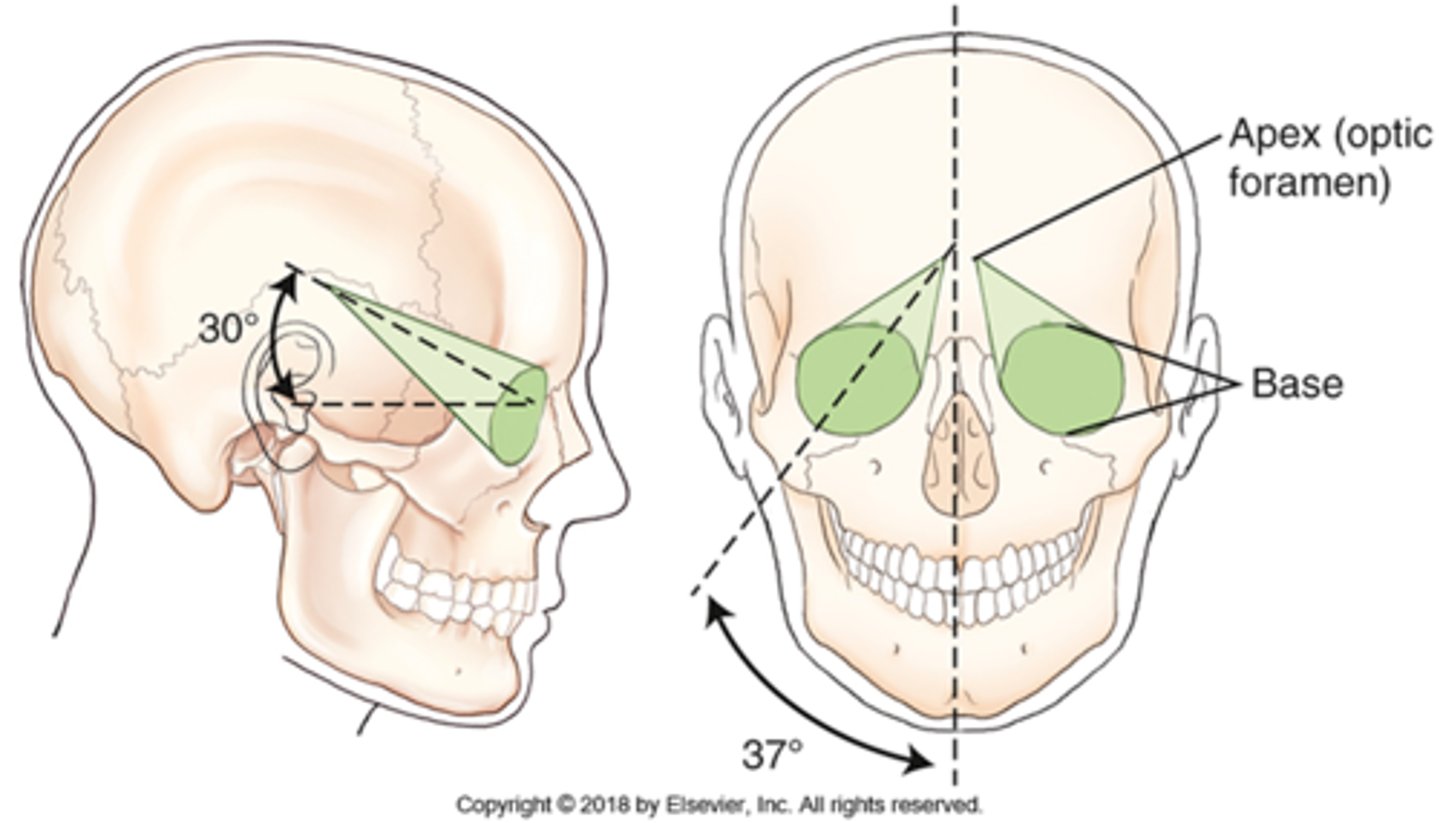
The posterior aspect of the orbit is termed the:
A) apex.
B) base.
C) sphenoid strut.
D) crown.
A) apex
pg. 398: each orbit is cone-shaped ... the rim of the orbit, which corresponds to the outer circular portion of the cone is called the base. The posterior portion of the cone, the apex, corresponds to the optic foramen, through which the optic nerve passes.
How many facial bones help make up the bony orbit?
A) Three
B) Four
C) Five
D) Seven
B) Four
pg. 398: the seven bones that make up each orbit include three cranial bones and four facial bones.
3 cranial bones: frontal, sphenoid, ethmoid
4 facial bones: maxilla, zygoma, lacrimal, palatine
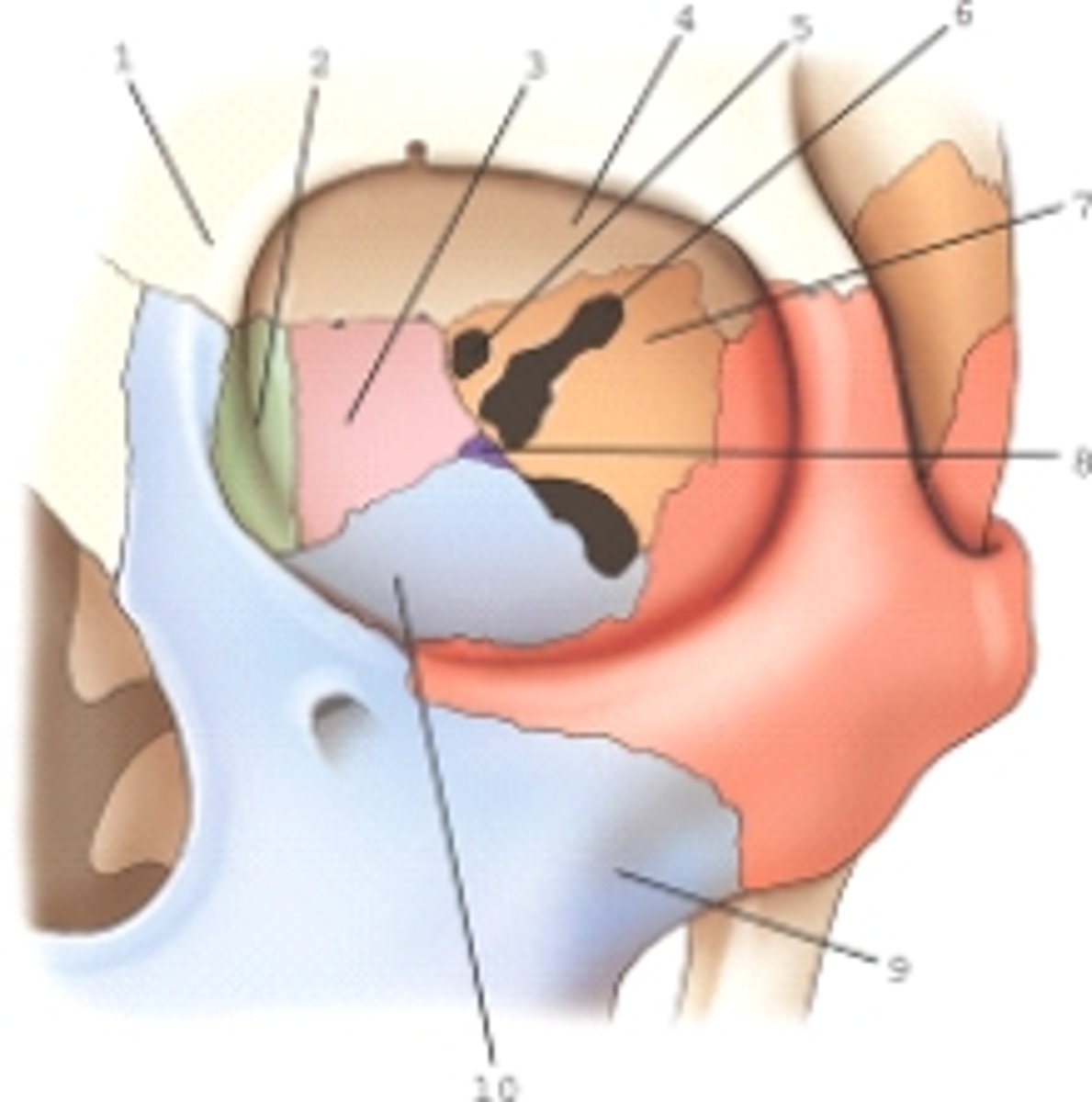
The bone labeled 2 is the:
A) lacrimal.
B) maxilla.
C) ethmoid.
D) palatine.
A) lacrimal.
pg. 398

Part 10 is part of which bone?
A) Maxilla
B) Sphenoid
C) Zygomatic
D) Ethmoid
A) Maxilla
pg. 398: orbital surface of maxilla

Part 7 is part of which bone?
A) Ethmoid
B) Lacrimal
C) Zygomatic
D) Sphenoid
D) Sphenoid
pg. 398

Part 3 is part of which bone?
A) Palatine
B) Sphenoid
C) Lacrimal
D) Ethmoid
D) Ethmoid
pg. 398

Number 6 is which of the following?
A) Inferior orbital fissure
B) Optic foramina
C) Superior orbital fissure
D) Lacrimal duct
C) Superior orbital fissure
pg. 399

Part 9 is part of which bone?
A) Zygomatic
B) Maxilla
C) Sphenoid
D) Ethmoid
B) Maxilla
pg. 398

The part labeled 8 is part of which bone?
A) Palatine
B) Sphenoid
C) Ethmoid
D) Zygomatic
A) Palatine
pg. 398-399
What is the only paranasal sinus not contained within a cranial bone?
A) Maxillary
B) Sphenoid
C) Ethmoid
D) Frontal
A) Maxillary
pg. 395: Only the maxillary sinus is part of the facial bone structure. The frontal, ethmoid, and sphenoid sinuses are contained within their respective cranial bones.
The term antrum of Highmore is an older term for the:
A) frontal sinuses.
B) ethmoid sinuses.
C) maxillary sinuses.
D) nasal cavity.
C) maxillary sinuses.
pg. An older term for maxillary sinus is antrum, an abbreviation for antrum of Highmore.
Which sinus often produces an air/fluid level indicating a basilar skull fracture?
A) Ethmoid
B) Maxillary
C) Sphenoid
D) Frontal
C) Sphenoid
pg. 396: because the sphenoid sinuses are so close to the base/floor of the cranium, sometimes pathologic processes make their presence known by their effect on these sinuses. An example is the demonstration of an air-fluid level within the sphenoid sinuses after skull trauma. This air-fluid level may provide evidence that the patient has a basal skull fracture and that either blood or CSF is leaking through the fracture into the sphenoid sinuses (sphenoid effusion)
Where are the ethmoid sinuses located within the ethmoid bone?
A) Perpendicular plate
B) Pterygoid processes
C) Cribriform plate
D) Lateral masses
D) Lateral masses
pg. 396: the ethmoid sinuses are contained within the lateral masses or labyrinths of the ethmoid bone.
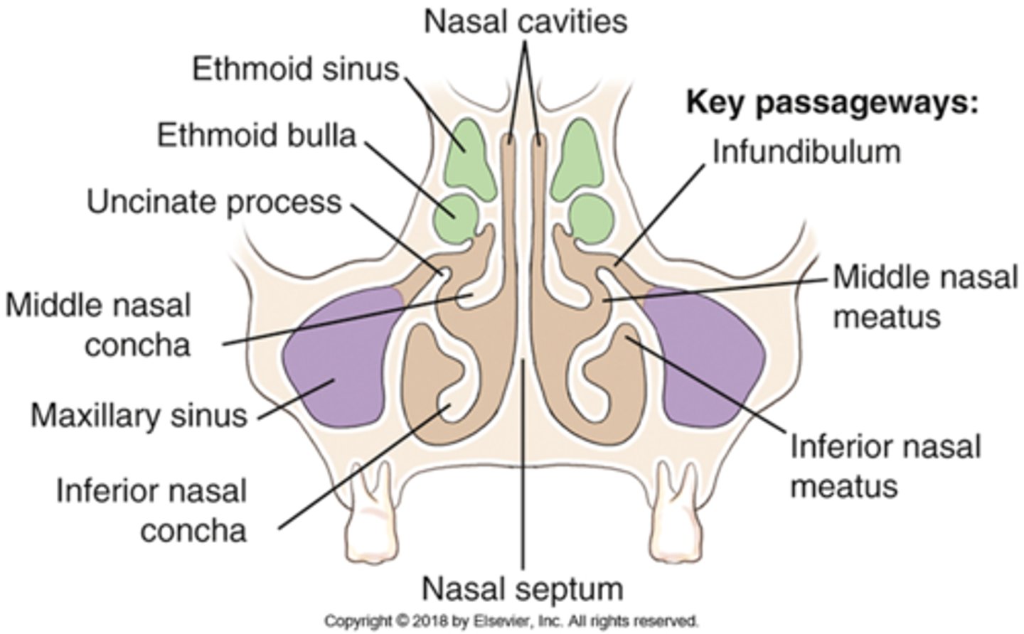
The ____ passageway drains the maxillary sinus into the middle nasal conchae.
A) ethmoid bulla
B) infundibulum
C) uncinate process
D) inferior nasal concha
B) infundibulum
pg. 396: the large maxillary sinus drains through the infundibulum passageway down through the middle nasal meatus.
The ____ sinuses develop last and are not fully developed until the teenage years.
A) ethmoid
B) sphenoid
C) nasal
D) maxillary
A) ethmoid
pg. 395: the ethmoid sinus develops last.
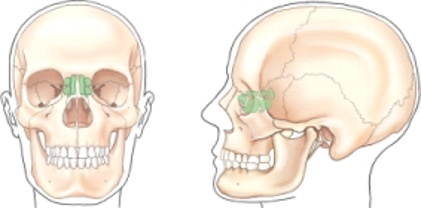
Which group of sinuses is shaded (screened) in the frontal and lateral views of the illustration below?
A) Maxillary
B) Sphenoid
C) Ethmoid
D) Frontal
C) Ethmoid
pg. 396: fig. 11.61
Which bone is involved with a tripod fracture?
A) Maxilla
B) Ethmoid
C) Temporal
D) Zygomatic
D) Zygomatic
pg. 404: a tripod fracture is caused by a blow to the cheek, resulting in fracture of the zygoma in three places (orbital process, maxillary process, and arch).
A fracture involving the facial bones where a blow to one side causes a fracture to the opposite side is termed a ____ fracture.
A) tripod
B) blow-out
C) Le Fort
D) contrecoup
D) contrecoup
pg. 404: a contrecoup fracture is a fracture to one side of a structure that is caused by an impact on the opposite side.
Which of the following imaging modalities is utilized to determine the degree of skeletal metastases especially in the cranium?
A) CT
B) MRI
C) Nuclear medicine
D) Radiography
C) Nuclear medicine
pg. 412: NM provides a senesitive screening procedure (radionuclide bone scan) for detection of skeletal metastases.
Which of the following imaging modalities should NOT be used to rule out a possible metal foreign body in the eye?
A) CT
B) MRI
C) Nuclear medicine
D) Radiography
B) MRI
pg. 404: foreign body of the eye refers to metal or other types of fragments in the eye.
The patient interview before an MRI procedure includes questions regarding history of a foreign object in the eye. Because the magnetic field causes the metal fragment to move, injury occurs due to the soft tissue.
Where is the CR centered for a lateral projection of the facial bones?
A) Outer canthus
B) Acanthion
C) Midway between the glabella and the EAM
D) Zygoma, midway between the EAM and the outer canthus
D) Zygoma, midway between the EAM and the outer canthus
pg. 419: center CR to zygoma, midway between outer canthus and EAM.
What is the angle between the OML and the plane of the IR for the parietoacanthial (Waters) projection?
A) 40°
B) 37°
C) 42°
D) 15° to 20°
B) 37°
pg. 420: adjust the head until MML line is perpendicular to the plane of the IR. OML forms a 37° angle with the table/upright imaging surface.
Where does the CR exit for a modified parietoacanthial (modified Waters) projection of the facial bones?
A) Nasion
B) Glabella
C) Acanthion
D) Midorbits
C) Acanthion
pg. 422: align CR perpendicular, centered to exit at the acanthion.